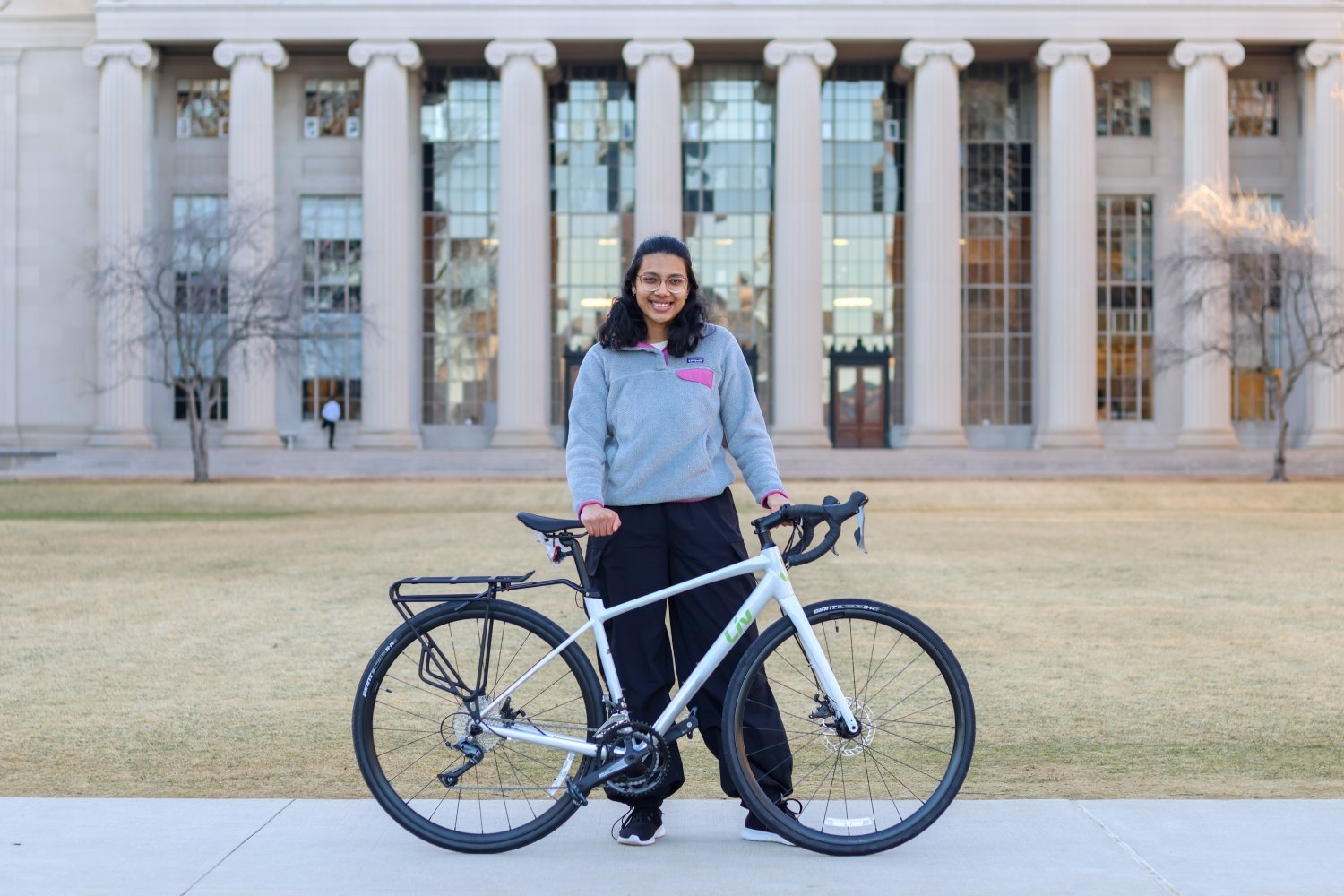
Amulya Aluru ’23, MEng ’24 and the MIT Spokes have spent the summer spreading science, over 3,000 miles on two wheels.
Lillian Eden | Department of Biology
August 22, 2024
Amulya Aluru ’23, MEng ’24, will head to the University of California at Berkeley for a PhD in molecular and cell biology PhD this fall. Aluru knows her undergraduate 6-7 major and MEng program, where she worked on a computational project in a biology lab, have prepared her for the next step of her academic journey.
“I’m a lot more comfortable with the unknown in terms of research — and also life,” she says. “While I’ve enjoyed what I’ve done so far, I think it’s equally valuable to try and explore new topics. I feel like there’s still a lot more for me to learn in biology.”
Unlike many of her peers, however, Aluru won’t reach the San Francisco Bay Area by car, plane, or train. She will arrive by bike — a journey she began in Washington just a few days after receiving her master’s degree.
Showing that science is accessible
Spokes is an MIT-based nonprofit that each year sends students on a transcontinental bike ride. Aluru worked for months with seven fellow MIT students on logistics and planning. Since setting out, the team has bonded over their love of memes and cycling-themed nicknames: Hank “Handlebar Hank” Stennes, Clelia “Climbing Cleo” Lacarriere, Varsha “Vroom Vroom Varsha” Sandadi, Rebecca “Railtrail Rebecca” Lizarde, JD “JDerailleur Hanger” Hagood, Sophia “Speedy Sophia” Wang, Amulya “Aero Amulya” Aluru, and Jessica “Joyride Jess” Xu. The support minivan, carrying food, luggage, and occasionally injured or sick cyclists, even earned its own nickname: “Chrissy”, short for Chrysler Pacifica.
“I really wanted to do something to challenge myself, but not in a strictly academic sense,” Aluru says of her decision to join the team and bike more than 3,000 miles this summer.
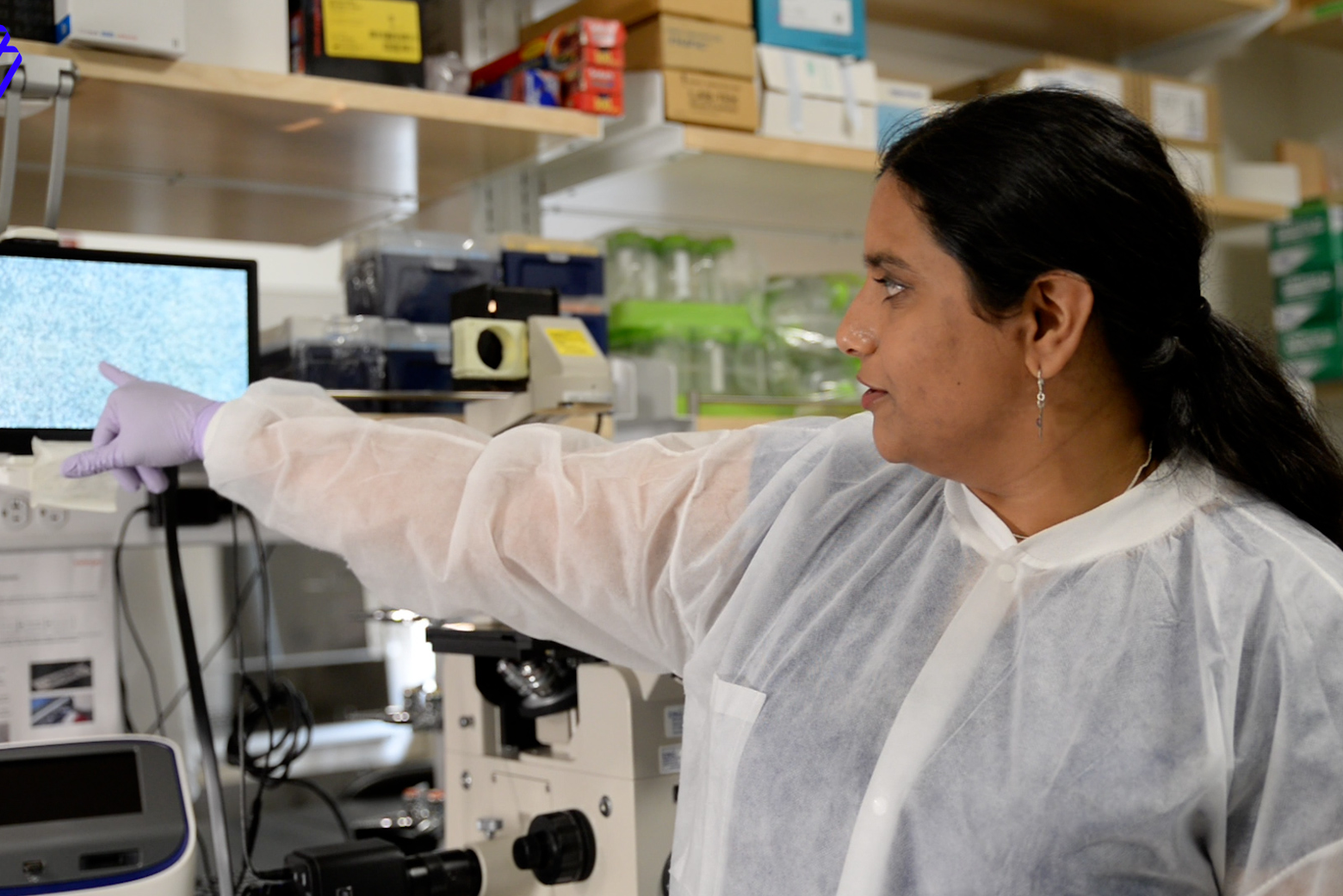
The Spokes team is not biking across the country solely to accomplish such a feat. Throughout their journey, they’ll be offering a variety of science demonstrations, including making concrete with Rice Krispies, demonstrating the physics of sound, using 3D printers, and, in Aluru’s case, extracting DNA from strawberries.
“We’re going to be in a lot of really different learning environments,” she says. “I hope to demonstrate that science can be accessible, even if you don’t have a lab at your disposal.”
These demonstrations have been held in venues such as a D.C. jail, a space camp, and libraries and youth centers across the country; their learning festivals were even featured on a local news channel in Kentucky.
Some derailments
The team was beset with challenges from the first day they started their journey. Aluru’s first day on the road involved driving to every bike shop and REI store in the D.C. metro area to purchase bike computers for navigation because the ones the team had already purchased would only display maps of Europe.
Four days in and four Chrysler Pacificas later — the first was unsafe due to bald tires, the second made a weird sound as they pulled out of the rental lot, and the third’s gas pedal stopped working over 50 miles away from the nearest rental agency — the team was back together again in Waynesboro, Virginia, for the first time since they’d set out.
Since then, they’ve had run-ins with local fauna — including mean dogs and a meaner turtle — attempted to repair a tubeless bike that was not, in fact, tubeless, and slept in Chrissy the minivan after their tents got soaked and blew away.
Although it hasn’t all been smooth riding, the team has made time for fun. They’ve perfected the art of eating a Clif bar while on two wheels, played around on monkey bars in Colorado, met up with Stanford Spokes, enjoyed pounds of ice cream, and downed gallons of lattes.
The team prioritized routes on bike trails, rather than highways, as much as possible. Their teaching activities are scheduled between visits to National Parks like Tahoe, Zion, Bryce Canyon, Arches, and touring and hiking places like Breaks Interstate Park, Mammoth Cave, and the Collegiate Peaks.
Aluru says she’s excited to see parts of the country she’s never visited before, and experience the terrain under her own power — except for breaks when it’s her turn to drive Chrissy.
Rolling with the ups and downs
Aluru was only a few weeks into her first Undergraduate Research Opportunities Program project in the late professor Angelika Amon’s lab when the Covid-19 pandemic hit, quickly transforming her wet lab project into a computational one. David Waterman, her postdoc mentor in the Amon Lab, was trained as a biologist, not a computational scientist. Luckily, Aluru had just taken two computer science classes.
“I was able to have a big hand in formulating my project and bouncing ideas off of him,” she recalls. “That helped me think about scientific questions, which I was able to apply when I came back to campus and started doing wet lab research again.”
When Aluru returned to campus, she began work in the Page Lab at the Whitehead Institute for Biomedical Research. She continued working there for the rest of her time at MIT, first as an undergraduate student and then as an MEng student.
The Page Lab’s work primarily concerns sex differences and how those differences play out in genetics, development, and disease — and the Department of Electronic Engineering and Computer Science, which oversees the MEng program, allows students to pursue computational projects across disciplines, no matter the department.
For her MEng work, Aluru looked at sex differences in human height, a continuation of a paper that the Page Lab published in 2019. Height is an easily observable human trait and, from previous research, is known to be sex-biased across at least five species. Genes that have sex-biased expression patterns, or expression patterns that are higher or lower in males compared to females, may play a role in establishing or maintaining these sex differences. Through statistical genetics, Aluru replicated the findings of the earlier paper and expanded them using newly published datasets.
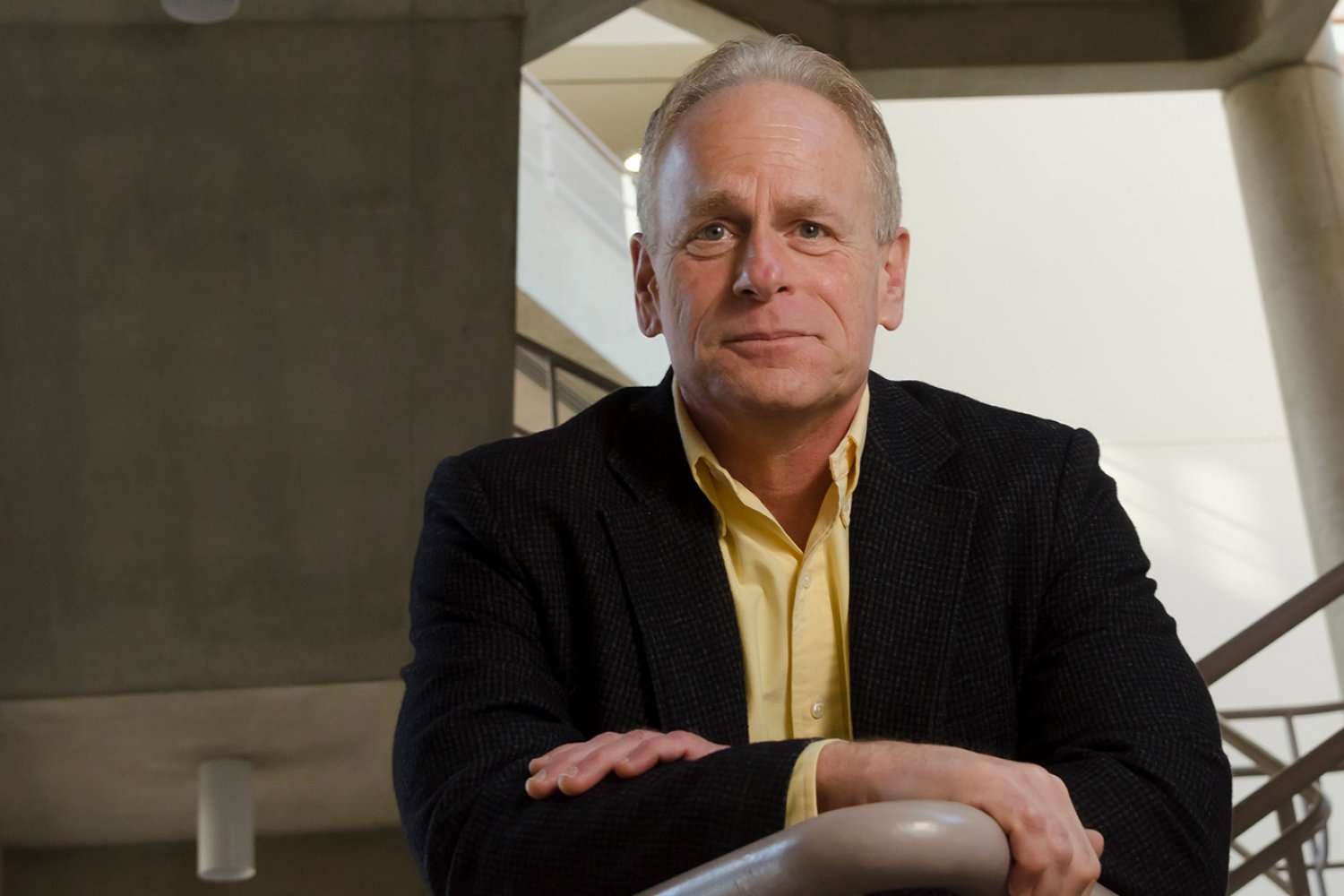
“Amulya has had an amazing journey in our department,” says David Page, professor of biology and core member of the Whitehead Institute. “There is simply no stopping her insatiable curiosity and zest for life.”
Working with the lab as a graduate student came with more day-to-day responsibility and independence than when she was an undergrad.
“It was a shift I quite appreciated,” Aluru says. “At times it was challenging, but I think it was a good challenge: learning how to structure my research on my own, while still getting a lot of support from lab members and my PI [principal investigator].”
Gearing up for the future
Since departing MIT, Aluru and the rest of the Spokes team have spent their nights camping, sleeping in churches, and staying with hosts. They enjoyed the longest day of the year in a surprisingly “Brooklyn chic” house, spent a lazy afternoon on a river, and pinky-promised to be in each other’s weddings. The team has also been hosted by, met up with, and run into MIT alums as they’ve crossed the country.
As Aluru looks to the future, she admits she’s not exactly sure what she’ll study — but when she reaches the West Coast, she knows she’s not leaving what she’s built through MIT far behind.
“There’s going to be a small MIT community even there — a lot of my friends are in San Francisco, and a few people I know are also going to be at Berkeley,” she says. “I have formed a community at MIT that I know will support me in all my future endeavors.”