Sriram “Sri” Srikant was known for insightful questions and irrepressible love of the pursuit of knowledge.
Lillian Eden | Department of Biology
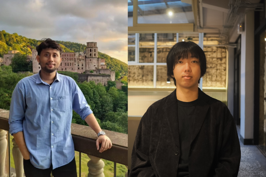
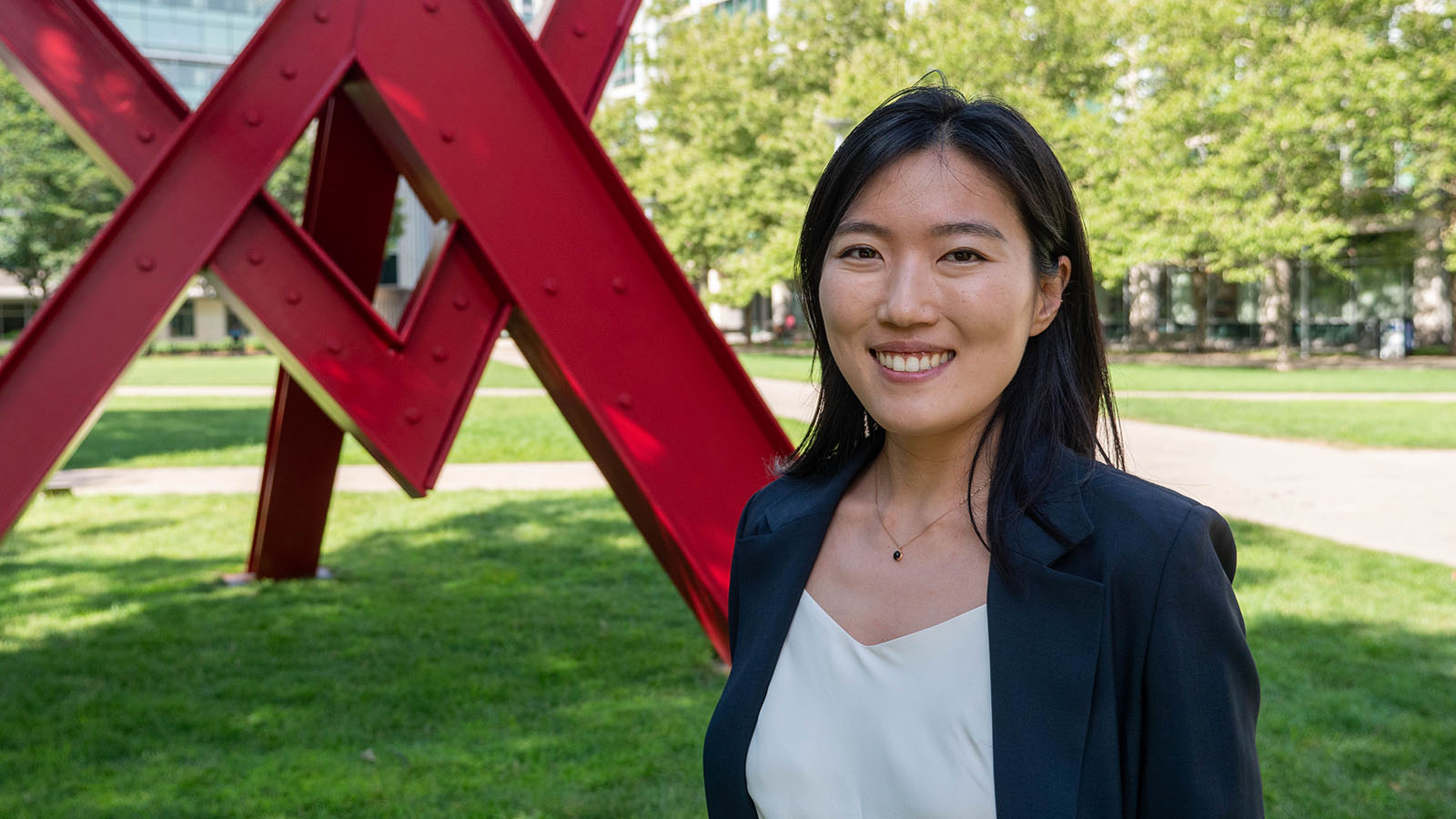
Sriram “Sri” Srikant, a postdoctoral Scholar in the Laub Lab in the Department of Biology at MIT, succumbed to cancer in March. He was 35.
Srikant received a degree in Chemical Engineering with a minor in chemistry from the Indian Institute of Technology Madras in 2011, and a PhD in Molecular and Cellular Biology from Harvard University in 2019. Among many accomplishments, Srikant was awarded an HHMI International Student Research Fellowship and a Peralta Prize for an outstanding dissertation proposal, both in 2013.
Srikant is described by mentors and colleagues alike as brilliant — a remarkable researcher who was both knowledgeable and approachable and whose enthusiasm was a bright beacon to all who had the chance to know him.
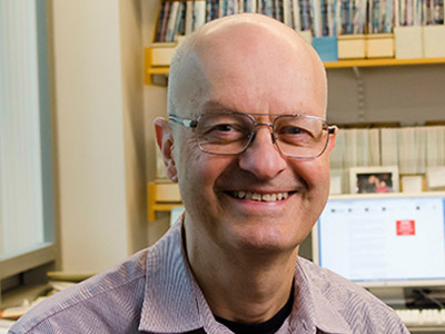
“There’s a blues line that I love, ‘Let the Midnight Special shine the ever lovin’ light on me,’” says Harvard College Professor Andrew Murray, one of Srikant’s thesis advisors. “For me, Sri was that Midnight Special, and we were lucky to have his ever lovin’ light shine on us.”
Academics are often equally motivated by a mix of a love of the work and a desire to succeed, whether it be by publications, grants, or high-impact findings. According to colleagues, however, Srikant’s passion came entirely from his need to know more.
“He told me once that ‘A life without science wouldn’t be worth living,’” said Dia Ghose, PhD ’24, a graduate student in the Laub Lab. “He wanted to move his career forward so he could keep doing science, but he didn’t care about impressing people. He just loved science and wanted to keep doing it.”
In the face of a terminal diagnosis, Srikant kept coming into the lab until his illness made it impossible. His marks on Building 68, however, remain — people are and will continue using the strains he built, the technique he developed, and the expertise he was so generous in sharing.
“There’s so many reminders of him, which is how it should be, because he contributed so much,” Ghose says. “He’s living on in the lab, and we’re still using everything that he gave us every day.”
The generosity of Sri Standard Time
As a graduate student at Harvard, Srikant pursued his thesis work in a joint PhD in the labs of both Murray and Professor of Molecular and Cellular Biology Rachelle Gaudet.
“The experiments in Rachelle’s lab failed utterly, and those in mine failed miserably, but gave enough glimmers of possibility for him to make a series of technical innovations to turn something that looked hopeless into a very nice paper,” recalls Murray. “There was no part of science he wasn’t curious about, there was nothing he wouldn’t discuss, and there was no technical challenge he wouldn’t take on.”
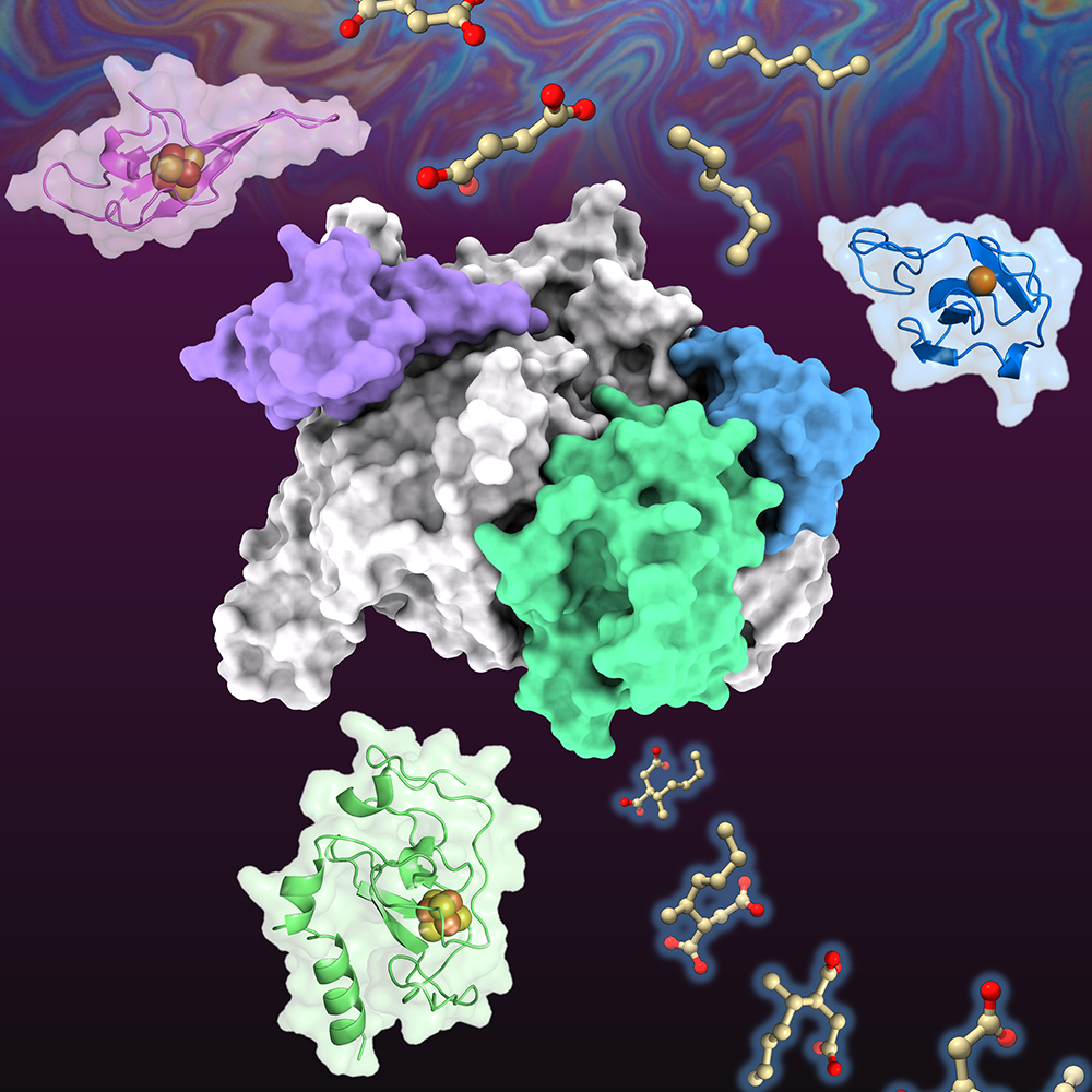
In the Laub Lab, Srikant developed an experimental evolution approach to studying phage, the viruses that infect bacteria. Srikant set up an experimental pipeline to explore how phages can evolve to overcome anti-phage defense systems in bacteria. He was also investigating the broader mechanisms of how phage genomes evolve, and the types of mutations they acquire. In the case of recombination between co-infecting phages, he was developing a new methodology to study exactly how recombination between different phages occurs.
The experimental evolution approach swept through not just the lab at MIT but across the world, and Srikant assisted other labs in implementing his process.
“He was this incredibly selfless, generous guy who was always willing to help out other people,” says Michael Laub, Salvador E. Luria Professor and HHMI Investigator. “He also had this incredible encyclopedic knowledge and memory about all aspects of phages, and he was constantly drawing on that to help people with their projects.”
Srikant was so generous with his time and expertise that he was usually on “SST” or “Sri standard time”—which was, often, running late. He would declare he was heading out or needed to start experiments, and then engage in hours-long conversations with lab mates on topics ranging from physics to visa issues.
Srikant’s hobbies included reading papers from other fields — he was, simply put, interested in the pursuit of knowledge. If he wasn’t an expert on some topic, he could spend hours studying it, just in case he could be helpful. After ChatGPT was released, lab mates joked that ChatGP-Sri was more knowledgeable, had more reliable answers, and was usually available 24/7, says Tong Zhang, PhD ’24, another graduate student in the Laub Lab.
Srikant’s sole area of ignorance was seemingly was pop culture. He didn’t know who Taylor Swift was, and only knew of Lady Gaga from the one time she wore a meat dress more than a decade ago—which, Ghose noted, was a rather niche reference.
Always curious, never quiet
Murray recalls an incident when he was flying from Boston to San Francisco with Srikant, discussing science every minute of the flight. Srikant was so passionate about the subject that his neighbor felt the need to shush him repeatedly, which Srikant took in stride, saying, with a smile, “People have been telling me to be quieter my entire life, and they’re probably right!”
From his first year at Harvard to his final days in the Laub Lab, Srikant was known for his boundless curiosity. Murray says that it’s a rare thing, after a department seminar, for students to ask questions, but Srikant would always put his hand up. That habit continued through graduate school and at science and lab meetings during his too-brief time at MIT.
“It was remarkable,” Laub says. “After any talk, he always had the most probing, incisive, and really helpful questions, across very broad fields.”
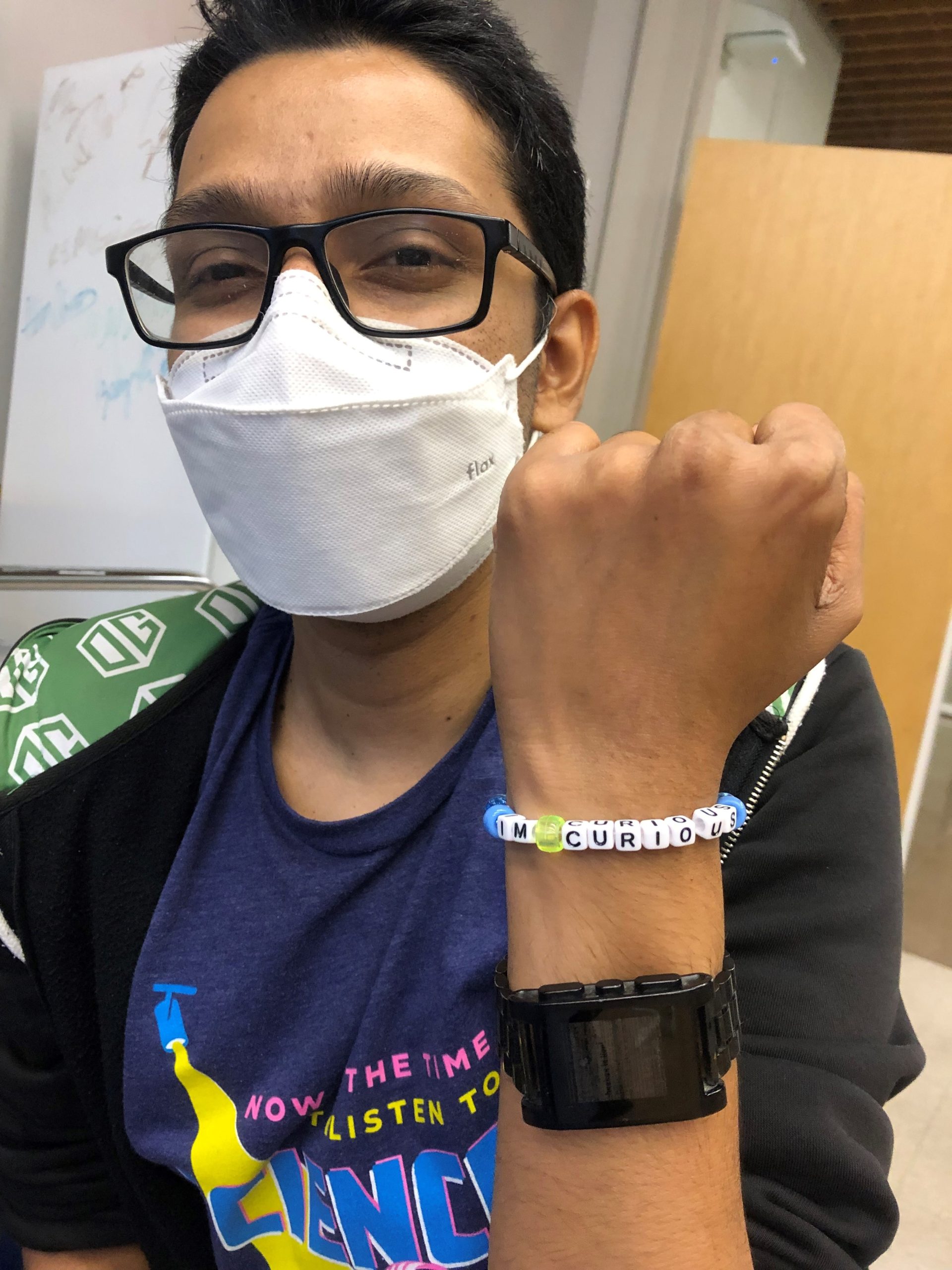
Every time he asked a question, whether it was in class during his time at Harvard or at the Building 68 research retreat on the cape, Srikant would begin with, “One of the things I’m curious about.” Ghose says the phrase became something akin to a meme in the lab, and Srikant even commemorated the colloquialism with a bracelet that read ‘I’m curious.’
“For a person that brilliant and knowledgeable, Sri was so special. His impact on me and others will last forever,” Zhang says. “I have always been, and I will continue, looking up to him, honoring his passion for science, his brilliance as a scientist, and his kindness and generosity as a great friend.”