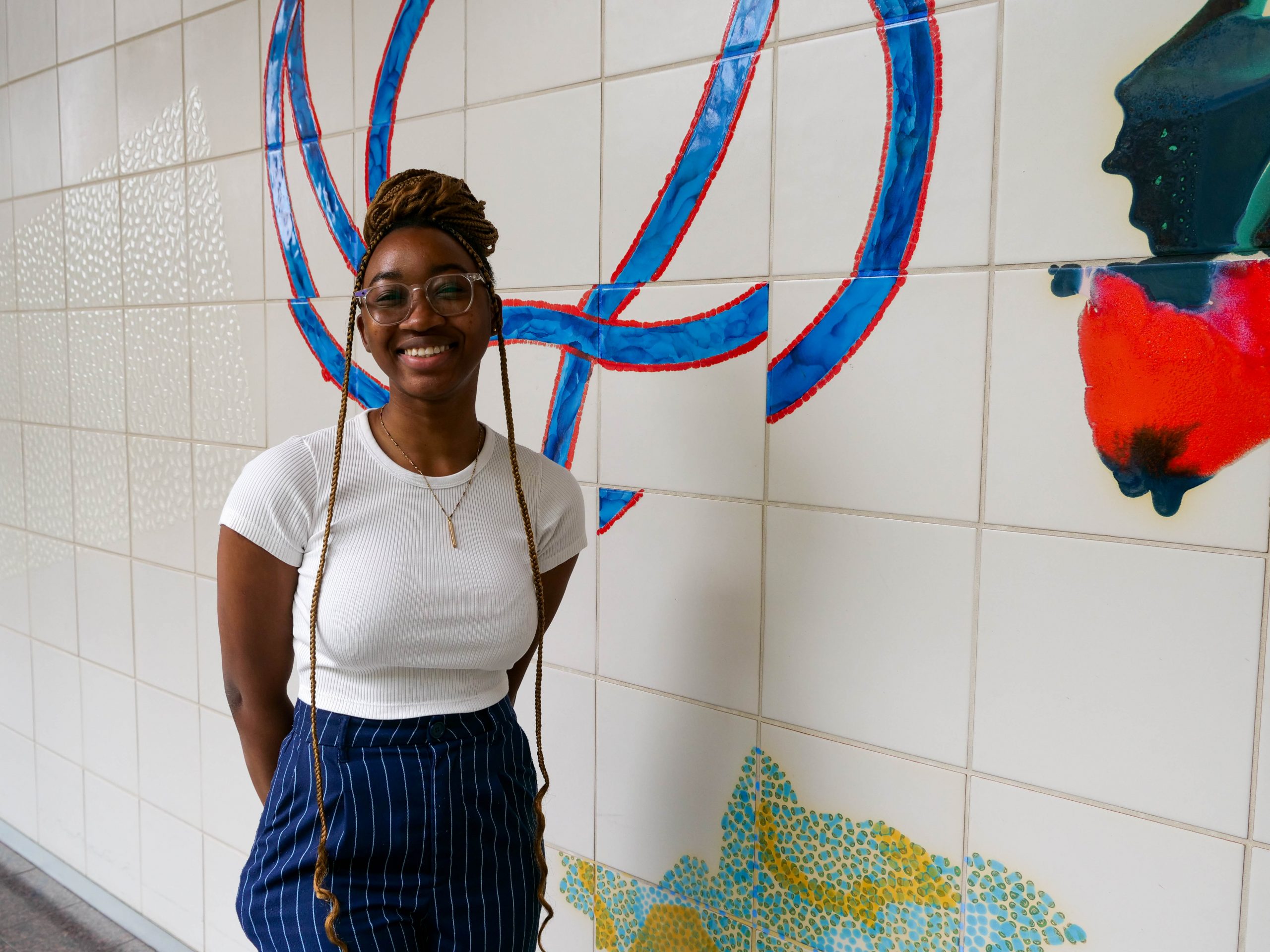
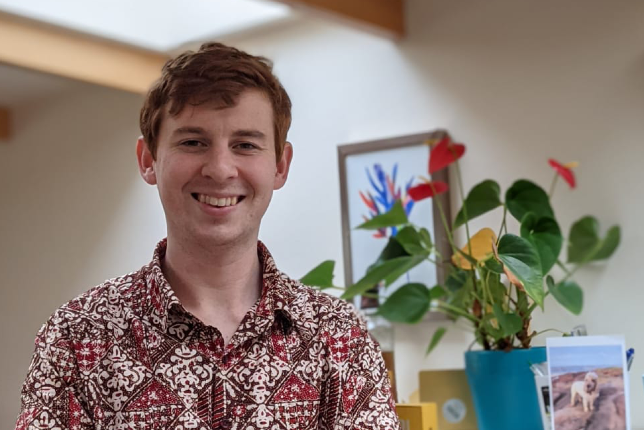
Cyrille Teforlack first stumbled across the work of MIT Professor of Biology Peter Reddien on YouTube while Teforlack was taking a cell biology class at Bethune-Cookman University, where he is now a rising senior. Teforlack became fascinated by Reddien’s work on regeneration in planarians, freshwater flatworms.
“That was how I figured out what kind of science I was interested in. I remember watching the video over and over,” Teforlack recalls. “I was like, ‘I have to figure out where this guy works.'”
The answer was, of course, at the Whitehead Institute, where Reddien is a Core Member and Associate Director. Teforlack spent the summer working in Reddien’s lab as part of the Bernard S. and Sophie G. Gould MIT Summer Research Program in Biology (BSG-MSRP-Bio). The program offers students the opportunity to work on cutting-edge research that isn’t available at their home institutions.
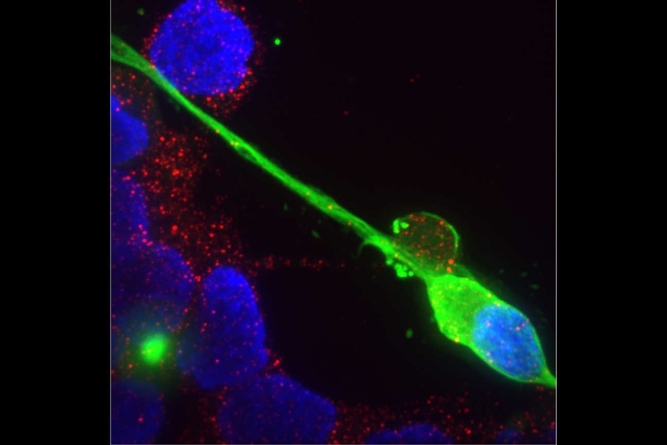
During the summer, Teforlack was working on eye regeneration and how different proteins and secreted factors affect the planarian’s cartoon-like crossed eyes. To understand the underlying requirements for regeneration, Teforlack used a technique called RNA interface (RNAi) to silence genes and see how it affected the planarian’s morphology when they regenerated.
The phenotypes Teforlack saw—he had a list of more than 100 candidate genes to work with—ranged from barely noticeable to strikingly defective. He studied regeneration by slicing off the head of the worm to see how the head regenerated and removing the eyes from the intact head to see how just that organ regrew.
“The eye has some connections to the brain and the rest of the body,” Teforlack explains. “But when you cut off their head, there’s no brain—but they can still regrow everything. It’s a no-brainer.”
However, some gene changes proved fatal or didn’t result in regeneration—the flatworms died and melted away. Teforlack didn’t realize that at first, however. There was one instance early in the program when he went to feed some worms—and was shocked to find they were missing.
“It was a really scary day for me in the lab,” Teforlack jokes. “I lost 12 worms, somehow. But I remember feeding them yesterday. How do you lose worms?”
Other phenotypes were more successful—showing atypical structures in the regenerated head or eyes. The optic cup of the eye regenerated in the wrong shape, for example, or separated in the middle as it regenerated.
Teforlack has been surprised by how much is still unknown about planarian regeneration. For example, a particular gene is expressed when the worm needs to regenerate wounded tissue on the half of the body facing the head, as opposed to the tail; it is unclear, however, how the planarian body detects which side the wound is on. .
“Even though it’s basic science, it’s still so intricate, and there’s so many little things that can build up to culminate in a bigger question,” Teforlack says.
Teforlack became known as “the worm guy” among his fellow students.
“Having a cool organism to talk about makes talking about it more exciting for myself, and for everyone else that’s listening,” he says. “This is something I never thought I would be a part of. It feels really great to be at a cutting-edge place doing really cool research.”
In addition to hands-on lab work, MSRP-Bio students often meet to discuss their work and do activities together. Teforlack says the program created plenty of opportunities to find community in his cohort, from arts and crafts to dodgeball.
The program also offers professional development activities like presentations from faculty including with the undergraduate and graduate officers Adam Martin and Mary Gehring to answer questions about applying for graduate school. Teforlack says he also found that faculty, despite their busy schedules, are always willing to take time out of their day for a chat.
“It’s been cool to meet all these different people and see the diversity of science that goes on and how many of them collaborate together on a variety of different projects,” Teforlack says. “This experience has helped me think like a scientist and value my own opinions. Being in an environment where your ideas are accepted, and you can learn from these scientists, has been really exciting.”
Teforlack worked closely with HHMI staff scientist Lucila Simone and graduate student Bryanna Canales. Canales herself participated in the program. As an MSRP-Bio student, Canales worked on metastasis in zebrafish in the lab of Daniel K. Ludwig Professor for Cancer Research, and Koch Institute Intramural Faculty Richard O. Hynes.
Canales says explaining her work to her peers during her time in MSRP-Bio was invaluable because it was more like teaching—she had to explain things in simple terms and found she was not the only one who sometimes struggled to do that.
“The program made me feel more comfortable talking to people that I could learn from,” Canales recalls. “The MSRP-Bio experience humanized the institution and the people here. Everyone here is really smart, but going through the process with the students in my cohort, it feels like less of a big deal if you don’t know something.”
Canales says she’s seen Teforlack’s confidence grow this summer, taking the initiative and staying one step ahead instead of asking what he should be doing.
“It’s been nice to know that I can do science by myself—more or less—and still feel accomplished and know that everything I’m doing is the correct step, and if it’s not, I know how to troubleshoot things,” Teforlack says.
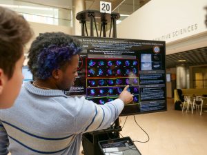
Teforlack’s work culminated in a poster session in August for MSRP-Bio students, where he showed some of the defective phenotypes he was characterizing and a short movie of the planarians eating the RNAi delivery system: liver.
“Cyrille showed a captivating movie of the small worms eating liver laced with double-stranded RNA that can downregulate specific genes,” says MIT Biology Department Head Amy Keating. “He also had beautiful images of the resulting phenotypes, which included disrupted optic cup structure. I always learn something new at the MSRP poster session!”
Long term, Teforlack plans to pursue a PhD in stem cell biology, and he says the program has reinforced that desire.
“It’s been cool to be around so many scientists,” Teforlack says. “Ten weeks isn’t enough time for anyone to learn anything perfectly. I’m excited to grow as a researcher.”
Although the MSRP-Bio program has come to a close for 2023, Teforlack’s time isn’t done in Reddien’s lab: he will return to continue his work in 2024.
“The MSRP program is a great opportunity for students to directly immerse in research here at MIT and to learn new concepts and methods,” Reddien says. “Cyrille was a terrific student and contributed a lot over the summer. I look forward to seeing his next steps with research into regeneration.”
Cancer drugs known as checkpoint blockade inhibitors have proven effective for some cancer patients. These drugs work by taking the brakes off the body’s T cell response, stimulating those immune cells to destroy tumors.
Some studies have shown that these drugs work better in patients whose tumors have a very large number of mutated proteins, which scientists believe is because those proteins offer plentiful targets for T cells to attack. However, for at least 50 percent of patients whose tumors show a high mutational burden, checkpoint blockade inhibitors don’t work at all.
A new study from MIT reveals a possible explanation for why that is. In a study of mice, the researchers found that measuring the diversity of mutations within a tumor generated much more accurate predictions of whether the treatment would succeed than measuring the overall number of mutations.
If validated in clinical trials, this information could help doctors to better determine which patients will benefit from checkpoint blockade inhibitors.
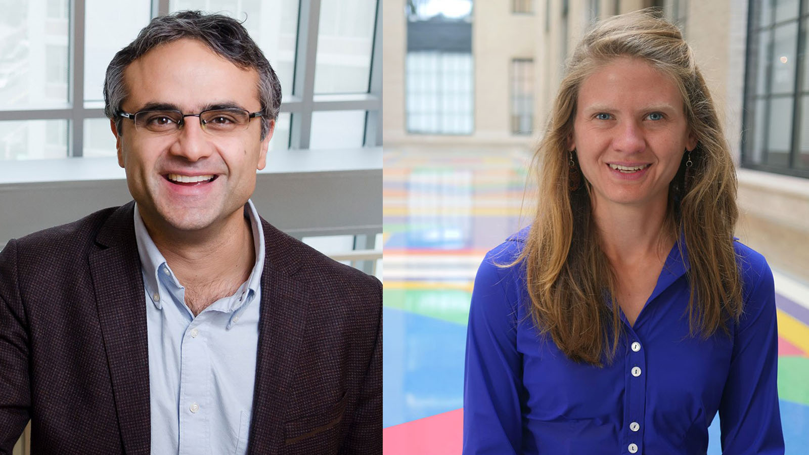
“While very powerful in the right settings, immune checkpoint therapies are not effective for all cancer patients. This work makes clear the role of genetic heterogeneity in cancer in determining the effectiveness of these treatments,” says Tyler Jacks, the David H. Koch Professor of Biology and a member of MIT’s Koch Institute for Cancer Research.
Jacks; Peter Westcott, a former MIT postdoc in the Jacks lab who is now an assistant professor at Cold Spring Harbor Laboratory; and Isidro Cortes-Ciriano, a research group leader at EMBL’s European Bioinformatics Institute (EMBL-EBI), are the senior authors of the paper, which appears today in Nature Genetics.
A diversity of mutations
Across all types of cancer, a small percentage of tumors have what is called a high tumor mutational burden (TMB), meaning they have a very large number of mutations in each cell. A subset of these tumors has defects related to DNA repair, most commonly in a repair system known as DNA mismatch repair.
Because these tumors have so many mutated proteins, they are believed to be good candidates for immunotherapy treatment, as they offer a plethora of potential targets for T cells to attack. Over the past few years, the FDA has approved a checkpoint blockade inhibitor called pembrolizumab, which activates T cells by blocking a protein called PD-1, to treat several types of tumors that have a high TMB.
However, subsequent studies of patients who received this drug found that more than half of them did not respond well or only showed short-lived responses, even though their tumors had a high mutational burden. The MIT team set out to explore why some patients respond better than others, by designing mouse models that closely mimic the progression of tumors with high TMB.
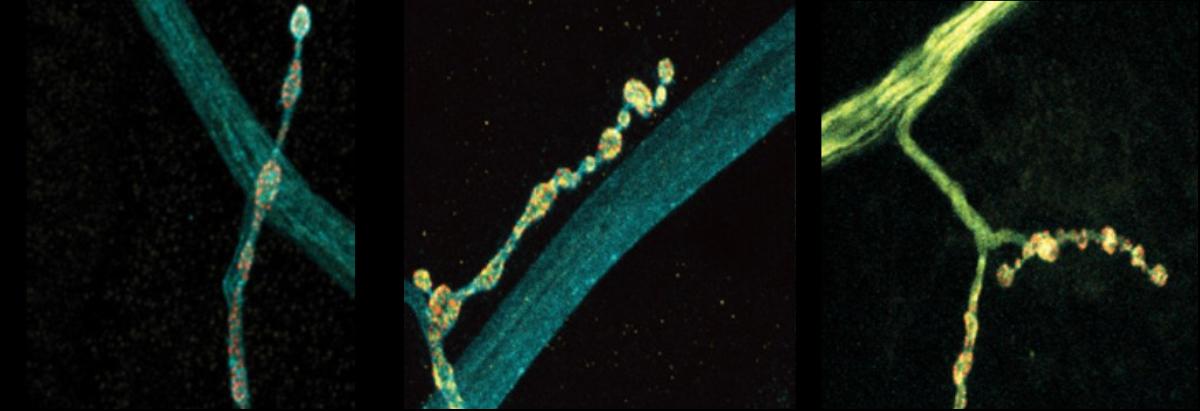
These mouse models carry mutations in genes that drive cancer development in the colon and lung, as well as a mutation that shuts down the DNA mismatch repair system in these tumors as they begin to develop. This causes the tumors to generate many additional mutations. When the researchers treated these mice with checkpoint blockade inhibitors, they were surprised to find that none of them responded well to the treatment.
“We verified that we were very efficiently inactivating the DNA repair pathway, resulting in lots of mutations. The tumors looked just like they look in human cancers, but they were not more infiltrated by T cells, and they were not responding to immunotherapy,” Westcott says.
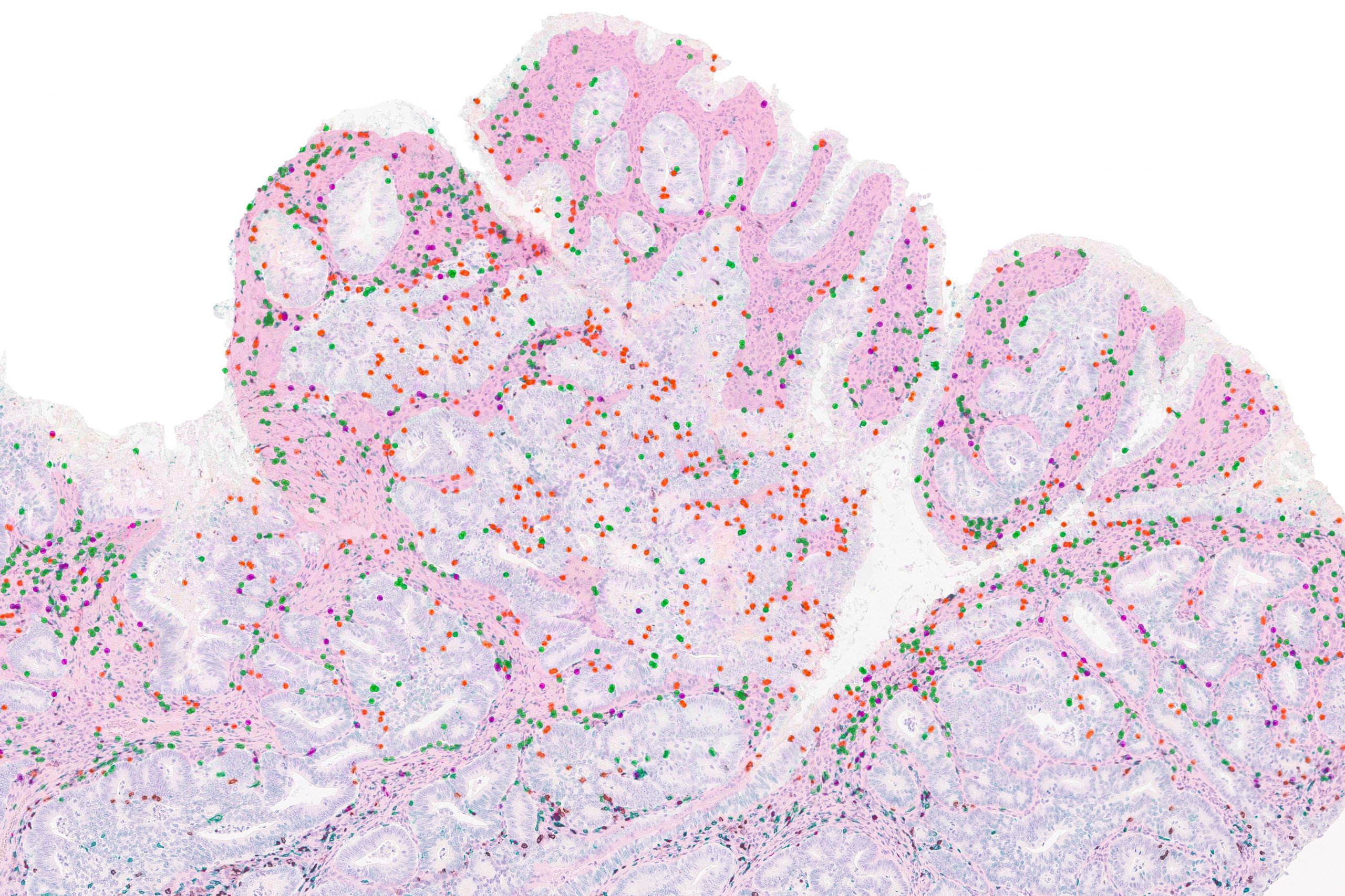
The researchers discovered that this lack of response appears to be the result of a phenomenon known as intratumoral heterogeneity. This means that, while the tumors have many mutations, each cell in the tumor tends to have different mutations than most of the other cells. As a result, each individual cancer mutation is “subclonal,” meaning that it is expressed in a minority of cells. (A “clonal” mutation is one that is expressed in all of the cells.)
In further experiments, the researchers explored what happened as they changed the heterogeneity of lung tumors in mice. They found that in tumors with clonal mutations, checkpoint blockade inhibitors were very effective. However, as they increased the heterogeneity by mixing tumor cells with different mutations, they found that the treatment became less effective.
“That shows us that intratumoral heterogeneity is actually confounding the immune response, and you really only get the strong immune checkpoint blockade responses when you have a clonal tumor,” Westcott says.
Failure to activate
It appears that this weak T cell response occurs because the T cells simply don’t see enough of any particular cancerous protein, or antigen, to become activated, the researchers say. When the researchers implanted mice with tumors that contained subclonal levels of proteins that normally induce a strong immune response, the T cells failed to become powerful enough to attack the tumor.
“You can have these potently immunogenic tumor cells that otherwise should lead to a profound T cell response, but at this low clonal fraction, they completely go stealth, and the immune system fails to recognize them,” Westcott says. “There’s not enough of the antigen that the T cells recognize, so they’re insufficiently primed and don’t acquire the ability to kill tumor cells.”
To see if these findings might extend to human patients, the researchers analyzed data from two small clinical trials of people who had been treated with checkpoint blockade inhibitors for either colorectal or stomach cancer. After analyzing the sequences of the patients’ tumors, they found that patients’ whose tumors were more homogeneous responded better to the treatment.
“Our understanding of cancer is improving all the time, and this translates into better patient outcomes,” Cortes-Ciriano says. “Survival rates following a cancer diagnosis have significantly improved in the past 20 years, thanks to advanced research and clinical studies. We know that each patient’s cancer is different and will require a tailored approach. Personalized medicine must take into account new research that is helping us understand why cancer treatments work for some patients but not all.”
The findings also suggest that treating patients with drugs that block the DNA mismatch repair pathway, in hopes of generating more mutations that T cells could target, may not help and could be harmful, the researchers say. One such drug is now in clinical trials.
“If you try to mutate an existing cancer, where you already have many cancer cells at the primary site and others that may have disseminated throughout the body, you’re going to create a super heterogeneous collection of cancer genomes. And what we showed is that with this high intratumoral heterogeneity, the T cell response is confused and there is absolutely no response to immune checkpoint therapy,” Westcott says.
The research was funded by the Koch Institute Support (core) Grant from the U.S. National Cancer Institute, the Howard Hughes Medical Institute, and a Damon Runyon Fellowship Award.
For 45 undergraduate students from institutions across the US, the summer was filled with days in the lab experiencing cutting-edge scientific research.
The Bernard S. and Sophie G. Gould MIT Summer research program in Biology (BSG-MSRP Bio) provides undergraduates the chance to work full-time in labs and see behind the curtain of the science—and life—they could have if they decided to pursue graduate studies in scientific research.
The program is offered in collaboration with MIT’s Department of Brain & Cognitive Sciences (BCS) and the Center for Brains, Minds, and Machines (CBMM), with students working in labs affiliated with the Biology Department, BCS, CBMM, the Department of Chemistry, and the Whitehead Institute.
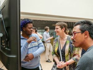
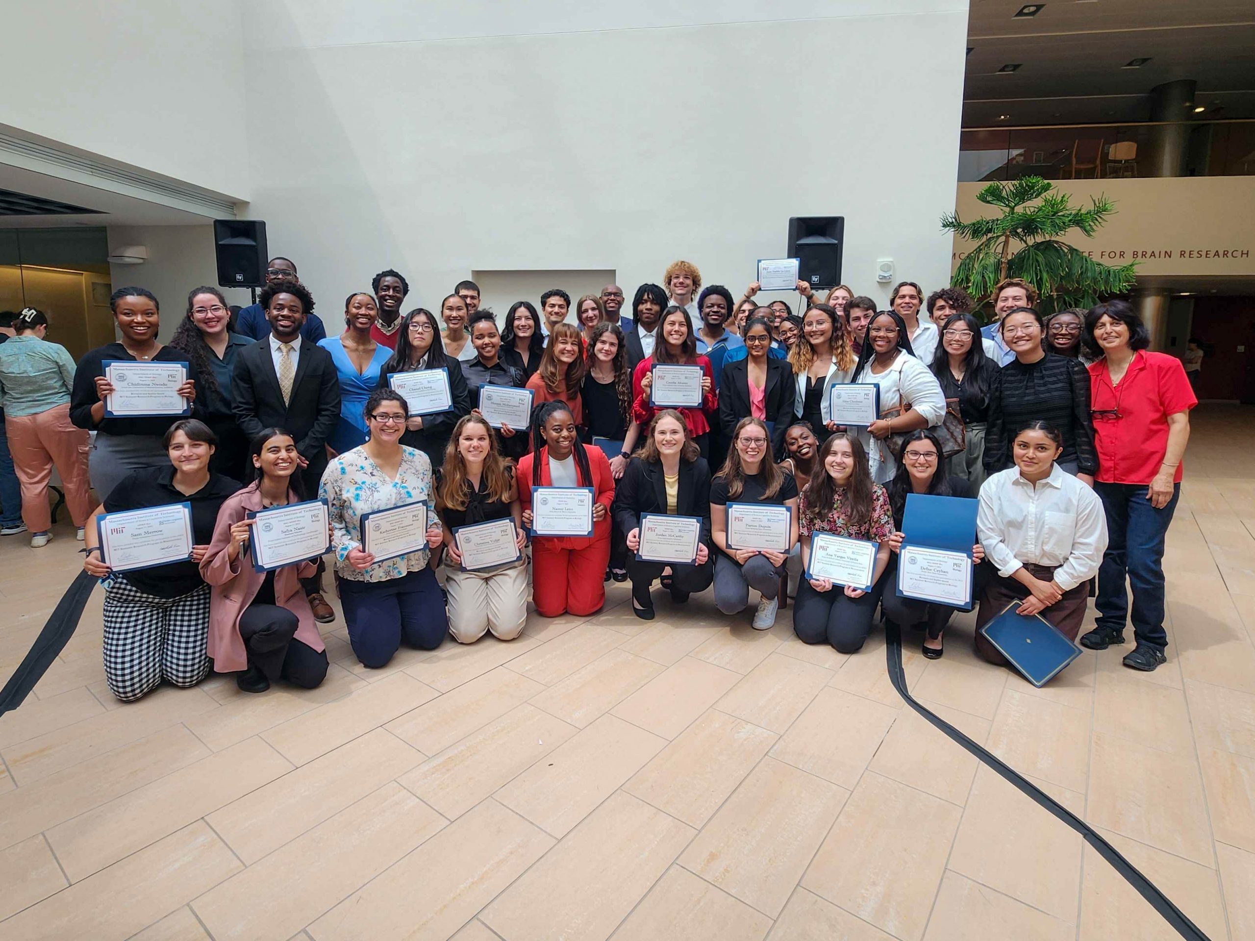
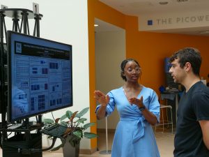
The students’ work culminated in a bustling poster session at the beginning of August, hosted by the Picower Institute for Learning and Memory.
“As always, the poster session was a lively and inspiring event, as students presented results that they sometimes obtained in a rush just days before,” says MIT Biology Department Head Amy Keating. “It was a challenge to see all the posters I wanted to, with people standing several rows deep around the speakers, eager to learn about the new science.”
The purpose of the MSRP-Bio program is to provide an intensive research experience to students who do not have access to cutting-edge research facilities at their home institutions– and to introduce them to MIT. The program offers professional development by bringing in faculty as guest speakers throughout the ten weeks of the program and provides resources like campus housing, stipends, mentoring, and trips around Massachusetts.
Since its inception, MSRP-Bio has left a mark both on the students and on the MIT community. For example, Associate Professor of Biology Joey Davis’ connection to the MSRP-Bio program threads through his career as a researcher.
The MSRP-Bio program launched in 2003, two years before Davis began as a graduate student at MIT. Some students in his graduate student cohort—some who are now his colleagues, including Associate Professor of Biology Eliezer Calo and Assistant Professor of Biology Francisco Sanchez-Rivera were among the initial participants of the MSRP-Bio program. Davis’ first graduate student attended the program, and so did three of his current graduate students. Two students who worked in his lab last summer as part of the program are also returning to pursue PhDs in the Department of Biology this fall.
“The MSRP students get to hang out with current graduate students and get a sense of the people that you can form lifelong relationships with,” Davis says. “What does it look like to be a scientist? What would my peers be like? It’s a pretty unique opportunity.”
Davis credits Senior Lecturer Mandana Sassanfar, who spearheads many outreach activities, for her expansive work identifying students who would thrive at MIT and giving them research experiences that aren’t available at their home institutions.
“It’s hard to identify folks that haven’t had these opportunities before, even though they are so, so capable,” Davis says. “It’s incredible what Mandana has been able to do.”
Since the program began, hundreds of students have participated. The majority went on to enroll in PhD or MD/PhD programs at MIT and other highly-ranked graduate programs nationwide. Almost two dozen are now faculty at various institutions across the U.S.
For some attendees, the poster session was a blast from the past. Chidera Okeke said her time in the lab of Professor of Biology Adam Martin as an MSRP-Bio student and Gould Fellow was what convinced her to apply to the PhD program. Okeke, a Fisk University alumna, is now a second-year graduate student at MIT in the lab of Class of 1922 Career Development Professor and Whitehead Institute Core Member Olivia Corradin.
“It was the only program I saw that really mirrored grad school and the day-to-day of what it would look like as a PhD student—lab meetings and extracurricular activities,” she says. “I also found out a lot about the application process. People were very transparent about it, and everyone was just genuinely nice.”
She noted, however, that 2021—the year she was a summer student—was the last year of physical posters. Students, since then, have displayed their posters on large TV screens with iPads that allow them to navigate their posters, and the posters include time reminders for when a session is drawing to a close.
“I honestly prefer the e-poster because I could zoom in,” says MSRP-Bio student Fareeda Abu-Juam, a College of Wooster undergraduate and novice computational biologist who worked in Davis’ lab this summer. She squeezed extra images onto her poster and used the zooming capabilities to better display them for people asking questions.
Because the attendees at the poster session come from different research backgrounds, Abu-Juam says it was an excellent opportunity to answer questions she hadn’t considered before.
“It’s great that so many people came out to support undergraduates. It’s nice to be in a place where they’re supporting us like that,” said Christina “CJ” Volpe, a student in the lab of Howard S. and Linda B. Stern Career Development Professor and Intramural Faculty at the Koch Institute Stefani Spranger. “I’ve never been at an institution where they’re doing cutting-edge research. Investigating something that has never been done before. It was an amazing experience. I don’t know how I’m going to go back to my home institution now.”
Cyrille Teforlack, an undergraduate from Bethune Cookman University who has been working on flatworm eye regeneration in the lab of Professor of Biology and Core Member and Associate Director of the Whitehead Institute Peter Reddien, says he’s had plenty of practice for the poster session. Between discussing their research informally with fellow 2023 MSRP-Bio students and presenting in lab meetings throughout the summer, MSRP-Bio students have many opportunities to build confidence in discussing their work.
“I’ve gotten really good feedback from people,” he says. “The questions they asked made me think about different questions to think about for my own project.”
Some students also had the opportunity to see what the work was like for newer faculty still in the process of setting up their labs—a helpful thing for those considering academia. MSRP-Bio student Nina Greeley spent the summer in new Whitehead Institute Fellow Lindsey Backman’s lab. Backman, who opened her lab at the Whitehead Institute to study the proteins of anaerobic bacteria in the human microbiome just last year, also participated in the MSRP-Bio program.
As for what advice Greeley would give to students doing poster sessions for the first time, Greeley had this to say: “People want to know why your work is relevant. Keep it simple. Explain what you did, the result, and how you think the lab will go in future directions.”