How a unique class of neurons may set the table for brain development

A new MIT study from the Nedivi Lab finds that somatostatin-expressing neurons follow a unique trajectory when forming connections in the brain’s visual cortex that may help establish the conditions needed for sensory experience to refine circuits.

David Orenstein | The Picower Institute for Learning and Memory
January 14, 2026

The way the brain develops can shape us throughout our lives, so neuroscientists are intensely curious about how it happens. A new study by researchers in The Picower Institute for Learning and Memory at MIT that focused on visual cortex development in mice, reveals that an important class of neurons follows a set of rules that while surprising, might just create the right conditions for circuit optimization.

During early brain development, multiple types of neurons emerge in the visual cortex (where the brain processes vision). Many are “excitatory,” driving the activity of brain circuits, and others are “inhibitory,” meaning they control that activity. Just like a car needs not only an engine and a gas pedal, but also a steering wheel and brakes, a healthy balance between excitation and inhibition is required for proper brain function. During a “critical period” of development in the visual cortex, soon after the eyes first open, excitatory and inhibitory neurons forge and edit millions of connections, or synapses, to adapt nascent circuits to the incoming flood of visual experience. Over many days, in other words, the brain optimizes its attunement to the world.

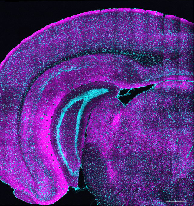
In the new study in The Journal of Neuroscience, a team led by MIT research scientist Josiah Boivin and Professor Elly Nedivi visually tracked somatostatin (SST)-expressing inhibitory neurons forging synapses with excitatory cells along their sprawling dendrite branches, illustrating the action before, during and after the critical period with unprecedented resolution. Several of the rules the SST cells appeared to follow were unexpected—for instance, unlike other cell types, their activity did not depend on visual input—but now that the scientists know these neurons’ unique trajectory, they have a new idea about how it may enable sensory activity to influence development: SST cells might help usher in the critical period by establishing the baseline level of inhibition needed to ensure that only certain types of sensory input will trigger circuit refinement.

“Why would you need part of the circuit that’s not really sensitive to experience? It could be that it’s setting things up for the experience-dependent components to do their thing,” said Nedivi, William R. and Linda R. Young Professor in The Picower Institute and MIT’s Departments of Biology and Brain and Cognitive Sciences.

Boivin added: “We don’t yet know whether SST neurons play a causal role in the opening of the critical period, but they are certainly in the right place at the right time to sculpt cortical circuitry at a crucial developmental stage.”

A unique trajectory

To visualize SST-to-excitatory synapse development, Nedivi and Boivin’s team used a genetic technique that pairs expression of synaptic proteins with fluorescent molecules to resolve the appearance of the “boutons” SST cells use to reach out to excitatory neurons. They then performed a technique called eMAP, developed by Kwanghun Chung’s lab in the Picower Institute, that expands and clears brain tissue to increase magnification, allowing super-resolution visualization of the actual synapses those boutons ultimately formed with excitatory cells along their dendrites. Co-author and postdoc Bettina Schmerl helped lead the eMAP work.

These new techniques revealed that SST bouton appearance and then synapse formation surged dramatically when the eyes opened and then as the critical period got underway. But while excitatory neurons during this timeframe are still maturing, first in the deepest layers of the cortex and later in its more superficial layers, the SST boutons blanketed all layers simultaneously, meaning that, perhaps counter intuitively, they sought to establish their inhibitory influence regardless of the maturation stage of their intended partners.

Many studies have shown that eye opening and the onset of visual experience sets in motion the development and elaboration of excitatory cells and another major inhibitory neuron type (parvalbumin-expressing cells). Raising mice in the dark for different lengths of time, for instance, can distinctly alter what happens with these cells. Not so for the SST neurons. The new study showed that varying lengths of darkness had no effect on the trajectory of SST bouton and synapse appearance; it remained invariant, suggesting it is pre-ordained by a genetic program or an age-related molecular signal, rather than experience.

Moreover, after the initial frenzy of synapse formation during development, many synapses are then edited, or pruned away, so that only the ones needed for appropriate sensory responses endure. Again, the SST boutons and synapses proved to be exempt from these redactions. Though the pace of new SST synapse formation slowed at the peak of the critical period, the net number of synapses never declined and even continued increasing into adulthood.

“While a lot of people think that the only difference between inhibition and excitation is their valence, this demonstrates that inhibition works by a totally different set of rules,” Nedivi said.

In all, while other cell types were tailoring their synaptic populations to incoming experience, the SST neurons appeared to provide an early but steady inhibitory influence across all layers of the cortex. After excitatory synapses have been pruned back by the time of adulthood, the continued upward trickle of SST inhibition may contribute to the increase in the inhibition to excitation ratio that still allows the adult brain to learn, but not as dramatically or as flexibly as during early childhood.

A platform for future studies

In addition to shedding light on typical brain development, Nedivi said, the study’s techniques can enable side-by-side comparisons in mouse models of neurodevelopmental disorders such as autism or epilepsy where aberrations of excitation and inhibition balance are implicated.

Future studies using the techniques can also look at how different cell types connect with each other in brain regions other than the visual cortex, she added.

Boivin, who will soon open his own lab as a faculty member at Amherst College, said he is eager to apply the work in new ways.

“I’m excited to continue investigating inhibitory synapse formation on genetically defined cell types in my future lab,” Boivin said. “I plan to focus on the development of limbic brain regions that regulate behaviors relevant to adolescent mental health.”

In addition to Nedivi, Boivin and Schmerl, the paper’s other authors are Kendyll Martin, and Chia-Fang Lee.

Funding for the study came from the National Institutes of Health, the Office of Naval Research and the Freedom Together Foundation.

New chemical method makes it easier to select desirable traits in crops

Whitehead Institute Member Mary Gehring and colleagues offer a new method for generating large-scale genetic changes without irradiation.

Mackenzie White | Whitehead Institute
January 8, 2026

Crops increasingly need to thrive in a broader range of conditions, including drought, salinity, and heat. Traditional plant breeding can select for desirable traits, but is limited by the genetic variation that already exists in plants. In many crops, domestication and long-term selection have narrowed genetic diversity, constraining efforts to develop new varieties.

To work around these limits, researchers have developed ways to introduce helpful traits, such as drought or salt tolerance, into plants through mutation breeding. This deliberately introduces random genetic changes into plants. Then researchers screen the genetically altered plants to see which have acquired useful traits. One widely used approach relies on radiation to generate structural variants—large-scale DNA changes that can affect multiple genes at once. However, irradiation introduces logistical and regulatory hurdles that restrict who can use it and which crops can be studied.

In a paper published in PLOS Genetics on December 18, Whitehead Institute Member Mary Gehring and colleagues offer a new method for generating large-scale genetic changes without irradiation.

Lead author Lindsey Bechen, the Gehring lab manager; Gehring; former postdoc P.R.V. Satyaki (now a faculty member at the University of Toronto); and their colleagues developed the approach by exposing germinating seeds to etoposide, a chemotherapy drug, during early growth.

The drug interferes with an enzyme that helps manage DNA structure during cell division. When cells attempt to repair the resulting breaks in their DNA, errors in the repair process can produce large-scale rearrangements in the genome. Seeds collected from treated plants carry these changes in a heritable form.

The process relies on standard laboratory tools: seeds are germinated on growth medium containing the drug, then transferred to soil to complete their life cycle.

“I was surprised at how efficient it was,” says Gehring, who is also a professor of biology at MIT and an HHMI Investigator. “The diversity of new traits that you could see just by looking at the plants in the first generation was extensive.”

The researchers demonstrated the method in Arabidopsis thaliana, a model plant widely used in genetic studies. Roughly two-thirds of treated plant lines showed visible differences, including changes in leaf shape, plant size, pigmentation, and fertility. Genetic analyses linked these traits to deletions, duplications, and rearrangements of DNA segments.

In several cases, the team linked specific plant traits to individual genetic changes. A dwarf plant with thick stems and unusual leaves carried a large change that disrupted a gene involved in leaf development. Another plant, marked by green-and-white mottled leaves, carried a deletion in the gene IMMUTANS—the same gene identified in radiation-induced mutants described more than 60 years ago.

Beyond Arabidopsis, Gehring’s lab is applying the technique to pigeon pea, a drought-tolerant legume and an important source of dietary protein in parts of Asia and Africa. Pigeon pea is an underutilized crop with the potential to become a staple crop—if its lack of genetic diversity, caused by a historical cultivation bottleneck, can be overcome. Often referred to as orphan crops, species like pigeon pea receive limited research attention and often lack the genetic variation needed for breeding improved varieties.

“All of the traits that we might want to see in pigeon pea are not present in the existing population,” says Gehring. “The idea is to do a large-scale mutation experiment to increase genetic diversity.”

The team, which includes Gehring lab postdoc Sonia Boor, is now screening treated pigeon pea lines for salt tolerance, a trait that shapes where crops can be grown and how they perform in saline soils. Although pigeon pea takes longer to grow than Arabidopsis, the researchers have reached the second generation and identified several lines that show promising responses under saline conditions.

The researchers’ chemical approach may also be beneficial for crops that are difficult to modify using gene-editing tools such as CRISPR. Although CRISPR enables precise genetic changes, it often relies on genetic transformation, a technically challenging step for many plant species.

“A lot of species that one works with, either in agriculture or horticulture, are not amenable to genetic transformation,” says Gehring.

The new method complements existing genetic tools rather than replacing them. By providing a more accessible alternative to irradiation, chemical mutation could expand the availability of large-scale genetic changes and novel plant varieties.

Looking ahead, Gehring’s lab plans to develop comprehensive collections of Arabidopsis mutants carrying well-characterized structural variants. Such resources could help researchers better understand how large-scale changes in genome structure influence plant development and performance, informing future efforts to study and enhance crops.

Bechen, L. L., Ahsan, N., Bahrainwala, A., Gehring, M., & Satyaki, P. R. (2025). A simple method to efficiently generate structural variation in plants. PLOS Genetics21(12). https://doi.org/10.1371/journal.pgen.1011977
High-fat diets make liver cells more likely to become cancerous

New research from the Yilmaz Lab suggests liver cells exposed to too much fat revert to an immature state that is more susceptible to cancer-causing mutations.

Anne Trafton | MIT News
December 22, 2025

One of the biggest risk factors for developing liver cancer is a high-fat diet. A new study from MIT reveals how a fatty diet rewires liver cells and makes them more prone to becoming cancerous.

The researchers found that in response to a high-fat diet, mature hepatocytes in the liver revert to an immature, stem-cell-like state. This helps them to survive the stressful conditions created by the high-fat diet, but in the long term, it makes them more likely to become cancerous.

“If cells are forced to deal with a stressor, such as a high-fat diet, over and over again, they will do things that will help them survive, but at the risk of increased susceptibility to tumorigenesis,” says Alex K. Shalek, director of the Institute for Medical Engineering and Sciences (IMES), the J. W. Kieckhefer Professor in IMES and the Department of Chemistry, and a member of the Koch Institute for Integrative Cancer Research at MIT, the Ragon Institute of MGH, MIT, and Harvard, and the Broad Institute of MIT and Harvard.

The researchers also identified several transcription factors that appear to control this reversion, which they believe could make good targets for drugs to help prevent tumor development in high-risk patients.

Shalek; Ömer Yilmaz, an MIT associate professor of biology and a member of the Koch Institute; and Wolfram Goessling, co-director of the Harvard-MIT Program in Health Sciences and Technology, are the senior authors of the study, which appears today in Cell. MIT graduate student Constantine Tzouanas, former MIT postdoc Jessica Shay, and Massachusetts General Brigham postdoc Marc Sherman are the co-first authors of the paper.

Cell reversion

A high-fat diet can lead to inflammation and buildup of fat in the liver, a condition known as steatotic liver disease. This disease, which can also be caused by a wide variety of long-term metabolic stresses such as high alcohol consumption, may lead to liver cirrhosis, liver failure, and eventually cancer.

In the new study, the researchers wanted to figure out just what happens in cells of the liver when exposed to a high-fat diet — in particular, which genes get turned on or off as the liver responds to this long-term stress.

To do that, the researchers fed mice a high-fat diet and performed single-cell RNA-sequencing of their liver cells at key timepoints as liver disease progressed. This allowed them to monitor gene expression changes that occurred as the mice advanced through liver inflammation, to tissue scarring and eventually cancer.

In the early stages of this progression, the researchers found that the high-fat diet prompted hepatocytes, the most abundant cell type in the liver, to turn on genes that help them survive the stressful environment. These include genes that make them more resistant to apoptosis and more likely to proliferate.

At the same time, those cells began to turn off some of the genes that are critical for normal hepatocyte function, including metabolic enzymes and secreted proteins.

“This really looks like a trade-off, prioritizing what’s good for the individual cell to stay alive in a stressful environment, at the expense of what the collective tissue should be doing,” Tzouanas says.

Some of these changes happened right away, while others, including a decline in metabolic enzyme production, shifted more gradually over a longer period. Nearly all of the mice on a high-fat diet ended up developing liver cancer by the end of the study.

When cells are in a more immature state, it appears that they are more likely to become cancerous if a mutation occurs later on, the researchers say.

“These cells have already turned on the same genes that they’re going to need to become cancerous. They’ve already shifted away from the mature identity that would otherwise drag down their ability to proliferate,” Tzouanas says. “Once a cell picks up the wrong mutation, then it’s really off to the races and they’ve already gotten a head start on some of those hallmarks of cancer.”

The researchers also identified several genes that appear to orchestrate the changes that revert hepatocytes to an immature state. While this study was going on, a drug targeting one of these genes (thyroid hormone receptor) was approved to treat a severe form of steatotic liver disease called MASH fibrosis. And, a drug activating an enzyme that they identified (HMGCS2) is now in clinical trials to treat steatotic liver disease.

Another possible target that the new study revealed is a transcription factor called SOX4, which is normally only active during fetal development and in a small number of adult tissues (but not the liver).

Cancer progression

After the researchers identified these changes in mice, they sought to discover if something similar might be happening in human patients with liver disease. To do that, they analyzed data from liver tissue samples removed from patients at different stages of the disease. They also looked at tissue from people who had liver disease but had not yet developed cancer.

Those studies revealed a similar pattern to what the researchers had seen in mice: The expression of genes needed for normal liver function decreased over time, while genes associated with immature states went up. Additionally, the researchers found that they could accurately predict patients’ survival outcomes based on an analysis of their gene expression patterns.

“Patients who had higher expression of these pro-cell-survival genes that are turned on with high-fat diet survived for less time after tumors developed,” Tzouanas says. “And if a patient has lower expression of genes that support the functions that the liver normally performs, they also survive for less time.”

While the mice in this study developed cancer within a year or so, the researchers estimate that in humans, the process likely extends over a longer span, possibly around 20 years. That will vary between individuals depending on their diet and other risk factors such as alcohol consumption or viral infections, which can also promote liver cells’ reversion to an immature state.

The researchers now plan to investigate whether any of the changes that occur in response to a high-fat diet can be reversed by going back to a normal diet, or by taking weight-loss drugs such as GLP-1 agonists. They also hope to study whether any of the transcription factors they identified could make good targets for drugs that could help prevent diseased liver tissue from becoming cancerous.

“We now have all these new molecular targets and a better understanding of what is underlying the biology, which could give us new angles to improve outcomes for patients,” Shalek says.

The research was funded, in part, by a Fannie and John Hertz Foundation Fellowship, a National Science Foundation Graduate Research Fellowship, the National Institutes of Health, and the MIT Stem Cell Initiative through Foundation MIT.

3 Questions with new faculty member Yunha Hwang: Using computation to study the world’s best single-celled chemists

The assistant professor utilizes microbial genomes to examine the language of biology. Her appointment reflects MIT’s commitment to exploring the intersection of genetics research and AI.

Lillian Eden | Department of Biology
December 15, 2025

Today, out of an estimated 1 trillion species on Earth, 99.999 percent are considered microbial — bacteria, archaea, viruses, and single-celled eukaryotes. For much of our planet’s history, microbes ruled the Earth, able to live and thrive in the most extreme of environments. Researchers have only just begun in the last few decades to contend with the diversity of microbes — it’s estimated that less than 1 percent of known genes have laboratory-validated functions. Computational approaches offer researchers the opportunity to strategically parse this truly astounding amount of information.

An environmental microbiologist and computer scientist by training, new MIT faculty member Yunha Hwang is interested in the novel biology revealed by the most diverse and prolific life form on Earth. In a shared faculty position as the Samuel A. Goldblith Career Development Professor in the Department of Biology, as well as an assistant professor at the Department of Electrical Engineering and Computer Science and the MIT Schwarzman College of Computing, Hwang is exploring the intersection of computation and biology.  

Q: What drew you to research microbes in extreme environments, and what are the challenges in studying them?

A: Extreme environments are great places to look for interesting biology. I wanted to be an astronaut growing up, and the closest thing to astrobiology is examining extreme environments on Earth. And the only thing that lives in those extreme environments are microbes. During a sampling expedition that I took part in off the coast of Mexico, we discovered a colorful microbial mat about 2 kilometers underwater that flourished because the bacteria breathed sulfur instead of oxygen — but none of the microbes I was hoping to study would grow in the lab.

The biggest challenge in studying microbes is that a majority of them cannot be cultivated, which means that the only way to study their biology is through a method called metagenomics. My latest work is genomic language modeling. We’re hoping to develop a computational system so we can probe the organism as much as possible “in silico,” just using sequence data. A genomic language model is technically a large language model, except the language is DNA as opposed to human language. It’s trained in a similar way, just in biological language as opposed to English or French. If our objective is to learn the language of biology, we should leverage the diversity of microbial genomes. Even though we have a lot of data, and even as more samples become available, we’ve just scratched the surface of microbial diversity.

Q: Given how diverse microbes are and how little we understand about them, how can studying microbes in silico, using genomic language modeling, advance our understanding of the microbial genome?

A: A genome is many millions of letters. A human cannot possibly look at that and make sense of it. We can program a machine, though, to segment data into pieces that are useful. That’s sort of how bioinformatics works with a single genome. But if you’re looking at a gram of soil, which can contain thousands of unique genomes, that’s just too much data to work with — a human and a computer together are necessary in order to grapple with that data.

During my PhD and master’s degree, we were only just discovering new genomes and new lineages that were so different from anything that had been characterized or grown in the lab. These were things that we just called “microbial dark matter.” When there are a lot of uncharacterized things, that’s where machine learning can be really useful, because we’re just looking for patterns — but that’s not the end goal. What we hope to do is to map these patterns to evolutionary relationships between each genome, each microbe, and each instance of life.

Previously, we’ve been thinking about proteins as a standalone entity — that gets us to a decent degree of information because proteins are related by homology, and therefore things that are evolutionarily related might have a similar function.

What is known about microbiology is that proteins are encoded into genomes, and the context in which that protein is bounded — what regions come before and after — is evolutionarily conserved, especially if there is a functional coupling. This makes total sense because when you have three proteins that need to be expressed together because they form a unit, then you might want them located right next to each other.

What I want to do is incorporate more of that genomic context in the way that we search for and annotate proteins and understand protein function, so that we can go beyond sequence or structural similarity to add contextual information to how we understand proteins and hypothesize about their functions.

Q: How can your research be applied to harnessing the functional potential of microbes?

A: Microbes are possibly the world’s best chemists. Leveraging microbial metabolism and biochemistry will lead to more sustainable and more efficient methods for producing new materials, new therapeutics, and new types of polymers.

But it’s not just about efficiency — microbes are doing chemistry we don’t even know how to think about. Understanding how microbes work, and being able to understand their genomic makeup and their functional capacity, will also be really important as we think about how our world and climate are changing. A majority of carbon sequestration and nutrient cycling is undertaken by microbes; if we don’t understand how a given microbe is able to fix nitrogen or carbon, then we will face difficulties in modeling the nutrient fluxes of the Earth.

On the more therapeutic side, infectious diseases are a real and growing threat. Understanding how microbes behave in diverse environments relative to the rest of our microbiome is really important as we think about the future and combating microbial pathogens.

Zuri Sullivan

Education

  • Undergraduate: AB, Molecular and Cellular Biology, Harvard University, 2012
  • Graduate: 2020, Yale University

Research Summary

In animals, host defense has two modes: antimicrobial programs, which kill pathogens directly; and sickness, a state of altered physiology and behavior that is actively generated by brain-immune system interactions. The lab is interested in (1) how and (2) why infections make us sick – the neuroimmune interactions that lead to sickness, and their impact on host fitness. Our goal is to understand the mechanistic basis of sickness as a host defense strategy.

Awards & Honors

Celebrating worm science

Time and again, an unassuming roundworm has illuminated aspects of biology with major consequences for human health.

Jennifer Michalowski | McGovern Institute
December 12, 2025

For decades, scientists with big questions about biology have found answers in a tiny worm. That worm–a millimeter-long creature called Caenorhabditis elegans–has helped researchers uncover fundamental features of how cells and organisms work. The impact of that work is enormous: Discoveries made using C. elegans have been recognized with four Nobel prizes and have led to the development of new treatments for human disease.

In a perspective piece published in the November 2025 issue of the journal PNAS, eleven biologists including Robert Horvitz, the David H. Koch (1962) Professor of Biology at MIT, celebrate Nobel Prize-winning advances made through research in C. elegans. The authors discuss how that work has led to advances for human health and highlight how a uniquely collaborative community among worm researchers has fueled the field.

MIT scientists are well represented in that community: The prominent worm biologists who coauthored the PNAS paper include former MIT graduate students Andy Fire and Paul Sternberg, now at Stanford University and the California Institute of Technology, and two past postdoctoral researchers in Horvitz’s lab, University of Massachusetts Medical School professor Victor Ambros and Massachusetts General Hospital investigator Gary Ruvkun. Ann Rougvie at the University of Minnesota is the paper’s corresponding author.

Early worm discoveries

“This tiny worm is beautiful—elegant both in its appearance and in its many contributions to our understanding of the biological universe in which we live,” says Horvitz, who in 2002 was awarded the Nobel Prize in Medicine along with colleagues Sydney Brenner and John Sulston for discoveries that helped explain how genes regulate programmed cell death and organ development. Horvitz is also a member of MIT’s McGovern Institute for Brain Research and Koch Institute for Integrative Cancer Research as well as an investigator at the Howard Hughes Medical Institute.

Those discoveries were among the early successes in C. elegans research, made by pioneering scientists who recognized the power of the microscopic roundworm. C. elegans offers many advantages for researchers: The worms are easy to grow and maintain in labs; their transparent bodies make cells and internal processes readily visible under a microscope; they are cellularly very simple (e.g., they have only 302 nerve cells, compared with about 100 billion in a human) and their genomes can be readily manipulated to study gene function.

Most importantly, many of the molecules and processes that operate in C. elegans have been retained throughout evolution, meaning discoveries made using the worm can have direct relevance to other organisms, including humans. “Many aspects of biology are ancient and evolutionarily conserved,” Horvitz explains. “Such shared mechanisms can be most readily revealed by analyzing organisms that are highly tractable in the laboratory.”

In the 1960s, Brenner, a molecular biologist who was curious about how animals’ nervous systems develop and function, recognized that C. elegans offered unique opportunities to study these processes. Once he began developing the worm into a model for laboratory studies, it did not take long for other biologists to join him to take advantage of the new system.

In the 1970s, the unique features of the worm allowed Sulston to track the transformation of a fertilized egg into an adult animal, tracing the origins of each of the adult worm’s 959 cells. His studies revealed that in every developing worm, cells divide and mature in predictable ways. He also learned that some of the cells created during development do not survive into adulthood and are instead eliminated by a process termed programmed cell death.

By seeking mutations that perturbed the process of programmed cell death, Horvitz and his colleagues identified key regulators of that process, which is sometimes referred to as apoptosis. These regulators, which both promote and oppose apoptosis, turned out to be vital for programmed cell death across the animal kingdom.

In humans, apoptosis shapes developing organs, refines brain circuits, and optimizes other tissue structures. It also modulates our immune systems and eliminates cells that are in danger of becoming cancerous. The human version of CED-9, the anti-apoptotic regulator that Horvitz’s team discovered in worms, is BCL-2. Researchers have shown that activating apoptotic cell death by blocking BCL-2 is an effective treatment for certain blood cancers. Today, researchers are also exploring new ways of treating immune disorders and neurodegenerative disease by manipulating apoptosis pathways.

Collaborative worm community

Horvitz and his colleagues’ discoveries about apoptosis helped demonstrate that understanding C. elegans biology has direct relevance to human biology and disease. Since then, a vibrant and closely connected community of worm biologists—including many who trained in Horvitz’s lab—has continued to carry out impactful work. In their PNAS article, Horvitz and his coauthors highlight that early work, as well as the Nobel Prize-winning work of:

  • Andrew Fire and Craig Mello, whose discovery of an RNA-based system of gene silencing led to powerful new tools to manipulate gene activity. The innate process they discovered in worms, known as RNA interference, is now used as the basis of six FDA-approved therapeutics for genetic disorders, silencing faulty genes to stop their harmful effects.
  • Martin Chalfie, who used a fluorescent protein made by jellyfish to visualize and track specific cells in C. elegans, helping launch the development of a set of tools that transformed biologists’ ability to observe molecules and processes that are important for both health and disease.
  • Victor Ambros and Gary Ruvkun, who discovered a class of molecules called microRNAs that regulate gene activity not just in worms, but in all multicellular organisms. This prize-winning work was started when Ambros and Ruvkun were postdoctoral researchers in Horvitz’s lab. Humans rely on more than 1,000 microRNAs to ensure our genes are used at the right times and places. Disruptions to microRNAs have been linked to neurological disorders, cancer, cardiovascular disease, and autoimmune disease, and researchers are now exploring how these small molecules might be used for diagnosis or treatment.

Horvitz and his coauthors stress that while the worm itself made these discoveries possible, so too did a host of resources that facilitate collaboration within the worm community and enable its scientists to build upon the work of others. Scientists who study C. elegans have embraced this open, collaborative spirit since the field’s earliest days, Horvitz says, citing the Worm Breeder’s Gazette, an early newsletter where scientists shared their observations, methods, and ideas.

Today, scientists who study C. elegans—whether the organism is the centerpiece of their lab or they are looking to supplement studies of other systems—contribute to and rely on online resources like WormAtlas and WormBase, as well as the Caenorhabditis Genetics Center, to share data and genetic tools. Horvitz says these resources have been crucial to his own lab’s work; his team uses them every day.

Just as molecules and processes discovered in C. elegans have pointed researchers toward important pathways in human cells, the worm has also been a vital proving ground for developing methods and approaches later deployed to study more complex organisms. For example, C. elegans, with its 302 neurons, was the first animal for which neuroscientists successfully mapped all of the connections of the nervous system. The resulting wiring diagram, or connectome, has guided countless experiments exploring how neurons work together to process information and control behavior. Informed by both the power and limitations of the C. elegans’ connectome, scientists are now mapping more complex circuitry, such as the 139,000-neuron brain of the fruit fly, whose connectome was completed in 2024.

C. elegans remains a mainstay of biological research, including in neuroscience. Scientists worldwide are using the worm to explore new questions about neural circuits, neurodegeneration, development, and disease. Horvitz’s lab continues to turn to C. elegans to investigate the genes that control animal development and behavior. His team is now using the worm to explore how animals develop a sense of time and transmit that information to their offspring.

Also at MIT, Steven Flavell’s team in the Department of Brain and Cognitive Sciences and the Picower Institute for Learning and Memory is using the worm to investigate how neural connectivity, activity, and modulation integrate internal states, such as hunger, with sensory information, such as the smell of food, to produce sometimes long-lasting behaviors. Flavell is Horvitz’s academic grandson, as Flavell trained with one of Horvitz’s postdoctoral trainees. As new technologies accelerate the pace of scientific discovery, Horvitz and his colleagues are confident that the humble worm will bring more unexpected insights.

Paper: “From nematode to Nobel: How community-shared resources fueled the rise of Caenorhabditis elegans as a research organism”

Alumni Feature: Carrie Muh, SB ’96, ’97, SM ’97

Muh came to MIT planning to pursue health policy, but ended up majoring in biology and political science, and earned a master's degree in political science before heading to Columbia University for medical school. Now she serves as the chief of pediatric neurosurgery and surgical director of the Pediatric Epilepsy Program at Maria Fareri Children’s Hospital and Westchester Medical Center in Valhalla, New York.

Kara Baskin | MIT Technology Review
December 8, 2025

Carrie Muh ’96, ’97, SM ’97 works in an office surrounded by letters from grateful parents. As the chief of pediatric neurosurgery and surgical director of the Pediatric Epilepsy Program at Maria Fareri Children’s Hospital and Westchester Medical Center in Valhalla, New York, Muh performs life-changing surgeries.

“I see parents who come into my office on their post­operative visit in tears because, for the first time, their child is able to talk or walk. Having a mom come in and say their child said ‘Mama’ for the first time is huge,” she says. Other patients can finally play sports after a lifetime of falls.

About 2% of kids have epilepsy, a neurological condition that can cause seizures, falls, and language issues. About 30% of pediatric epilepsy patients are resistant to the drugs available to treat the condition, but in some cases surgery can help. “Surgery can be such a huge game-changer. Even if it can’t cure them, it can significantly improve quality of life,” she says.

Muh came to MIT planning to pursue health policy. She majored in both biology and political science and then earned a master’s degree in political science. But after a summer interning at the White House, she saw a stronger opportunity for influence as a physician.

As a medical student at Columbia University, Muh got to observe the transplant of a heart from a child who had passed away to another child in need. That sparked her interest in pediatric surgery. “I was able to watch a surgical team save a child’s life,” she remembers.

She took a gap year during medical school to conduct brain tumor research at Columbia, shadowing neurosurgical residents and observing the precise poetry of their surgery. “I absolutely knew that was for me,” she says, adding that the need was also compelling. “There aren’t enough pediatric epilepsy surgery specialists in the country.”

Now patients often travel to Muh for laser ablation, which destroys the part of the brain responsible for seizures without damaging nearby healthy tissue. In other cases, she installs a vagal-nerve stimulator in a child’s chest, which can make seizures less frequent and intense. An additional option is to outfit a child’s brain with EEG electrodes to pinpoint areas of seizure activity; then she can treat those precise areas. For some children, a responsive neurostimulator—“a pacemaker for the brain,” she calls it—can stop a seizure in its tracks.

“Most of my research for the last five years has been on new ways to use technology to help more patients,” she says—younger people and those who have not traditionally been considered candidates for these devices.

Despite her workload, Muh finds time for Yankees games and Broadway plays with her three children. She also travels internationally to care for vulnerable patients. In April 2024, she performed some of the first pediatric epilepsy surgeries with deep brain stimulation in Ukraine. She was also scheduled to head to Kenya for similar work in September of this year.

But wherever she travels, she maintains strong ties to MIT as class secretary and as a former Undergraduate Association president. This reflects her outgoing nature, though she once doubted if she would fit in with the Institute’s intensely engineering-focused culture.

“My dad had gone to MIT and always told me how amazing it was. I loved engineering and science from a young age, so he thought I would obviously love MIT. But I didn’t know if I was ‘techy’ enough to go,” she jokes, even though in high school she did research at NASA’s Student Space and Biology program while juggling sports and theater commitments.

When she toured campus, though, she was hooked.

“I made lifelong friends at MIT and actually met my husband at the wedding of one of my sorority sisters,” she says. “I discovered MIT was a welcoming, open place. I tell my kids now: ‘I’m proud to be a nerd!’ Cool, passionate people are proud of the work they do and the things they love.”

Alumni Spotlight: Michael Franklin ’88

Franklin describes himself as an overachiever, so perhaps it’s not surprising that when he set out to become an educational counselor, one of the MIT alums who volunteers to interview applicants for undergraduate admission, he quickly started racking up record numbers.

Kathryn M. O'Neill | Slice of MIT
December 4, 2025

Michael Franklin ’88 describes himself as an overachiever. So perhaps it’s not surprising that when he set out to become an educational counselor (EC)—an MIT alum who volunteers to interview applicants for undergraduate admission—he quickly started racking up record numbers.

In his first year as an EC, Franklin did 96 interviews—a lot but not quite the most anyone conducted for the 2023–’24 admission cycle. The following year, he redoubled his efforts and earned the top spot. He did it again for students hoping to enter in 2025–’26, interviewing a whopping 160 candidates—nearly twice as many as the No. 2 interviewer.

Interviewing for MIT is a passion he shares with his wife, Debbie Birnby ’91, who conducted 44 interviews herself for students applying for this year. “We started doing this, and it turned out to be just amazing talking to people,” Franklin says. “There’s this glow about students when they talk about what they really like to do, and I enjoy seeing that.”

Birnby agrees. “You hear bad stuff on the news, and then you see young people and you have hope for the future,” she says. “They have so much energy and enthusiasm.”

A Huge Volunteer Corps

Educational counselors form one of the largest groups of MIT volunteers, with more than 7,500 people signed up during the 2024–’25 interview cycle alone. Many—like Franklin and Birnby—love it enough to come back year after year. Currently, MIT has more than 3,500 ECs who have volunteered for over five years and more than 2,000 who have been interviewing for over 10 years. Five ECs have been interviewing for over 50 years.

All play a vital role by helping MIT Admissions get a more holistic view of the candidates, according to Yi Tso ’85, the staff member who runs the EC program as director of the Educational Council. The average EC completes just about six interviews each year. So Franklin and Birnby—who also produce very informative reports on candidates, Tso emphasizes—really stand out: “They are clearly among our super-superstar volunteers.”

The couple’s large interview numbers are, in part, an accident of geography. ECs typically interview candidates who live near them, but when Franklin and Birnby decided to start interviewing in 2022, they were living in an area of Maryland without many MIT applicants. As a result, they took on interviews with “overflow” candidates—those without access to a local EC. They could conduct these interviews easily online, so the pair—who were both newly retired (Franklin was a software developer; Birnby was in lab technical service)—quickly got into a groove and just kept going.

Two years later, they moved back to the Boston area, “partly because we kept telling people how great Boston was, so we started believing it,” Franklin jokes. Since the area has a robust group of ECs, the couple—who by then had been named regional coordinators for the EC program in Boston—continued to interview students from the overflow list.

The Personal Touch

ECs start their work with very little information—just the student’s name, high school, and contact information—and EC guidelines recommend that they spend 30 to 60 minutes with each student. Birnby says she typically spends about an hour and a half. Franklin often takes even more time; he admits he happily spoke for four hours with one enthusiastic candidate. “You meet all these interesting people,” he explains, noting that he and his wife have heard students discuss a full range of interests and ambitions, including everything from competing in the sailing Junior Olympics to launching a national-scale desalination project.

ECs also answer questions from applicants, and both Franklin and Birnby say most students are eager to learn more about campus culture. “A lot of people don’t have a good idea about how weird and wonderful MIT is. It’s a really weird place in a totally good way,” Franklin says. He likes to tell students about the Banana Lounge, the Pirate Certificate, the Baker House piano drop, and other quirky traditions.

Both Franklin and Birnby hope they can help students find out if MIT will be a good fit for them—because that’s at the heart of why they care enough to give back to the Institute themselves. “At MIT I felt I had found my people. I fit there,” says Birnby, who was a biology major while Franklin studied political science. (She says they knew each other when they were both at the Institute but didn’t become a couple until decades later.)

Of course, most candidates ECs interview do not ultimately gain admission. Consider that for the 2025–’26 year, MIT admitted 1,334 undergraduates out of a competitive field of 29,282 applicants. Still, Franklin and Birnby have been able to congratulate several students each year. Today there are MIT students from all over the world—from North Carolina to Kyrgyzstan—who can say they were interviewed by one of them.

Mentors and Friends

Franklin and Birnby have made a point of keeping in touch with many of these students, who now count them as mentors and friends. The pair begin by congratulating students as soon as they can see who has been accepted, which is posted online. “We can’t see results until they see. So we’re like, check already!” Birnby says.

In the fall, they welcome the new students. Then they invite their admitted interviewees from all classes—a group that now numbers 55—to various gatherings throughout the year. In 2024, for example, the pair hosted 10 students for Thanksgiving at their house in Somerville.

“When I came to MIT, it felt so reassuring to know I always had someone to talk to and ask questions of during my MIT journey,” says Yumn Elameer ’28, whom Franklin interviewed. “I’m so grateful to have gotten Mike as an interviewer, to have gained him as a friend and as someone I know will always be there for help, a good laugh, or advice.”

Ron Vale

Education

  • Graduate: PhD, 1985, Stanford University
  • Undergraduate: BA, 1980, Biology and Chemistry, College of Creative Studies, University of California Santa Barbara

Research Summary

The Vale lab uses microscopy, along with biochemical and genetic approaches, to peer into the secret lives of cells and understand how they move, divide, transport materials, and process information. The lab has focused for many years on microtubule-based motor proteins, kinesin and dynein, aiming to understand how they generate movement and transport specific cargos inside of cells. The laboratory also has investigated biochemical mechanisms involved in immune cell signaling. A new area of interest is studying how cells adapt to harsh conditions and stressors such as episodes of heat, cold or drought.

Awards

  • American Association for Cancer Research, Fellow, 2025
  • Royal Society, Foreign Member, 2023
  • Gairdner Award in Biomedical Research, 2019
  • Shaw Prize in Life Sciences and Medicine, 2017
  • Distinguished Scientist of the Marine Biological Laboratory, 2016
  • National Academy of Medicine, Member, 2014
  • Albert Lasker Award for Basic Medical Research, 2012
  • Wiley Prize for Biomedical Sciences, 2012
  • American Academy of Arts and Sciences, Fellow, 2002
  • National Academy of Sciences, Member, 2001
RNA editing study finds many ways for neurons to diversify

When MIT neurobiologists including Troy Littleton tracked how more than 200 motor neurons in fruit flies each edited their RNA, they cataloged hundreds of target sites and widely varying editing rates. Scores of edits altered proteins involved in neural communication and function.

David Orenstein | The Picower Institute for Learning and Memory
November 20, 2025

All starting from the same DNA, neurons ultimately take on individual characteristics in the brain and body. Differences in which genes they transcribe into RNA help determine which type of neuron they become, and from there, a new MIT study shows, individual cells edit a selection of sites in those RNA transcripts, each at their own widely varying rates.

The new study surveyed the whole landscape of RNA editing in more than 200 individual cells commonly used as models of fundamental neural biology: tonic and phasic motor neurons of the fruit fly. One of the main findings is that most sites were edited at rates between the “all or nothing” extremes many scientists have assumed based on more limited studies in mammals, said senior author Troy Littleton, Menicon Professor in the Departments of Biology and Brain and Cognitive Sciences. The resulting dataset and analyses published in eLife set the table for discoveries about how RNA editing affects neural function and what enzymes implement those edits.

“We have this ‘alphabet’ now for RNA editing in these neurons,” Littleton said. “We know which genes are edited in these neurons so we can go in and begin to ask questions as to what is that editing doing to the neuron at the most interesting targets.”

Andres Crane, who earned his PhD in Littleton’s lab based on this work, is the study’s lead author.

From a genome of about 15,000 genes, Littleton and Crane’s team found, the neurons made hundreds of edits in transcripts from hundreds of genes. For example, the team documented “canonical” edits of 316 sites in 210 genes. Canonical means that the edits were made by the well-studied enzyme ADAR, which is also found in mammals including humans. Of the 316 edits, 175 occurred in regions that encode the contents of proteins. Analysis indeed suggested 60 are likely to significantly alter amino acids. But they also found 141 more editing sites in areas that don’t code for proteins but instead affect their production, which means they could affect protein levels, rather than their contents.

The team also found many “non-canonical” edits that ADAR didn’t make. That’s important, Littleton said, because that information could aid in discovering more enzymes involved in RNA editing, potentially across species. That, in turn, could expand the possibilities for future genetic therapies.

“In the future, if we can begin to understand in flies what the enzymes are that make these other non-canonical edits, it would give us broader coverage for thinking about doing things like repairing human genomes where a mutation has broken a protein of interest,” Littleton said.

Moreover, by looking specifically at fly larvae, the team found many edits that were specific to juveniles vs. adults, suggesting potential significance during development. And because they looked at full gene transcripts of individual neurons, the team was also able to find editing targets that had not been cataloged before.

Widely varying rates

Some of the most heavily edited RNAs were from genes that make critical contributions to neural circuit communication such as neurotransmitter release, and the channels that cells form to regulate the flow of chemical ions that vary their electrical properties. The study identified 27 sites in 18 genes that were edited more than 90 percent of the time.

Yet neurons sometimes varied quite widely in whether they would edit a site, which suggests that even neurons of the same type can still take on significant degrees of individuality.

“Some neurons displayed ~100 percent editing at certain sites, while others displayed no editing for the same target,” the team wrote in eLife. “Such dramatic differences in editing rate at specific target sites is likely to contribute to the heterogeneous features observed within the same neuronal population.”

On average, any given site was edited about two-thirds of the time, and most sites were edited within a range well between all or nothing extremes.

“The vast majority of editing events we found were somewhere between 20% and 70%,” Littleton said. “We were seeing mixed ratios of edited and unedited transcripts within a single cell.”

Also, the more a gene was expressed, the less editing it experienced, suggesting that ADAR could only keep up so much with its editing opportunities.

Potential impacts on function

One of the key questions the data enables scientists to ask is what impact RNA edits have on the function of the cells. In a 2023 study, Littleton’s lab began to tackle this question by looking at just two edits they found in the most heavily edited gene: Complexin. Complexin’s protein product restrains release of the neurotransmitter glutamate, making it a key regulator of neural circuit communication. They found that by mixing and matching edits, neurons produced up to eight different versions of the protein with significant effects on their glutamate release and synaptic electrical current. But in the new study, the team reports 13 more edits in Complexin that are yet to be studied.

Littleton said he’s intrigued by another key protein, called Arc1, that the study shows experienced a non-canonical edit. Arc is a vitally important gene in “synaptic plasticity,” which is the property neurons have of adjusting the strength or presence of their “synapse” circuit connections in response to nervous system activity. Such neural nimbleness is hypothesized to be the basis of how the brain can responsively encode new information in learning and memory. Notably, Arc1 editing fails to occur in fruit flies that model Alzheimer’s disease.

Littleton said the lab is now working hard to understand how the RNA edits they’ve documented affect function in the fly motor neurons.

In addition to Crane and Littleton, the study’s other authors are Michiko Inouye and Suresh Jetti.

The National Institutes of Health, The Freedom Together Foundation and The Picower Institute for Learning and Memory provided support for the study.

Research:

Andrés B CraneMichiko O InouyeSuresh K JettiJ Troy Littleton (2025) A stochastic RNA editing process targets a select number of sites in individual Drosophila glutamatergic motoneurons eLife 14:RP108282.
https://doi.org/10.7554/eLife.108282.2