DNA shaper steers nervous system development

A team in the Horvitz Lab have discovered that cohesin, a protein complex that helps shape the structure of the genome, is also critical for establishing the identity of some neurons. This could help scientists find a way to treat rare developmental disorders caused by mutations to the cohesin complex.

Jennifer Michalowski | McGovern Institute
August 3, 2026

A functional nervous system depends on the cooperation of many kinds of cells. So as developing organisms build their nervous systems, their neurons must take on different forms and functions to fulfill their designated roles. That carefully orchestrated process gives rise to thousands of different cell types in the human brain.

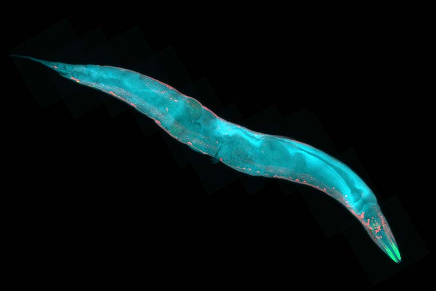
In the tiny worm known as C. elegans, the nervous system is far simpler, comprising a mere 118 classes of neurons.

At MIT, scientists in H. Robert Horvitz’s lab are studying the worms to learn about how nervous systems develop. Horvitz is the David H. Koch Professor of Biology at MIT, an Investigator at the McGovern Institute for Brain Research at MIT, and an Investigator at the Howard Hughes Medical Institute. His team has just discovered that a protein complex called cohesin, which helps shape the three-dimensional structure of the genome in both worms and humans, is critical for establishing some neurons’ identities as development unfolds.

The findings, reported July 31, 2026, in the journal Science Advances, could help scientists find a way to treat a rare developmental disorder called Cornelia de Lange syndrome, which is caused by mutations that interrupt the cohesin complex.

Model organism

Postdoctoral researcher Dongyeop Lee explains that C. elegans is a powerful model for studying neurodevelopment not just because its nervous system has been comprehensively mapped, but also because of the ease and speed with which scientists can study the function of its genes.

Because many of the worm’s genes have been retained through evolution, findings from studies of C. elegans often reveal important aspects of human biology. The current study began with worms that, because of a genetic mutation, make too many neurons of a certain type.

Adrenergic neurons, named for the kind of neurotransmitter they use to communicate with other neurons, are vital for enabling worms to respond to both their environment and their own internal state. Normally, C. elegans has just two pairs of adrenergic neurons: two RIM neurons and two RIC neurons. But the worms Lee studied had extras of both.

Takashi Hirose, a former member of the Horvitz lab, first observed this change in 2007.

Lee later continued the study and discovered that worms carrying a mutation in a gene called coh-1 have extra adrenergic neurons. The coh-1 gene encodes one part of the cohesin complex.

When Lee tested other mutations that disrupt cohesin, he found the same effect: Worms without fully functional cohesin had too many RIM neurons and too many RIC neurons.

Molecular switch

With a series of experiments designed to tease apart how cohesin impacts neurons’ identities, Lee discovered that cohesin cooperates with a gene-regulating protein called EOR-1 (known in humans as PLZF) to direct some neurons to develop into neurons that communicate with the inhibitory neurotransmitter GABA.

By reorganizing the structure of the genome, cohesin can change the way gene regulators like EOR-1 interact with DNA. Lee’s experiments showed that when either cohesin or EOR-1 couldn’t do its job, cells that should have become GABA-producing neurons become adrenergic neurons instead.

“What we found is that there are two alternative possible fates of certain neurons, and cohesin acts as a molecular switch that decides one of the possible neuronal fates,” Lee explains. “This means the structure of genomic DNA in the nucleus is important for neuronal fate determination.”

Disease connection

Lee adds that extra adrenergic neurons were not the only abnormality he observed in worms with cohesin mutations. Cohesin is important for shaping cells and tissues throughout the body. “The mutants have severe developmental defects,” Lee says. “They grow slowly. They don’t move well, and they also have defects in reproduction.”

Notably, the problems Lee saw in the worms echo aspects of Cornelia de Lange syndrome, a rare genetic disorder that impacts physical, cognitive, and behavioral development. Cornelia de Lange syndrome can be caused by mutations in cohesin genes, and Lee says that the discovery of how cohesin mutations affect worm development and behavior opens new opportunities to study the disease and search for potential therapeutic targets in C. elegans.

The Horvitz lab already has some promising leads. Taking advantage of the quick genetic screens that are possible in worms, Lee has found additional mutations that can counteract impaired cohesin, improving the health of worms with cohesin mutations. The team is now working to identify the genes where these suppressor mutations occur, so they can investigate whether they might make good therapeutic targets in humans.

Meanwhile, the team is also exploring a potential role for cohesin in shaping the fates of other neuron types, as well as searching broadly for additional molecules that work with cohesin to guide development. “We expect we have opened up a new biology,” Lee says. “This paper is just the beginning.”

Paper.

Douglas Lauffenburger named Institute Professor

Lauffenburger described being named an Institute Professor as “an honor that is especially gratifying because it recognizes the extraordinary impact of our unique MIT biological engineering department. I’ve been blessed with the rare opportunity to help create something revolutionary, here in this remarkable institution.”

Anne Trafton | Jennifer Chu | MIT News
July 20, 2026

A physician and neuroscientist who studies how anesthesia affects the brain; a leader in aerospace engineering, policy, and education; and the founding head of MIT’s Department of Biological Engineering have been awarded MIT’s highest faculty honor: the title of Institute Professor.

With the appointments of Emery Brown, Daniel Hastings SM ’78, PhD ’80, and Douglas Lauffenburger, there are now 12 Institute Professors at MIT, along with 10 Institute Professors Emeriti.

The appointments, which took effect July 1, were announced today in an email to the faculty from Sally Kornbluth, MIT’s president; Anantha Chandrakasan, MIT provost; and Roger Levy, chair of the faculty and a professor of brain and cognitive sciences.

Emery Brown

Brown, who has been a member of the MIT faculty since 2005, says he is “tremendously honored” to be appointed as an Institute Professor.

“It’s a pleasure to know that your colleagues hold you in such high esteem and that the work that you’re doing is valued,” says Brown, who is the Edward Hood Taplin Professor of Medical Engineering and Computational Neuroscience, an investigator at The Picower Institute for Learning and Memory, and a professor in the Department of Brain and Cognitive Sciences and the Institute for Medical Engineering and Science. “When you look down the list of people who have had this title, it’s an amazing group.”

After graduating from Harvard University with a bachelor’s degree in applied mathematics in 1978, Brown earned a PhD in statistics, also from Harvard, and an MD from Harvard Medical School. Since 1992, he has been a member of the Harvard Medical School faculty, and until recently he was a practicing anesthesiologist at Massachusetts General Hospital.

Throughout his career, Brown has made contributions in several different areas of neuroscience. In the early stages of his research career, he developed statistical methods to characterize the properties of the human circadian clock. He showed how light exposure can shift the phase of the human clock, depending on the circadian phase during which the light is administered. He also developed methods to demonstrate, from analyses of physiological data collected under special low-light conditions, that the intrinsic period of the human clock, like that of other species, is closer to 24 hours and not 25. Brown also measured the impact of shift work schedules that were designed using circadian physiology.

Later, he developed new statistical techniques and signal processing methods to analyze data collected in systems neuroscience experiments. As part of this work, he devised algorithms to decode the position of an animal in its environment by reading the activity of a small group of place cell neurons in the animal’s brain.

Joining MIT’s faculty just over 20 years ago represented an “inflection point” in his career, Brown says.

“I was an anesthesiologist doing statistical research, interested in neuroscience, and MIT allowed me to tie all those together,” he says. “I could work with colleagues who could help me understand the neuroscience of anesthesia, have another outlet for the statistical research that I was doing, and also more direct interactions with undergraduates and grad students.”

Over the past two decades, Brown has applied statistical techniques to studying what happens to the brain under anesthesia. His work has revealed how drugs such as propofol alter the brain’s intrinsic oscillations, which can be seen with electroencephalography (EEG).

During the awake state, these oscillations usually have high frequencies and low amplitudes, but as anesthetic drugs are given, they shift to low frequencies and high amplitudes. These changes disrupt normal communication between different brain regions, leading to loss of consciousness.

Brown has also shown that these EEG oscillations can be used to monitor whether a patient is too deeply unconscious, and he has developed a closed-loop anesthesia delivery system that can monitor these oscillations in real-time and guide anesthetic dosing during surgery.

In 2024, Brown was presented with the National Medal of Science. Among his other awards, he is also a recipient of a National Institute of Health Director’s Pioneer Award, the Gruber Prize in Neuroscience, and the Swartz Prize for Computational and Theoretical Neuroscience. He one of a small group of researchers to be an elected member of all three National Academies of Medicine, Sciences, and Engineering, as well as the National Academy of Inventors.

From 2012 to 2022, he served as co-director of the Harvard-MIT Program in Health Sciences and Technology. He has also played an instrumental role in several important efforts at MIT, including the 2010 Report on the Initiative for Faculty Race and Diversity, and the founding of the MIT Institute for Data, Systems, and Society (IDSS) in 2015.

Outside of his work at MIT, Brown served on President Obama’s Brain Initiative Working Group, as well as the National Academy of Sciences Committee on Women in Science and Engineering and the Council of the National Institutes of Neurological Disorders and Stroke.

Brown is also known for his commitment to teaching and mentoring students. In 2024, he was named a recipient of MIT’s “Committed to Caring” award — an honor given by MIT’s Office of Graduate Education to faculty members who have served as exceptional mentors to graduate students.

Daniel Hastings

When Hastings, the Cecil (1923) and Ida Green Professor in Education, was notified of the new distinction, it came as a total surprise.

“The people who were there will tell you that I could not believe it at first,” he says. “I never thought of myself as being in the same league as some of the Institute Professors I knew.”

Hastings grew up in England and Jamaica, and developed an early fascination with space, as a fan of the fictional “Star Trek,” and later “Star Wars” and “Stargate” (he’s seen every episode and movie of all three franchises), as well as the very real NASA Apollo program.

After receiving a bachelor’s degree in mathematics from Oxford University, he enrolled at MIT, earning his master’s degree in 1978 and PhD in 1980, both in aeronautics and astronautics. In 1985 he joined the faculty as an assistant professor and was promoted to full professor in 1993.

Throughout his tenure, Hastings has made significant and lasting impacts in astronautical engineering, particularly through his studies in space plasma environment interactions, electric propulsion, and space systems architecture.

His early research on the physical interactions between plasma and spacecraft, for which he co-wrote the definitive text (“Spacecraft Environment Interactions,” published in 1996), enabled the safe operation of solar panels on spacecraft today. Prior to Hastings’ work, high voltage solar arrays on satellites often experienced catastrophic arcing — a dangerous jumping of electrical current from one panel to another. These failures turned out to be a result of interactions with the surrounding space plasma.

Hastings developed theories to characterize these interactions. His theories informed NASA’s design of the solar panels to power the International Space Station, which are still in operation today. His work also established guidelines across the aerospace industry on the design of resilient solar panels and ways to handle issues once in orbit.

In his studies of electric propulsion, Hastings characterized the fundamental physical interactions between ion engine plumes and spacecraft systems. His work was pivotal in incorporating ion propulsion systems into many commercial satellites and deep space probes and helped to push what was an experimental technology into mainstream use in space propulsion.

In his more recent work, Hastings has explored the concept of flexible and distributed space architectures. He and his students are developing models for spacecraft that can serve purposes beyond their original mission intent. For instance, a spacecraft may incorporate a port that could serve as a waystation for future satellites to dock and refuel. Such a flexible and distributed system could help to support future missions to the moon and Mars.

In recognition of his research contributions, Hastings received the AIAA Losey Atmospheric Sciences Award in 2002, was elected to the National Academy of Engineering in 2017, and was recognized as an honorary fellow of the American Institute of Aeronautics and Astronautics (AIAA) in 2021.

Throughout his career, Hastings has taken on numerous leadership roles, at the national, international, and Institute levels. Shortly after becoming full professor, he served as associate department head of research in MIT’s Department of Aeronautics and Astronautics (AeroAstro). He then took a two-year leave from the Institute to serve as chief scientist of the U.S. Air Force. During that time, he advised the Air Force chief of staff and secretary and successfully strengthened investments in space research in the U.S.  Air Force space program.

Hastings has served as an advisor on multiple expert panels and boards, including as the chair of the Air Force Scientific Advisory Board, and as a member of the NASA Advisory Council, the National Science Board, the Intelligence Science Board, and most recently, the Defense Science Board and User Advisory Group of the National Space Council. He has also chaired multiple National Research Council studies and advised the space and engineering industries in various capacities, including serving on the boards of the Aerospace Corporation, Draper, and Blue Origin. He has just finished a two-year term as president of the American Institute of Aeronautics and Astronautics.

At MIT, Hastings has stepped up to serve in pivotal leadership posts. From 2000 to 2005, he served as the director of MIT’s Technology and Policy Program, then director of the Engineering Systems Division. From 2006 to 2013, as dean for undergraduate education, he helped to develop initiatives in equity, financial aid, and curriculum development, and strengthened international education and study abroad programs during a nationally challenging economic period. He received the Gordon Y. Billard Award in 2013 for his work on international education. In 2014 he began a five-year term as director of the Singapore-MIT Alliance for Research and Technology, during which he worked to reinforce MIT’s global collaborations. And from 2019 to 2023 he served as head of AeroAstro, supporting new research and educational initiatives as he navigated the department through the global pandemic.

Hastings has also worked in multiple capacities to make the Institute a more welcoming and inclusive community. He has served as associate dean of engineering for diversity, equity, and inclusion (2021-2023), Institute Community and Equity Officer (interim, 2023-2024), and co-chair of the MIT Values Statement Committee, as well as vice chancellor for undergraduate and graduate education (interim, 2024-2025).

“MIT has been a great place for me,” Hastings reflects. “It has a mission to address some of the most pressing problems in the world. It is a high-energy place. This is a place that I am excited to work in and I want to give back to make it better.”

Douglas Lauffenburger

Lauffenburger, who is the Ford Professor of Biological Engineering, Chemical Engineering, and Biology, was the central founder of MIT’s Department of Biological Engineering, which he chaired from its inception in 1998 until 2019.

Before coming to MIT, Lauffenburger earned his undergraduate degree from the University of Illinois at Urbana-Champaign in 1975 and a PhD from the University of Minnesota at the Twin Cities in 1979, both in chemical engineering.

While in graduate school, he became fascinated by the biological sciences. Early in his career, as a faculty member at the University of Pennsylvania and at the University of Illinois, his research and teaching straddled the line between chemical engineering and cell biology. Due to his unique background, MIT recruited Lauffenburger in the late 1990s to launch its new Department of Biological Engineering.

At the time, many universities had programs in biomedical engineering — an interdisciplinary field that applies techniques from electrical, chemical, or mechanical engineering to medical problems. Lauffenburger envisioned a distinct discipline of biological engineering, in which engineers would pursue an understanding of how biological systems function at the level of molecular and cellular mechanisms, with the goal of manipulating them to create new technologies for applications across medicine, energy, the environment, nutrition, and manufacturing.

“What was clear to me was that because biological systems comprise molecular processes, which are integrated in very complex ways, a true engineering analysis and design approach ought to be useful in moving it beyond mere tinkering and trial-and-error,” he says. “We needed to develop engineering frameworks for biology based on design principles, models, and predictions.”

As department head, Lauffenburger guided the development of new curricula at both graduate and undergraduate levels, and recruited faculty members whose work spanned engineering, molecular and cellular biology, microbiology, and immunology. The new department began offering graduate degrees in the late 1990s, and an undergraduate major beginning in 2005. Since its inception, the program has served as a model for similar programs at many other institutions worldwide.

Lauffenburger described being named an Institute Professor as “an honor that is especially gratifying because it recognizes the extraordinary impact of our unique MIT biological engineering department. I’ve been blessed with the rare opportunity to help create something revolutionary, here in this remarkable institution.”

Lauffenburger also played key roles in launching new interdisciplinary programs within MIT and with other institutions, including the Center for Biomedical Engineering, the Computational and Systems Biology Initiative, the DuPont-MIT Alliance, and the Cambridge-MIT Initiative.

His research has touched on many areas of biological science, including molecular cell biology, systems biology, and computational biology. Much of his work focuses on unraveling cell signaling mechanisms, using a combination of computational modeling and quantitative experiments. This work has shed light on processes such as cell proliferation, death, adhesion, and migration.

In the field of systems biology, he has created computational models across a spectrum of mathematical approaches, which can be used to identify drug targets and patient stratification strategies for a variety of diseases, including cancer and chronic inflammation, and predict the efficacy of drugs against those targets.

In 2021, he and Linda Griffith, the School of Engineering Professor of Teaching Innovation at MIT, were jointly awarded the Bernard M. Gordon Prize for Innovation in Engineering and Technology Education, the most prestigious engineering education award in the United States.

Lauffenburger is an elected member of the National Academy of Engineering and the American Academy of Arts and Sciences. He is a fellow of the American Association for the Advancement of Science, a founding fellow of the American Institute for Medical and Biological Engineering, and has served as president of the Biomedical Engineering Society.

Why are some bacterial genes high in purines?

In certain species of bacteria, the answer lies in shielding RNA transcripts from a quality-control factor called Rho. Understanding the requirements for expressible sequences is critical for expression engineering of therapeutic agents.

Lillian Eden | Department of Biology
July 2, 2026

In the study of bacteria, a longstanding dogma held that two molecular machines — RNA polymerase, which leads the way in transcribing DNA into RNA, and ribosomes, which bring up the rear translating RNA into proteins — worked so closely in tandem that they were effectively attached.

This close coupling of transcription and translation in bacteria was thought to be fundamental to gene expression in part because the trailing ribosome could shield nascent gene products from an effective and omnipresent quality-control protein called Rho.

In bacteria that exhibit something called runaway transcription, however, the polymerase instead speeds ahead, unhitched from its protective ribosome. Inexplicably, however, in bacteria that exhibit this runaway transcription, such as Bacillus subtilis, Rho targeted primarily noncoding, useless RNA products.

New research from the Department of Biology reveals that the secret to Rho’s quality-control specificity lies in the sequence composition of nucleotide bases that make up coding strands of DNA.

“We started with a hypothesis that Rho was regulated by sequence, but the fact that the sequence alone was enough to protect any gene in the entire B. subtilis genome from Rho was really surprising,” says Julia Dierksheide PhD ’26, a graduate student in the Li Lab and first author of a paper recently published in Nature Microbiology. “That’s a really diverse range of sequences — what sequence feature is shared by every single gene in the genome?”

Barricading with bias

Rho serves as a termination factor, meaning that it is a crucial mechanism for preventing bacteria from wasting precious resources by making RNA transcripts that serve no purpose.

All the information a bacterial cell needs is encoded in its DNA, which is made up of two strands of nucleic acids. These strands twist together to form a double helix, with genetic information codified in pairs of bases: purines guanine and adenine are matched with pyrimidines cytosine and thymine, respectively. Any sequence that gives rise to RNA transcripts is stored in complement to a parallel, noncoding strand, meaning that a large portion of genetic material is transcriptionally useless.

Coding DNA strands in certain bacteria were known to be significantly higher in purines guanine and adenine compared to the rest of the bacterial genome. The researchers found that this purine bias alone shields productive mRNA transcripts from Rho-mediated termination.

“I love having a big, complicated dataset and trying to reduce that to biological meaning,” Dierksheide says. “It seems like Rho itself has been broadly shaping the evolution of the B. subtilis genome to create these sequence composition biases.”

Bacterial species that, over generations, have lost Rho no longer exhibit this strong purine bias.

Rho also serves as a regulatory factor in bacteria becoming motile, forming biofilms, or sporulating, all of which are critical for biology and survival. The purine bias could also provide a layer of protection against the insertion of foreign DNA, for example, when a viral bacteriophage infects bacteria.

“Bacteria exist as single cells, so everything that they do, they have to do through gene expression,” Dierksheide says. “Understanding the fundamental details about how gene expression works, how a cell encodes all the information it needs to survive in the nucleotide sequence of the genome, is really exciting.”

Future directions

Although the exact mechanism underlying Rho’s specificity remains unclear, these results crack an underlying code in the composition of bacterial genomes.

Dierksheide said she hoped to perform a similar screen to characterize Rho’s specificity in Escherichia coli, which diverged from B. subtilis on the evolutionary tree an estimated 2 billion years ago and still exhibits coupled transcription-translation, where the transcribing RNA polymerase is closely followed by a translating ribosome.

The high sequence specificity of B. subtilis Rho is crucial for the protection of its runaway RNA polymerase, in which that molecular machine speeds ahead of the ribosome. A systematic comparison to E. coli Rho could help reveal how this heightened stringency arose.

This information will be critical for engineering diverse bacterial species for applications including the production of therapeutic agents. Other bacterial species, such as B. subtilis, may be better models for this process because they have abundant secretion pathways, according to Dierksheide, making it much easier to produce and isolate proteins in large quantities.

“Our findings reveal an important criterion for successful sequence design that must be considered in expression engineering,” says associate department head, associate professor of biology, and Howard Hughes Medical Institute investigator Gene-Wei Li, the lead author of the study. “There are so many cryptic messages in the genome, like the purine bias, and we are just beginning to be able to decipher what they mean.”

Two MIT faculty members named 2026 Pew Biomedical Scholars

Cell biologist Whitney Henry and immunologist Harikesh Wong will receive four years of flexible funding to advance early-career research on ferroptosis and immune decision-making.

Nina Tamburello | Nikolay Kolev | Koch Institute | Ragon Institute
June 29, 2026

Whitney Henry and Harikesh Wong have been named 2026 Pew Scholars in the Biomedical Sciences. The Pew Charitable Trusts announced the 21-member class of early-career researchers, which includes the two MIT scientists as well as two alumni, on June 16. Each scholar will receive four years of funding to pursue cutting-edge research into human health and disease. Xin Gu PhD ’22 of Dana-Farber Cancer Institute and Christina Tringides ’15 of Rice University were also selected as scholars.

Henry, the Robert A. Swanson (1969) Career Development Professor of Life Sciences and a faculty member at the Koch Institute for Integrative Cancer Research, will use the Pew scholarship to examine how a stress-induced cell death program called ferroptosis contributes to injury and regeneration in the liver. Wong, assistant professor of biology at MIT and core member at the Ragon Institute of Mass General Brigham, MIT, and Harvard, will use his award to investigate how groups of immune cells reach a “communal decision” about whether to tolerate or attack a particular target.

Whitney Henry

Henry’s research centers on ferroptosis — an iron-dependent form of regulated cell death — and its role in shaping cell fate and tissue remodeling. Her lab investigates why some cells can withstand stress while others cross the threshold for ferroptosis, focusing on the molecular, metabolic, and tissue-level cues that shape ferroptosis vulnerability. The work draws on chemical biology, metabolomics, functional genomics, and in vivo models. By defining the mechanisms that govern ferroptosis susceptibility, Henry’s group aims not only to identify novel therapies that target the most dangerous subpopulations of cancer cells, those that are highly metastatic and resistant to conventional treatment, but also to advance understanding of diseases in which ferroptosis drives tissue injury, fibrosis, or impaired repair.

Harikesh Wong

Wong investigates how groups of cells organize into networks that collectively process information and control immune responses within tissues. These networks must continually balance the body’s need to protect itself against pathogens and tumors with the need to preserve healthy tissue function. Combining the tools of immunology with high-resolution fluorescence microscopy, computational modeling, and gene manipulation, his lab seeks to map, model, and manipulate the cell-cell interactions that govern these decisions within intact tissues, revealing how subtle changes in multicellular organization and communication can shift immune responses toward pathogen clearance and tolerance, or toward autoimmunity, chronic inflammation, and cancer.

Pew scholars are chosen from applicants nominated by leading academic institutions across the United States. This year’s class of 21 was selected from 211 nominees. The incoming scholars join a legacy of more than 1,000 scientists supported by the program since 1985. During their time as scholars, they will meet annually with fellow Pew-funded scientists to build connections across a wide variety of disciplines.

“Scientific discovery is moving at a rapid pace, and now more than ever we need curious and creative researchers leading the charge,” says Lee Niswander, a 1995 Pew scholar and chair of the program’s national advisory committee. “These new biomedical scholars are prepared to meet that challenge, and I look forward to watching their research unfold.”

Raised in the “Kitchen”

Karen O’Leary, lab associate and acting supervisor in the Glassware Sterilization Facility, has become a cornerstone of the department’s operations.

Samantha Edelen | Department of Biology
June 25, 2026

Early mornings in the halls of Building 68 feature the sounds of rolling wheels on big metal carts, the rattling of glassware, the whooshing of faucets, and the clanking of autoclaves.

These aren’t the sounds of researchers at work, but rather those of keeping the labs sterilized and stocked with the sundries of research: pipette tips, test tubes, flasks, petri dishes, and more.

Orchestrating this sunrise cacophony and the staff that undertakes it is Karen O’Leary, lab associate and acting supervisor in the Glassware Sterilization Facility, also known as the “kitchen.”

Thanks, in part, to O’Leary’s proactivity and hard work, the kitchen staff were recently recognized with an MIT Excellence Award in 2025 for exceptional contributions in service of the community.

“My goal is to get the scientists everything they need to do their research,” O’Leary says. “I’m good at what I do.”

O’Leary admits she did not always possess such confidence. In almost 40 years at MIT, O’Leary has grown into this critical role for the department, and the department itself has evolved, moving into a brand-new building and away from previously standard practices like submerging equipment in acid for sterilization.

From rookie to running the show

On Sept. 7, 1987, Karen O’Leary joined the MIT community as a staff member for the first time. The 18-year-old was fresh from vocational high school, where she studied cosmetology but felt too shy to pursue that as a career. She was also nervous about joining a research institution.

“When I started, I didn’t even know what a beaker was,” she recalls.

Too embarrassed to admit in her interview that she couldn’t remember her brand-new home phone number, “I just made one up.” Fortunately, this didn’t prevent her from getting the job, where she worked under the mentorship of Thelma Watkins, who would retire in 1996 after 21 years at MIT. Watkins was critical for instilling a good work ethic and boosting O’Leary’s confidence.

“She taught me to show up every day, and work hard, and laugh,” O’Leary says.

Even now, O’Leary continues to bring joy to that daily diligence, for herself and for her staff.

“Karen is always on top of things,” says longtime friend and fellow Lab Associate AnnMarie Budhai. “She doesn’t refuse work and always goes above and beyond.”

Facilities and Operations Manager Cesar Duarte says that O’Leary’s long tenure, support, and knowledge have been invaluable as he transitioned into his role in Building 68 starting in 2023.

“Karen is one of those people who makes everything around her run more smoothly and more pleasantly,” Duarte says.

Better, faster, safer

Although some might consider it drudgery, O’Leary says that washing glassware is her favorite task.

“I like that when I wash, I can see the job is complete at the end of the day,” she says.

Although washing glassware is a perennial task, safety and efficiency have come a long way in the past 38 years. More-effective autoclaves and dishwashers have eliminated steps like steaming to dissolve agar solvents before autoclaving, and scrubbing individual test tubes before washing.

O’Leary was working for the department in 2011 when Building 68 piloted a new approach to MIT’s management of regulated medical waste (RMW), such as petri dishes, blood, and needles — the new system, which is cheaper and produces less waste, is now used by all departments at MIT that produce RMW.

“EHS [the Environment, Health and Safety Office] has come really far — I’m glad we got away from acid,” O’Leary notes of the bygone era of submerging glass pipettes for sterilization. “Back then, no one knew of a better way.”

Other tasks include cleaning velvets, which are used for replicating bacterial colonies on petri dishes, and pouring agar plates.

“Everyone knows how to do almost every job, so we can take turns doing different tasks,” O’Leary says. “If you get sick, there’s always someone to cover.”

All in the family

For O’Leary, kinship with MIT has spanned generations. O’Leary was raised in Weymouth, Massachusetts, by a father who worked at MIT as a supervisor in the sheet metal shop. Having raised children of her own, now grown, O’Leary came to greatly appreciate the flexibility her job has granted her.

“I’ve had great work-family balance here,” she says. Even though she’s often at work more than an hour before the researchers that the kitchen serves, “The hours are great, and with MIT Health right across the street, it was easy to take everyone to doctors’ appointments.”

She’s also gained a chosen family at MIT, spending breaks at work taking long walks along the Charles River, “talking about anything and everything” with colleagues like Budhai and Lab Aide Janet Katin.

“We really grew up together,” she says.

Working at MIT has provided O’Leary with support and community, and she’d like to pay it forward. In addition to strolling with colleagues, she hits the gym to help maintain the energy required for her highly active work.

“I don’t like sitting around,” she says.

In addition to maintaining her stamina at work, she hopes that taking care of herself will keep her actively involved if she ever has grandchildren, and enable her to help neighborhood kids when she someday retires.

“I owe a lot to MIT,” she says. “I have been allowed to work hard and get satisfaction and have been appreciated and given space to care for my family.”

O’Leary returns this care to the Department of Biology in spades.

“It’s an understatement to say that Biology is lucky to have her,” says Duarte. “Karen’s overflowing energy, attention to detail, and care for the Biology research community are nothing short of amazing.”

Harriet having it all

From Boston to Moscow and across the U.S., Harriet Latham Robinson SM ’61, PhD ’65 has balanced an exciting career at the forefront of molecular biology with family, friends, and adventure.

Lillian Eden | Department of Biology
June 12, 2026

In winter 1997, at age 60, when many researchers might be looking forward to retirement, Harriet Latham Robinson SM ’61, PhD ’65 was pursuing a faculty position as the chief of microbiology and immunology at the Yerkes National Primate Research Center at Emory University in Atlanta, Georgia.

She got the job.

There, she would also co-found GeoVax, a biotechnology company, based on her preclinical research, including work on developing an HIV-1 vaccine.

Often, as the only woman in a room throughout much of her career, and in the still-developing and male-dominated field of molecular biology, her colleagues were referred to as “doctor” or “professor” at scientific symposia and committee meetings.

“In contrast,” she recalls, “I was Harriet.”

Becoming a scientist

Robinson was born in 1938, the second of four children, to a mother, Ruth, and a father, Allen, from Ohio and Connecticut, respectively. After finishing grammar school, she attended the Girls’ Latin School, a public magnet school for college-bound young women. Although the school offered only two classes in science — one semester of chemistry and a health class — Robinson credits her time there for inspiring a lifelong love of learning, especially history and languages.

“At our 50th and 60th high school reunions, I was struck by what my Girls’ Latin school classmates had done with their lives,” she says. “We had become not only wives, mothers, teachers, and nurses we were supposed to become, but also physicians, lawyers, professors, politicians, and businesswomen.”

Robinson pursued her undergraduate studies at Swarthmore College, where she intended to study political science. After an introductory biology course, however, she switched her major. Despite the shift, a love of languages persisted: Robinson took Russian and, the summer after her senior year of college, served as a Russian-English speaking guide at the 1959 American National Exhibition in Moscow. Despite mounting tensions between the United States and the Soviet Union, she served again in a similar role from September 1961 to January 1962 for a traveling transportation exhibition in Russia and Ukraine, where she was stationed by a Ford Thunderbird, wearing a TWA stewardess uniform.

“We were true entertainment, as well as education, and I worked to do my best to answer questions about America,” she says. “I was most surprised by the pride the Russian people took in the post-World War II accomplishments of their country.”

Robinson might not have had a career in science at all had it not been for a dean at Radcliffe College who recognized Robinson’s interest in science. Robinson had thought it appropriate, as a young lady, to pursue marriage and to only further her education to become a teacher or nurse. Seeking permission to take chemistry instead of education courses to fulfill requirements for getting a teaching degree, she was referred to a dean who considered it perfectly appropriate for a young woman to pursue another career. Robinson recalls that the dean declared, “My dear, you want to be a scientist.”

The foundation for a career

Robinson was soon accepted at MIT and was offered a fellowship to teach in an introductory biology lab to help pay her way. She returned from Moscow just five days before the start of a master’s program in biochemistry. In the Department of Biology at MIT, there were only a handful of women, no female faculty, and few ladies’ rooms in 1959.

It was there that she met Walter “Wally” J.K. Tannenberg, a onetime partner but lifelong friend and companion, an MD taking courses at MIT. He wasn’t “at all taken aback by my becoming an educated woman,” Robinson says. He taught her to ski, and they sailed his lightening, the Ondine, in circles around Robinson’s parents’ comparatively slow motor sailor, the Palometa.

Their breakup just before the winter holidays in 1963 precipitated her reentry to graduate school, to pursue her thesis work in the lab of Jim Darnell; she threw herself into studies to sit a qualifying exam less than a month after reentry.

“A Bell Labs physicist who had just joined the Darnell Lab opined that any concept in biology could be mastered in two weeks,” Robinson says. “Much to everyone’s amazement, I not only passed my qualifying exam, but did much better than expected.”

It was at the University of California at Berkeley during her postdoctoral work that she met her husband. Although the marriage would not last the test of time, Robinson and her husband were blessed with three boys, each 13 months apart.

Robinson knew that she wanted to take time away from her career to stay home with her children before they entered primary school. As a graduate student at MIT, to prepare for both having a career and pursuing motherhood, Robinson hired a housekeeper and committed to being in the lab for only a typical 9 a.m. to 5 p.m. workday. If she were to compete with her male counterparts and be with her children, she needed to be able to get things done while working short hours.

Robinson successfully completed her thesis work in just over two years.

“The difference between bearing children and rising up professional ladders is that you can start up the professional ladder after you are 40,” she advises. “Such is more problematic for having children.”

Robinson’s thesis work at MIT concerned how DNA, which is identical in all cells of an organism, produces different cell types from the same genetic blueprint. She explored this question through the lens of messenger RNA, a gene product that determines which DNA sequences are expressed in a cell. Later, her work on cancer-causing viruses in chickens would help lay the groundwork for gaining insight into genes that can cause tumors to form.

“In contrast to becoming a wife, becoming a PhD from MIT did not falter, but rather provided me with the foundations for a career I loved in which I used molecular biology and chickens to study the genetic basis of cancer and pioneered the use of DNA as a new method of vaccination,” Robinson says.

Cancer-causing viruses

Robinson, supported by an National Science Foundation fellowship, pursued postdoc training at the University of California at Berkeley, in the lab of Harry Rubin. The Rubin Lab specialized in work on a virus known to cause cancer: the Rous sarcoma virus, which causes rapid tumor onset when introduced into chickens. RNA, it had recently been discovered, was the underlying genetic cause of tumors developing in chickens exposed to the Rous sarcoma virus. It cannot, however, do this deadly work without co-infection with something called a helper virus — in this case, avian leukosis virus.

Both Rous sarcoma virus and its helper viruses were retroviruses, which can make DNA copies from RNA sequences, a departure from the previously accepted dogma that DNA is only transcribed into RNA, and not the other way around.

Robinson joined the Worcester Foundation for Biomedical Research in 1977, where she continued research on Rous helper viruses and had the opportunity to run her own lab for the first time. In 1998, she was recruited to be a professor of pathology at the University of Massachusetts Medical Center. While there, she conducted pioneering studies on the use of DNA for vaccination and worked on developing an AIDS vaccine.

In 1999, she moved again, this time to step into the role of chief of microbiology and immunology at the Yerkes National Primate Research Center at Emory University, where she began testing her candidate HIV vaccines in primates. While at the University of Massachusetts and Emory, Robinson and her lab used DNA vaccines, both with and without a poxvirus booster vaccine provided by Bernie Moss at the National Institutes of Health, to immunize animals against influenza, HIV, measles, and Ebola.

“From the early days of DNA vaccines, I had wanted to start a company to help move DNA vaccines from bench to bedside,” she says.

Thus, GeoVax, short for “Georgia Vaccines,” was born. Robinson co-founded it with Don Hildebrand in 2001 after her move to Yerkes; Robinson would serve as chief scientific officer and a member of the board of directors during her tenure at the company.

GeoVax successfully moved Robinson’s candidate AIDS vaccine into human clinical trials. These trials were stopped due to the generally poor performance of HIV vaccines in clinical trials, compared to the outstanding therapeutic potential of more recently developed anti-HIV drugs. GeoVax, however, continues to work on vaccines for Mpox, Covid-19, and Ebola, and has expanded its scope to include a cancer treatment.

A well-deserved retirement 

After rounds of good-natured roasting from colleagues at Emory University and GeoVax, Robinson retired and has been enjoying returning to Palo Alto, California, where her oldest son, Bill, and his wife now live.

Ultimately, Robinson hopes that her story can encourage everyone, especially young women, not to let pursuing a challenging and enriching career prevent them from realizing the dream of having a family.

“I have had a wonderful life, far exceeding what I ever could have anticipated,” Robinson says. “I have had international adventure, the romance of a man who truly loved me, the joy of motherhood, and the warmth, wonder, and adventure of family and friends, and last, but not least, the exhilaration of a career in molecular biology.”

Advancing stem cell research and building the next generation of biologists

Biology PhD student Giselle Valdes (Reddien Lab) studies stem cell regeneration while encouraging aspiring students and researchers.

Stefanie Koperniak | Division of Graduate and Undergraduate Education
June 11, 2026

As an undergraduate at Florida International University, Giselle Valdes tackled rigorous studies in the school’s Honors College while simultaneously caring for family members with medical needs.

“I think that the choice to pursue any field in the space of biology and medical research was entirely shaped by having to be there for my family,” says Valdes.

As a McNair Scholar and biomedical engineering major who also did extensive research in biochemistry, she leaned more toward undergraduate courses in mechanical and electrical engineering that were geared primarily toward equipping students to build medical devices. She began to shift her research interests more firmly into biology, however, the summer before her senior year in 2018. She spent 10 weeks on the MIT campus as a participant in the Bernard S. and Sophie G. Gould MIT Summer Research Program in Biology (BSG-MSRP-Bio), working in the lab of Associate Professor Eliezer Calo PhD ’11, also a former BSG-MSRP-Bio participant. The Calo Lab focuses on ribosomes, small cellular particles that translate RNA into proteins, and looks at how mutations in ribosome development can lead to disorders.

After working in Calo’s lab, she could see herself as a biology graduate student at MIT. In January 2019, she attended the MIT biology department’s Quantitative Methods Workshop, a weeklong, intensive workshop designed to introduce non-MIT undergraduates to tools and programming languages used to analyze experimental data in biology and neuroscience. While there, she was elated to receive an email from the department inviting her to interview for the PhD program. She was accepted and began her doctoral studies in the fall of that year.

“When I think about my experiences at MIT, both as an undergraduate in MIT programs and as a PhD candidate in biology, I think about all the great mentors who have helped me along the way,” says Valdes. “I’ve also really valued the richly collaborative community, and being able to take a lot of risks in how I address the questions I have the opportunity to pursue.”

Researching stem cell regeneration

Since she came from a biomedical engineering background, Valdes spent the first year of the biology doctoral program taking foundational biology courses and working in different labs to decide which type of research she wanted to do. She gravitated toward cell and developmental biology and joined the lab of Professor Peter Reddien, associate director of the Whitehead Institute for Biomedical Research. Valdes was awarded an MIT Fund for the Future of Science Fellowship to support her research.

“Giselle is doing terrific work on a fundamental problem related to adult stem cells and regeneration — how do progenitor cells choose what cell types to make? Fate choice in progenitors is typically studied in embryogenesis, and how it occurs in the context of adult regeneration is poorly understood and very important to address,” says Reddien.

Valdes has worked extensively with stem cells in highly regenerative flatworms, called planaria. analyzing the process of “cell fate choice,” or how cells determine which specific cell types and functions to develop. To date, Valdes, Reddien, and other researchers have studied “neighborhoods” of neoblasts (adult stem cells) and their fate choices, finding that different neighboring stem cells often chose different fate options — suggesting that cell fate choices are largely made by processes autonomous to individual cells.

Her current research aims to better understand the driving mechanism for cell fate choice, both within planaria and an additional model system: the evolutionarily distant acoel Hofstenia miamia.

“A lot of the things I’m doing in my current project have involved developing techniques that didn’t previously exist in our model organism,” says Valdes.

Working on model systems with limitations in the toolkit traditionally available to more well-established systems, such as transgenics, has allowed her to be creative in the techniques she applies to determine how stem cells choose what to become. It has also opened doors to collaborations, such as one with Ye Zhang of the Manalis Lab in the biological engineering department (now an assistant professor of biomedical engineering at Virginia Tech), that have allowed Valdes and team to sort neoblasts in novel ways based on their morphology, and better relate that to their dynamic state.

In summer 2024, Valdes mentored a BSG-MSRP-Bio student who now works with her on a current research project.

“She’s been with me as a technical assistant in the lab now for over a year, and we’ve been able to work on one of my projects together,” says Valdes. “It’s been exciting to come full circle in this way.”

Teaching and mentoring, near and far

In addition to her research, Valdes devotes a lot of her time to teaching and mentoring, both for MIT biology students and younger students discovering an interest in STEM.

“It’s been so rewarding to have a lot of opportunities to do for others what has been done for me,” she says.

Valdes has worked with secondary students both locally and abroad. She participates in the biology department’s developmental biology lab for high-school students and teaches in an annual biology lecture series for high schoolers. She has worked with the Enroot program from Cambridge Community Services, acting as a direct mentor to local high-school and community college students. At the Whitehead Institute’s Expedition: Bio program, for middle- and high-school students, she runs a planarian workshop. And she gives lab tours through the Whitehead Discovery Lab initiative, engaging in discussion with local high-school students.

Valdes has also assisted with a hackathon for Sprouting, a social impact venture providing STEM education opportunities to under-resourced communities in Puerto Rico. Sprouting was launched by Taylor Baum, a doctoral student in MIT’s Department of Electrical Engineering and Computer Science. Valdes taught coding essentials to Spanish-speaking middle- and high-school students in Puerto Rico.

“That was really emotional,” says Valdes. “The parents were so grateful, and there were kids who were clearly brilliant and gifted. They were able to really take off with the tools that we gave them.”

In her department, Valdes has been a teaching assistant for classes 7.003 (Applied Molecular Biology Laboratory) and 7.03 (Genetics). She is also a teaching assistant for the Quantitative Methods Workshop and teaches a Python module to students in the program.

“Giselle may be quite small in physical stature, yet she dominates the room when she speaks, and commands the full attention of an audience of 80 students when giving a lecture,” says Mandana Sassanfar, a biology senior lecturer and director of outreach who runs the Quantitative Methods Workshop. “She is highly respected both for her knowledge and the way she interacts with people. She is extremely approachable, very generous with her time, and always very supportive and encouraging. She is a wonderful mentor, teacher, and scientist.”

Valdes says she is always happy to help mentor undergrads and graduate students. She is co-founder and coordinator of the MIT Biology Application Assistance Program (BAAP), which aims to demystify the graduate school application process and offer interested applicants the tools and direct mentorship necessary for putting together a successful application. She also helped to coordinate, and has been an active participant in, the MIT BioPals Program, a student-organized peer mentorship initiative within the department that connects incoming first-year graduate students with senior graduate students. During the Covid-19 pandemic, this program provided critical support and social connection for new students navigating remote learning and social distancing.

After she completes her doctoral program, she envisions pursuing a postdoc and, ultimately, a faculty role, citing her passion for both academic research and teaching.

“My goal is to stay in academia in some way,” says Valdes. “I love mentorship and curiosity-driven science.”

Enzymes that assemble into droplets can speed up cellular reactions

MIT biologists find highly concentrated droplets can help cells keep enzymes organized and control growth signals.

Anne Trafton | MIT News
June 1, 2026

Within the past decade, biologists have discovered that one strategy cells use to keep their contents organized is a phenomenon known as phase separation.

Similar to the way oil forms droplets that float in a vinegar solution, proteins inside cells can phase separate to form highly concentrated droplets that keep them organized within the cell. In a new study, MIT researchers have now shown that this droplet formation is critical for controlling the function of a class of enzymes called kinases.

The researchers found that condensing into droplets optimizes the biochemical conditions needed for kinases to catalyze reactions, allowing them to more rapidly activate cell signaling pathways. In some cases, droplet formation can even change which reactions the kinases perform.

“Many biological molecules have this propensity to spontaneously separate. We were really interested in asking, if we have these kinases forming droplets, what is the consequence of that in the context of signaling?” says Lindsay Case, an assistant professor of biology at MIT and the senior author of the study.

Learning more about how these droplets form could help researchers design drugs that target kinases, some of which can be overactive in cancer cells.

“Understanding the chemistry of these compartments, and what molecules go into them and what molecules don’t go into them, could help us design drugs that better localize to their target of interest,” Case says.

Nicholas Lea, an MIT graduate student, is the lead author of the paper, which appears today in Cell Reports.

Forming droplets

Since her days as a graduate student, Case has been studying how the physical organization of molecules inside cells affects their function. As a postdoc, she began studying how phase separation might affect a signaling pathway that allows cells to sense when they’re attached to their environment, so they can respond appropriately.

Some of the proteins in this pathway are kinases, which activate other proteins by adding phosphate groups to them. Kinases can also activate themselves through a process called autophosphorylation.

“Inside of the cell, you have these kinase molecules that are responsible for carrying a signal through the cell, and we know that the organization of these molecules changes. When the information is present, they’re organized in a different way than when the information is not present,” Case says. “We think that having the right molecules in the right place is incredibly important for the right biochemistry to occur.”

Phase separation is one of the methods that cells appear to use for this organization. The most familiar example of phase separation can be seen in a salad dressing, where oil forms droplets to minimize contact with water-based vinegar. Proteins can phase separate when they are highly concentrated, leading them to self-assemble into dense droplets floating in the cell’s cytoplasm.

Case hypothesized that this phase separation, which brings kinases together at a high density, might help cells to boost the enzymes’ activity because they are more likely to bump into and phosphorylate each other.

In this study, Case and Lea set out to test that hypothesis, focusing on an enzyme called focal adhesion kinase (FAK). This kinase, which becomes activated when cells attach to their surrounding environment, activates pro-growth and pro-survival signals. In cancer cells, this signaling pathway can go awry, allowing cells to proliferate even when they detach from their original locations.

Scientists already knew that when cells are properly attached to their environment, that adhesion signal causes FAK to accumulate at the cell membrane. In the new study, the MIT team mimicked that effect by overexpressing FAK in cells. These cells were floating freely in a solution, not attached to any surface. Even so, the high concentration of FAK caused the kinase to phase separate into droplets, which turned on the pro-growth signal.

“It was surprising that just by condensing this protein into a droplet, you can actually turn on a signaling pathway that should be turned off,” Case says. “If FAK concentration is too high, you’re always getting these droplets and you’re always signaling, regardless of what the receptors that are supposed to be controlling this are doing.”

The findings suggest that in cancer cells, overexpression of FAK may lead to phase separation, which then helps to drive cancer progression and metastasis.

“It may be that for some kinases, you’re not supposed to form these droplets in the cytoplasm because it leads to this always-on signal, and then the cells no longer listen to the information coming from the environment,” Case says.

Interfering with FAK’s ability to form droplets could offer a new strategy for cancer drug development, she says.

Controlling reactions

The researchers also studied two other kinases, Mst2 and Abl. They found that these enzymes could also phase separate at high concentrations, and that this increased their activity. While phase separation of FAK in the cytoplasm may occur only in cancerous cells, for Mst2, it appears to be a strategy that healthy cells use to control a signaling pathway called Hippo, which promotes cell growth and survival.

Additionally, for both Mst2 and Abl, the researchers discovered that phase separation can lead the enzymes to phosphorylate additional targets, which may lead them to activate different signaling pathways.

“It’s not just that you’re getting faster phosphorylation, but in those cases, the patterns of what is actually getting phosphorylated were very different inside of the droplet compared to what might be happening in a non-droplet context,” Case says. “The kinase is able to phosphorylate amino acid residues beyond the set of canonical sites that have been described before.”

The researchers also found that when these droplets form, they attract high concentrations of ATP, the molecule that kinases use as a source of phosphate. This occurs because kinases tend to contain floppy sections containing many positively charged amino acids, which attract negatively charged ATP.

Using a machine-learning model, the researchers predicted that about 45 percent of the 500 kinases found in human cells would have the ability to form droplets like those seen in this study. Those kinases were also more likely to be highly positively charged, which could help them to recruit ATP into the droplets.

In future work, Case hopes to explore the possibility of designing drugs that could mimic ATP’s ability to be attracted into droplets within a cell, which could help reduce negative side effects of the drugs.

“By localizing drugs to the compartment where your target localizes, that could reduce off-target effects by concentrating the drug with the target of interest and reducing interactions with other molecules,” Case says.

The research was funded by a Searle Scholars Program Award, the U.S. Air Force Office of Scientific Research, the National Institutes of Health, the Royal G. and Mae H. Westaway Family Memorial Fund, and a David H. Koch Graduate Fellowship.

Scientists map which genes are active in a developing seed to build hardier crops

Many of the basic biological processes that allow seeds of global food staples like wheat, rice, and corn, to grow, transport nutrients, and develop useful traits like withstanding heat and drought are not yet fully understood. A new gene expression map of seed development offers a framework to better understand, and even guide, seed development to improve crop productivity.

Shafaq Zia | Whitehead Institute
May 19, 2026

Seeds like wheat, rice, and corn are at the center of the global food supply and provide most of the daily calories consumed worldwide. But despite their importance, scientists still do not fully understand many of the basic biological processes that allow these seeds to grow, transport nutrients, and develop traits that determine crop resiliency.

With fluctuating environmental conditions and other stressors threatening agriculture, there is a need to develop hardier crops better able to withstand heat, drought, and changing soil conditions. Scientists are increasingly looking to understand the hidden biology of seed development that could one day help them achieve this.

Now, researchers in the lab of Mary Gehring have created a detailed gene expression “map” of seed development in Arabidopsis thaliana, a small flowering plant in the mustard family that is widely used to study plant biology and is closely related to major crops like canola.

This map, also known as a transcriptional atlas, shows which genes are turned on or off in different cell types as the seed develops. Active genes make messenger RNA (mRNA) that guides the production of proteins necessary for cellular processes. By tracking which genes are active where, researchers can better understand the role each cell type plays across different stages of seed development.

The work, published May 21 in Nature Plants, offers scientists new clues about how plants coordinate key biological processes tied to agriculturally significant traits, including seed size and nutrient storage.

“Seeds are fundamental to sustaining human life,” says Caroline (Carly) Martin, lead author of the paper and a graduate student in the Gehring Lab. “By building this atlas, we now have a framework researchers can use to start asking much more precise questions about how seeds develop and if those processes might eventually be improved in different crops.”

Unlike previous atlases of Arabidopsis, which do not distinguish many cell types due to technological limitations, the new atlas provides a more complete and higher resolution view of the developing seed. The researchers have captured seed development at three precisely timed stages after pollination when the plant embryo, the nutrient-rich tissue that feeds it (called the endosperm), and the surrounding tissues from the mother plant rapidly grow and reorganize. Using this dataset, they have identified where genes that regulate how seeds grow and store nutrients are active.

The researchers have found a small group of cells near the plant embryo that activate genes involved in producing brassinosteroids, plant hormones that regulate growth. Previous studies had shown that disrupting the production of this hormone can reduce seed size, but it was not known where within the developing seed the hormone is made.

The new data shows that these hormone-producing cells sit directly next to cells in the endosperm that might respond to the hormone. This close arrangement suggests the two cell types may work together to help fine-tune seed size.

The atlas has also revealed that the endosperm, which nourishes the embryo during development and later becomes the edible portion of many staple crops, contains far more specialized cell types than previously understood by researchers.

The team has identified a small “founder” population of cells that may help establish a key region of the endosperm located at the boundary where nutrients enter the seed from the mother plant.

Because the amount and timing of resources supplied by the mother plant determine how much energy the seed can store, this region of the endosperm helps shape the seed’s nutritional profile. These reserves — oils, starches, and proteins — are essential for both seed development and human nutrition.

These findings, taken together, could allow researchers to better understand — and even guide — seed development to improve crop productivity.

“We’re already seeing that seed filling in many crops is vulnerable to heat stress,” says Gehring, who is also a professor of biology at MIT and an investigator at the Howard Hughes Medical Institute (HHMI). “If we are to solve the humanitarian crises of food insecurity and malnutrition, we need to understand, at a fundamental level, how seeds of different crops form, store nutrients, and survive environmental stress.”

Caroline A. Martin, Kylee R. Cogdill, Alesandra L. Pusey, and Mary Gehring. “A transcriptional atlas of early Arabidopsis seed development suggests mechanisms for inter-tissue coordination.” Nature Plants, May 21, 2026. https://doi.org/10.1038/s41477-026-02295-8

How tissues tune immune responses to match the threat

Organs which interface with the outside world, like the lungs, skin, and intestines, must balance responding quickly to threats while also avoiding triggering unnecessary inflammation. A new study has found that immune sensitivity in the communities of epithelial cells that line the lungs is not evenly distributed, with cells deeper in the tissue more likely to sound the alarm in response to a threat such as viruses, microbes, allergens, and other particles.

Mackenzie White | Whitehead Institute
May 14, 2026

Barrier organs that form boundaries between the body and the outside environment, such as the lungs, skin, and intestines, face a difficult balancing act. They must respond quickly to threats such as infection, but they also need to avoid triggering unnecessary inflammation that can damage the tissue. A new study led by Whitehead Institute member Pulin Li and graduate student in her lab Diep Nguyen reveals one way the lung manages that tradeoff.

Published on May 15 in Cell Systems, the research found that immune sensitivity is not evenly distributed across the lung. Instead, it arranges in tiers: cells at the outer surface respond cautiously, while cells deeper in the tissue are more likely to sound the alarm when a threat breaks through.

“The central question was how tissues balance the benefits and harmful effects of immune activation when they face different degrees of danger or stress,” says Li, who is also a professor of biology at MIT. “Too little immune activation leaves the tissue unprotected, but too much can create inflammation and damage.”

The team focused on the lung, where epithelial cells line the airways and air sacs and form a physical barrier between the body and the outside world. These cells sit at the point of first contact with inhaled viruses, microbes, allergens, and other particles. For that reason, they are often thought of as front-line defenders.

But the new study suggests that the lung’s outermost defenders are deliberately cautious.

Using mouse models of influenza infection and imaging methods that allowed them to measure infection and immune responses in individual cells, the researchers found that epithelial cells were the least likely to respond to infection by producing interferons, signaling proteins that help alert the immune system. Cells deeper in the tissue, especially endothelial cells that line blood vessels, were much more likely to respond.

This arrangement suggests that the lung uses location as a clue to the seriousness of a threat. A stimulus that remains at the surface may not require a large immune response. But when infection breaches the epithelial barrier and reaches deeper tissue, the lung treats that as a more dangerous threat and activates a stronger defense.

“A less severe threat only requires a lower level of immune response,” says Nguyen. “As a threat goes deeper into the tissue, the inner cell types can encode that information and indicate that the threat has invaded further.”

The researchers traced these differences in sensitivity, in part, to immune-sensing proteins called pattern recognition receptors. These receptors detect molecular signs of infection or damage. One receptor, RIG-I, helps cells recognize viral RNA. Epithelial cells had relatively low levels of RIG-I and related sensors, while deeper stromal cells had higher levels.

That lower sensitivity appears to protect the lung from unnecessary damage. When the researchers increased RIG-I levels in lung epithelial cells in mice, the animals mounted a stronger immune response to a non-infectious inflammatory trigger. But the heightened response caused more tissue damage and interfered with repair.

The finding helps explain why the lung’s surface cells may be tuned not to overreact. The lung constantly encounters harmless or low-level irritants. If epithelial cells responded too readily, they could turn minor disturbances into damaging false alarms.

The researchers also found evidence that similar patterns may exist in other barrier organs, including the intestine and trachea. That raises the possibility that spatially tiered immune sensing is a broader strategy for protecting organs that face the outside world.

“One impact of this work is that it helps us look at an old question in a new way: how do tissues balance protection with tissue damage?” says Nguyen. “We can start to understand that when we look at the building blocks of the tissue and how they work together.”

Li says the work also reflects the value of studying tissues as communities of cells rather than collections of identical responders.

“To understand physiology, you have to take a multicellular approach,” she says. “Thinking about tissues as communities of cells can reveal new insights into how they function.”

Diep H. Nguyen, Jiakun Tian, Sean-Luc Shanahan, Connie Kangni Wang, Tyler Jacks, Xiao Wang, and Pulin Li. “A tissue-scale strategy for sensing threats in barrier organs.” Cell Systems, May 14, 2026. https://doi.org/10.1016/j.cels.2026.101611