
DNA shaper steers nervous system development

A team in the Horvitz Lab have discovered that cohesin, a protein complex that helps shape the structure of the genome, is also critical for establishing the identity of some neurons. This could help scientists find a way to treat rare developmental disorders caused by mutations to the cohesin complex.

Jennifer Michalowski | McGovern Institute
August 3, 2026

A functional nervous system depends on the cooperation of many kinds of cells. So as developing organisms build their nervous systems, their neurons must take on different forms and functions to fulfill their designated roles. That carefully orchestrated process gives rise to thousands of different cell types in the human brain.

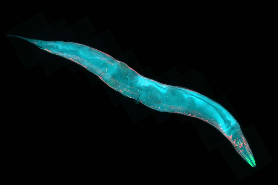
In the tiny worm known as C. elegans, the nervous system is far simpler, comprising a mere 118 classes of neurons.

At MIT, scientists in H. Robert Horvitz’s lab are studying the worms to learn about how nervous systems develop. Horvitz is the David H. Koch Professor of Biology at MIT, an Investigator at the McGovern Institute for Brain Research at MIT, and an Investigator at the Howard Hughes Medical Institute. His team has just discovered that a protein complex called cohesin, which helps shape the three-dimensional structure of the genome in both worms and humans, is critical for establishing some neurons’ identities as development unfolds.

The findings, reported July 31, 2026, in the journal Science Advances, could help scientists find a way to treat a rare developmental disorder called Cornelia de Lange syndrome, which is caused by mutations that interrupt the cohesin complex.

Model organism

Postdoctoral researcher Dongyeop Lee explains that C. elegans is a powerful model for studying neurodevelopment not just because its nervous system has been comprehensively mapped, but also because of the ease and speed with which scientists can study the function of its genes.

Because many of the worm’s genes have been retained through evolution, findings from studies of C. elegans often reveal important aspects of human biology. The current study began with worms that, because of a genetic mutation, make too many neurons of a certain type.

Adrenergic neurons, named for the kind of neurotransmitter they use to communicate with other neurons, are vital for enabling worms to respond to both their environment and their own internal state. Normally, C. elegans has just two pairs of adrenergic neurons: two RIM neurons and two RIC neurons. But the worms Lee studied had extras of both.

Takashi Hirose, a former member of the Horvitz lab, first observed this change in 2007.

Lee later continued the study and discovered that worms carrying a mutation in a gene called coh-1 have extra adrenergic neurons. The coh-1 gene encodes one part of the cohesin complex.

When Lee tested other mutations that disrupt cohesin, he found the same effect: Worms without fully functional cohesin had too many RIM neurons and too many RIC neurons.

Molecular switch

With a series of experiments designed to tease apart how cohesin impacts neurons’ identities, Lee discovered that cohesin cooperates with a gene-regulating protein called EOR-1 (known in humans as PLZF) to direct some neurons to develop into neurons that communicate with the inhibitory neurotransmitter GABA.

By reorganizing the structure of the genome, cohesin can change the way gene regulators like EOR-1 interact with DNA. Lee’s experiments showed that when either cohesin or EOR-1 couldn’t do its job, cells that should have become GABA-producing neurons become adrenergic neurons instead.

“What we found is that there are two alternative possible fates of certain neurons, and cohesin acts as a molecular switch that decides one of the possible neuronal fates,” Lee explains. “This means the structure of genomic DNA in the nucleus is important for neuronal fate determination.”

Disease connection

Lee adds that extra adrenergic neurons were not the only abnormality he observed in worms with cohesin mutations. Cohesin is important for shaping cells and tissues throughout the body. “The mutants have severe developmental defects,” Lee says. “They grow slowly. They don’t move well, and they also have defects in reproduction.”

Notably, the problems Lee saw in the worms echo aspects of Cornelia de Lange syndrome, a rare genetic disorder that impacts physical, cognitive, and behavioral development. Cornelia de Lange syndrome can be caused by mutations in cohesin genes, and Lee says that the discovery of how cohesin mutations affect worm development and behavior opens new opportunities to study the disease and search for potential therapeutic targets in C. elegans.

The Horvitz lab already has some promising leads. Taking advantage of the quick genetic screens that are possible in worms, Lee has found additional mutations that can counteract impaired cohesin, improving the health of worms with cohesin mutations. The team is now working to identify the genes where these suppressor mutations occur, so they can investigate whether they might make good therapeutic targets in humans.

Meanwhile, the team is also exploring a potential role for cohesin in shaping the fates of other neuron types, as well as searching broadly for additional molecules that work with cohesin to guide development. “We expect we have opened up a new biology,” Lee says. “This paper is just the beginning.”
Paper.

A cell protector collaborates with a killer

New research from the Horvitz Lab reveals what it takes for a protein that is best known for protecting cells against death to take on the opposite role.

Jennifer Michalowski | McGovern Institute
November 1, 2024

From early development to old age, cell death is a part of life. Without enough of a critical type of cell death known as apoptosis, animals wind up with too many cells, which can set the stage for cancer or autoimmune disease. But careful control is essential, because when apoptosis eliminates the wrong cells, the effects can be just as dire, helping to drive many kinds of neurodegenerative disease.

By studying the microscopic roundworm Caenorhabditis elegans—which was honored with its fourth Nobel Prize last month—scientists at MIT’s McGovern Institute have begun to unravel a longstanding mystery about the factors that control apoptosis: how a protein capable of preventing programmed cell death can also promote it. Their study, led by McGovern Investigator Robert Horvitz and reported October 9, 2024, in the journal Science Advances, sheds light on the process of cell death in both health and disease.

“These findings, by graduate student Nolan Tucker and former graduate student, now MIT faculty colleague, Peter Reddien, have revealed that a protein interaction long thought to block apoptosis in C. elegans, likely instead has the opposite effect,” says Horvitz, who shared the 2002 Nobel Prize for discovering and characterizing the genes controlling cell death in C. elegans.

Mechanisms of cell death

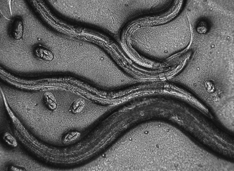
Horvitz, Tucker, Reddien and colleagues have provided foundational insights in the field of apoptosis by using C. elegans to analyze the mechanisms that drive apoptosis as well as the mechanisms that determine how cells ensure apoptosis happens when and where it should. Unlike humans and other mammals, which depend on dozens of proteins to control apoptosis, these worms use just a few. And when things go awry, it’s easy to tell: When there’s not enough apoptosis, researchers can see that there are too many cells inside the worms’ translucent bodies. And when there’s too much, the worms lack certain biological functions or, in more extreme cases, can’t reproduce or die during embryonic development.

Work in the Horvitz lab defined the roles of many of the genes and proteins that control apoptosis in worms. These regulators proved to have counterparts in human cells, and for that reason studies of worms have helped reveal how human cells govern cell death and pointed toward potential targets for treating disease.

A protein’s dual role

Three of C. elegans’ primary regulators of apoptosis actively promote cell death, whereas just one, CED-9, reins in the apoptosis-promoting proteins to keep cells alive. As early as the 1990s, however, Horvitz and colleagues recognized that CED-9 was not exclusively a protector of cells. Their experiments indicated that the protector protein also plays a role in promoting cell death. But while researchers thought they knew how CED-9 protected against apoptosis, its pro-apoptotic role was more puzzling.

CED-9’s dual role means that mutations in the gene that encode it can impact apoptosis in multiple ways. Most ced-9 mutations interfere with the protein’s ability to protect against cell death and result in excess cell death. Conversely, mutations that abnormally activate ced-9 cause too little cell death, just like mutations that inactivate any of the three killer genes.

An atypical ced-9 mutation, identified by Reddien when he was a PhD student in Horvitz’s lab, hinted at how CED-9 promotes cell death. That mutation altered the part of the CED-9 protein that interacts with the protein CED-4, which is proapoptotic. Since the mutation specifically leads to a reduction in apoptosis, this suggested that CED-9 might need to interact with CED-4 to promote cell death.

The idea was particularly intriguing because researchers had long thought that CED-9’s interaction with CED-4 had exactly the opposite effect: In the canonical model, CED-9 anchors CED-4 to cells’ mitochondria, sequestering the CED-4 killer protein and preventing it from associating with and activating another key killer, the CED-3 protein —thereby preventing apoptosis.

To test the hypothesis that CED-9’s interactions with the killer CED-4 protein enhance apoptosis, the team needed more evidence. So graduate student Nolan Tucker used CRISPR gene editing tools to create more worms with mutations in CED-9, each one targeting a different spot in the CED-4-binding region. Then he examined the worms. “What I saw with this particular class of mutations was extra cells and viability,” he says—clear signs that the altered CED-9 was still protecting against cell death, but could no longer promote it. “Those observations strongly supported the hypothesis that the ability to bind CED-4 is needed for the pro-apoptotic function of CED-9,” Tucker explains. Their observations also suggested that, contrary to earlier thinking, CED-9 doesn’t need to bind with CED-4 to protect against apoptosis.

When he looked inside the cells of the mutant worms, Tucker found additional evidence that these mutations prevented CED-9’s ability to interact with CED-4. When both CED-9 and CED-4 are intact, CED-4 appears associated with cells’ mitochondria. But in the presence of these mutations, CED-4 was instead at the edge of the cell nucleus. CED-9’s ability to bind CED-4 to mitochondria appeared to be necessary to promote apoptosis, not to protect against it.

Looking ahead

While the team’s findings begin to explain a long-unanswered question about one of the primary regulators of apoptosis, they raise new ones, as well. “I think that this main pathway of apoptosis has been seen by a lot of people as more or less settled science. Our findings should change that view,” Tucker says.

The researchers see important parallels between their findings from this study of worms and what’s known about cell death pathways in mammals. The mammalian counterpart to CED-9 is a protein called BCL-2, mutations in which can lead to cancer.  BCL-2, like CED-9, can both promote and protect against apoptosis. As with CED-9, the pro-apoptotic function of BCL-2 has been mysterious. In mammals, too, mitochondria play a key role in activating apoptosis. The Horvitz lab’s discovery opens opportunities to better understand how apoptosis is regulated not only in worms but also in humans, and how dysregulation of apoptosis in humans can lead to such disorders as cancer, autoimmune disease and neurodegeneration.