Why are some bacterial genes high in purines?

In certain species of bacteria, the answer lies in shielding RNA transcripts from a quality-control factor called Rho. Understanding the requirements for expressible sequences is critical for expression engineering of therapeutic agents.

Lillian Eden | Department of Biology
July 2, 2026

In the study of bacteria, a longstanding dogma held that two molecular machines — RNA polymerase, which leads the way in transcribing DNA into RNA, and ribosomes, which bring up the rear translating RNA into proteins — worked so closely in tandem that they were effectively attached.

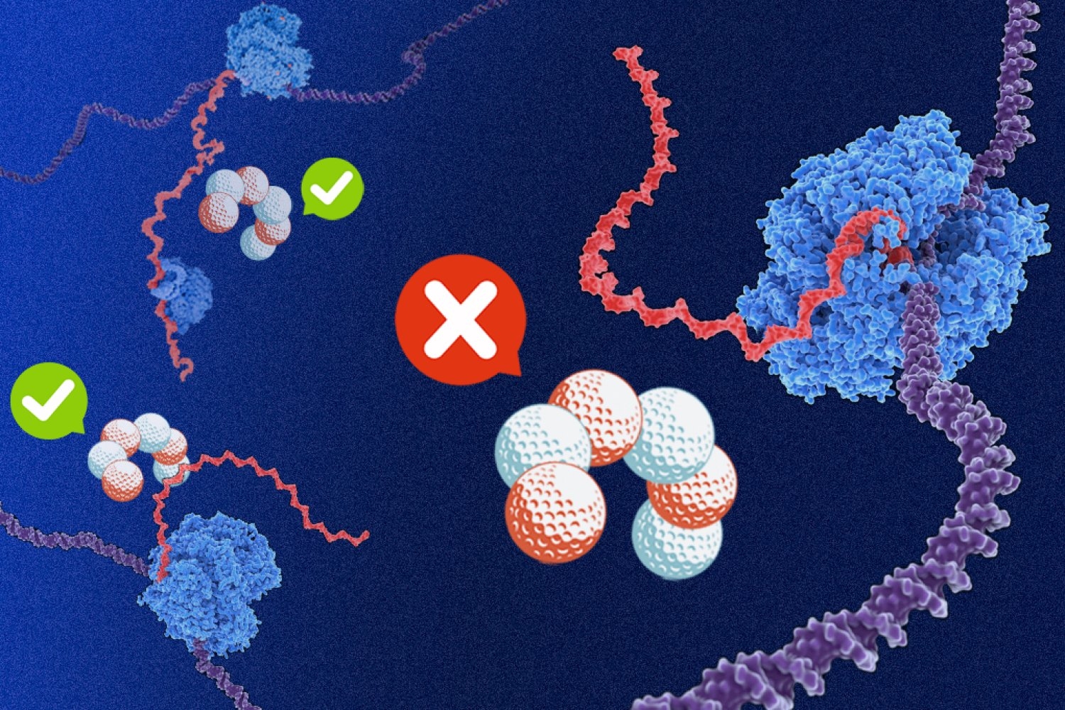
This close coupling of transcription and translation in bacteria was thought to be fundamental to gene expression in part because the trailing ribosome could shield nascent gene products from an effective and omnipresent quality-control protein called Rho.

In bacteria that exhibit something called runaway transcription, however, the polymerase instead speeds ahead, unhitched from its protective ribosome. Inexplicably, however, in bacteria that exhibit this runaway transcription, such as Bacillus subtilis, Rho targeted primarily noncoding, useless RNA products.

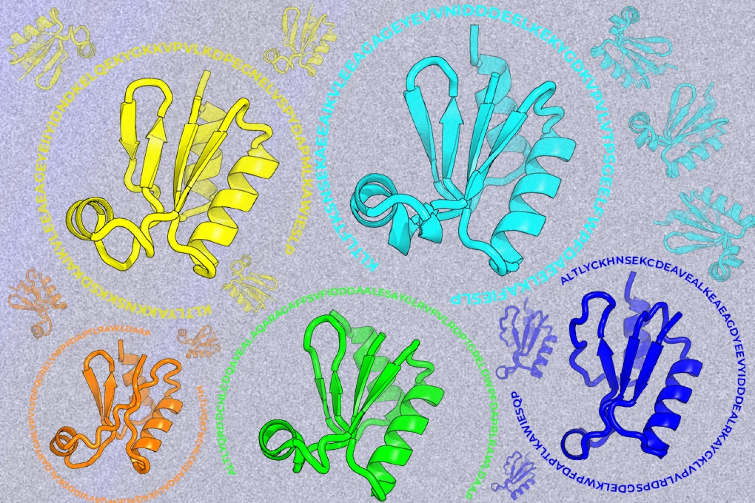
New research from the Department of Biology reveals that the secret to Rho’s quality-control specificity lies in the sequence composition of nucleotide bases that make up coding strands of DNA.

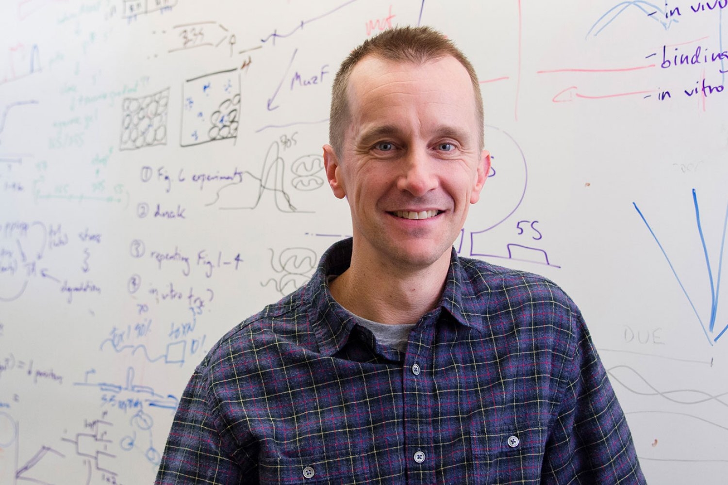
“We started with a hypothesis that Rho was regulated by sequence, but the fact that the sequence alone was enough to protect any gene in the entire B. subtilis genome from Rho was really surprising,” says Julia Dierksheide PhD ’26, a graduate student in the Li Lab and first author of a paper recently published in Nature Microbiology. “That’s a really diverse range of sequences — what sequence feature is shared by every single gene in the genome?”

Barricading with bias

Rho serves as a termination factor, meaning that it is a crucial mechanism for preventing bacteria from wasting precious resources by making RNA transcripts that serve no purpose.

All the information a bacterial cell needs is encoded in its DNA, which is made up of two strands of nucleic acids. These strands twist together to form a double helix, with genetic information codified in pairs of bases: purines guanine and adenine are matched with pyrimidines cytosine and thymine, respectively. Any sequence that gives rise to RNA transcripts is stored in complement to a parallel, noncoding strand, meaning that a large portion of genetic material is transcriptionally useless.

Coding DNA strands in certain bacteria were known to be significantly higher in purines guanine and adenine compared to the rest of the bacterial genome. The researchers found that this purine bias alone shields productive mRNA transcripts from Rho-mediated termination.

“I love having a big, complicated dataset and trying to reduce that to biological meaning,” Dierksheide says. “It seems like Rho itself has been broadly shaping the evolution of the B. subtilis genome to create these sequence composition biases.”

Bacterial species that, over generations, have lost Rho no longer exhibit this strong purine bias.

Rho also serves as a regulatory factor in bacteria becoming motile, forming biofilms, or sporulating, all of which are critical for biology and survival. The purine bias could also provide a layer of protection against the insertion of foreign DNA, for example, when a viral bacteriophage infects bacteria.

“Bacteria exist as single cells, so everything that they do, they have to do through gene expression,” Dierksheide says. “Understanding the fundamental details about how gene expression works, how a cell encodes all the information it needs to survive in the nucleotide sequence of the genome, is really exciting.”

Future directions

Although the exact mechanism underlying Rho’s specificity remains unclear, these results crack an underlying code in the composition of bacterial genomes.

Dierksheide said she hoped to perform a similar screen to characterize Rho’s specificity in Escherichia coli, which diverged from B. subtilis on the evolutionary tree an estimated 2 billion years ago and still exhibits coupled transcription-translation, where the transcribing RNA polymerase is closely followed by a translating ribosome.

The high sequence specificity of B. subtilis Rho is crucial for the protection of its runaway RNA polymerase, in which that molecular machine speeds ahead of the ribosome. A systematic comparison to E. coli Rho could help reveal how this heightened stringency arose.

This information will be critical for engineering diverse bacterial species for applications including the production of therapeutic agents. Other bacterial species, such as B. subtilis, may be better models for this process because they have abundant secretion pathways, according to Dierksheide, making it much easier to produce and isolate proteins in large quantities.

“Our findings reveal an important criterion for successful sequence design that must be considered in expression engineering,” says associate department head, associate professor of biology, and Howard Hughes Medical Institute investigator Gene-Wei Li, the lead author of the study. “There are so many cryptic messages in the genome, like the purine bias, and we are just beginning to be able to decipher what they mean.”

Raised in the “Kitchen”

Karen O’Leary, lab associate and acting supervisor in the Glassware Sterilization Facility, has become a cornerstone of the department’s operations.

Samantha Edelen | Department of Biology
June 25, 2026

Early mornings in the halls of Building 68 feature the sounds of rolling wheels on big metal carts, the rattling of glassware, the whooshing of faucets, and the clanking of autoclaves.

These aren’t the sounds of researchers at work, but rather those of keeping the labs sterilized and stocked with the sundries of research: pipette tips, test tubes, flasks, petri dishes, and more.

Orchestrating this sunrise cacophony and the staff that undertakes it is Karen O’Leary, lab associate and acting supervisor in the Glassware Sterilization Facility, also known as the “kitchen.”

Thanks, in part, to O’Leary’s proactivity and hard work, the kitchen staff were recently recognized with an MIT Excellence Award in 2025 for exceptional contributions in service of the community.

“My goal is to get the scientists everything they need to do their research,” O’Leary says. “I’m good at what I do.”

O’Leary admits she did not always possess such confidence. In almost 40 years at MIT, O’Leary has grown into this critical role for the department, and the department itself has evolved, moving into a brand-new building and away from previously standard practices like submerging equipment in acid for sterilization.

From rookie to running the show

On Sept. 7, 1987, Karen O’Leary joined the MIT community as a staff member for the first time. The 18-year-old was fresh from vocational high school, where she studied cosmetology but felt too shy to pursue that as a career. She was also nervous about joining a research institution.

“When I started, I didn’t even know what a beaker was,” she recalls.

Too embarrassed to admit in her interview that she couldn’t remember her brand-new home phone number, “I just made one up.” Fortunately, this didn’t prevent her from getting the job, where she worked under the mentorship of Thelma Watkins, who would retire in 1996 after 21 years at MIT. Watkins was critical for instilling a good work ethic and boosting O’Leary’s confidence.

“She taught me to show up every day, and work hard, and laugh,” O’Leary says.

Even now, O’Leary continues to bring joy to that daily diligence, for herself and for her staff.

“Karen is always on top of things,” says longtime friend and fellow Lab Associate AnnMarie Budhai. “She doesn’t refuse work and always goes above and beyond.”

Facilities and Operations Manager Cesar Duarte says that O’Leary’s long tenure, support, and knowledge have been invaluable as he transitioned into his role in Building 68 starting in 2023.

“Karen is one of those people who makes everything around her run more smoothly and more pleasantly,” Duarte says.

Better, faster, safer

Although some might consider it drudgery, O’Leary says that washing glassware is her favorite task.

“I like that when I wash, I can see the job is complete at the end of the day,” she says.

Although washing glassware is a perennial task, safety and efficiency have come a long way in the past 38 years. More-effective autoclaves and dishwashers have eliminated steps like steaming to dissolve agar solvents before autoclaving, and scrubbing individual test tubes before washing.

O’Leary was working for the department in 2011 when Building 68 piloted a new approach to MIT’s management of regulated medical waste (RMW), such as petri dishes, blood, and needles — the new system, which is cheaper and produces less waste, is now used by all departments at MIT that produce RMW.

“EHS [the Environment, Health and Safety Office] has come really far — I’m glad we got away from acid,” O’Leary notes of the bygone era of submerging glass pipettes for sterilization. “Back then, no one knew of a better way.”

Other tasks include cleaning velvets, which are used for replicating bacterial colonies on petri dishes, and pouring agar plates.

“Everyone knows how to do almost every job, so we can take turns doing different tasks,” O’Leary says. “If you get sick, there’s always someone to cover.”

All in the family

For O’Leary, kinship with MIT has spanned generations. O’Leary was raised in Weymouth, Massachusetts, by a father who worked at MIT as a supervisor in the sheet metal shop. Having raised children of her own, now grown, O’Leary came to greatly appreciate the flexibility her job has granted her.

“I’ve had great work-family balance here,” she says. Even though she’s often at work more than an hour before the researchers that the kitchen serves, “The hours are great, and with MIT Health right across the street, it was easy to take everyone to doctors’ appointments.”

She’s also gained a chosen family at MIT, spending breaks at work taking long walks along the Charles River, “talking about anything and everything” with colleagues like Budhai and Lab Aide Janet Katin.

“We really grew up together,” she says.

Working at MIT has provided O’Leary with support and community, and she’d like to pay it forward. In addition to strolling with colleagues, she hits the gym to help maintain the energy required for her highly active work.

“I don’t like sitting around,” she says.

In addition to maintaining her stamina at work, she hopes that taking care of herself will keep her actively involved if she ever has grandchildren, and enable her to help neighborhood kids when she someday retires.

“I owe a lot to MIT,” she says. “I have been allowed to work hard and get satisfaction and have been appreciated and given space to care for my family.”

O’Leary returns this care to the Department of Biology in spades.

“It’s an understatement to say that Biology is lucky to have her,” says Duarte. “Karen’s overflowing energy, attention to detail, and care for the Biology research community are nothing short of amazing.”

Enzymes that assemble into droplets can speed up cellular reactions

MIT biologists find highly concentrated droplets can help cells keep enzymes organized and control growth signals.

Anne Trafton | MIT News
June 1, 2026

Within the past decade, biologists have discovered that one strategy cells use to keep their contents organized is a phenomenon known as phase separation.

Similar to the way oil forms droplets that float in a vinegar solution, proteins inside cells can phase separate to form highly concentrated droplets that keep them organized within the cell. In a new study, MIT researchers have now shown that this droplet formation is critical for controlling the function of a class of enzymes called kinases.

The researchers found that condensing into droplets optimizes the biochemical conditions needed for kinases to catalyze reactions, allowing them to more rapidly activate cell signaling pathways. In some cases, droplet formation can even change which reactions the kinases perform.

“Many biological molecules have this propensity to spontaneously separate. We were really interested in asking, if we have these kinases forming droplets, what is the consequence of that in the context of signaling?” says Lindsay Case, an assistant professor of biology at MIT and the senior author of the study.

Learning more about how these droplets form could help researchers design drugs that target kinases, some of which can be overactive in cancer cells.

“Understanding the chemistry of these compartments, and what molecules go into them and what molecules don’t go into them, could help us design drugs that better localize to their target of interest,” Case says.

Nicholas Lea, an MIT graduate student, is the lead author of the paper, which appears today in Cell Reports.

Forming droplets

Since her days as a graduate student, Case has been studying how the physical organization of molecules inside cells affects their function. As a postdoc, she began studying how phase separation might affect a signaling pathway that allows cells to sense when they’re attached to their environment, so they can respond appropriately.

Some of the proteins in this pathway are kinases, which activate other proteins by adding phosphate groups to them. Kinases can also activate themselves through a process called autophosphorylation.

“Inside of the cell, you have these kinase molecules that are responsible for carrying a signal through the cell, and we know that the organization of these molecules changes. When the information is present, they’re organized in a different way than when the information is not present,” Case says. “We think that having the right molecules in the right place is incredibly important for the right biochemistry to occur.”

Phase separation is one of the methods that cells appear to use for this organization. The most familiar example of phase separation can be seen in a salad dressing, where oil forms droplets to minimize contact with water-based vinegar. Proteins can phase separate when they are highly concentrated, leading them to self-assemble into dense droplets floating in the cell’s cytoplasm.

Case hypothesized that this phase separation, which brings kinases together at a high density, might help cells to boost the enzymes’ activity because they are more likely to bump into and phosphorylate each other.

In this study, Case and Lea set out to test that hypothesis, focusing on an enzyme called focal adhesion kinase (FAK). This kinase, which becomes activated when cells attach to their surrounding environment, activates pro-growth and pro-survival signals. In cancer cells, this signaling pathway can go awry, allowing cells to proliferate even when they detach from their original locations.

Scientists already knew that when cells are properly attached to their environment, that adhesion signal causes FAK to accumulate at the cell membrane. In the new study, the MIT team mimicked that effect by overexpressing FAK in cells. These cells were floating freely in a solution, not attached to any surface. Even so, the high concentration of FAK caused the kinase to phase separate into droplets, which turned on the pro-growth signal.

“It was surprising that just by condensing this protein into a droplet, you can actually turn on a signaling pathway that should be turned off,” Case says. “If FAK concentration is too high, you’re always getting these droplets and you’re always signaling, regardless of what the receptors that are supposed to be controlling this are doing.”

The findings suggest that in cancer cells, overexpression of FAK may lead to phase separation, which then helps to drive cancer progression and metastasis.

“It may be that for some kinases, you’re not supposed to form these droplets in the cytoplasm because it leads to this always-on signal, and then the cells no longer listen to the information coming from the environment,” Case says.

Interfering with FAK’s ability to form droplets could offer a new strategy for cancer drug development, she says.

Controlling reactions

The researchers also studied two other kinases, Mst2 and Abl. They found that these enzymes could also phase separate at high concentrations, and that this increased their activity. While phase separation of FAK in the cytoplasm may occur only in cancerous cells, for Mst2, it appears to be a strategy that healthy cells use to control a signaling pathway called Hippo, which promotes cell growth and survival.

Additionally, for both Mst2 and Abl, the researchers discovered that phase separation can lead the enzymes to phosphorylate additional targets, which may lead them to activate different signaling pathways.

“It’s not just that you’re getting faster phosphorylation, but in those cases, the patterns of what is actually getting phosphorylated were very different inside of the droplet compared to what might be happening in a non-droplet context,” Case says. “The kinase is able to phosphorylate amino acid residues beyond the set of canonical sites that have been described before.”

The researchers also found that when these droplets form, they attract high concentrations of ATP, the molecule that kinases use as a source of phosphate. This occurs because kinases tend to contain floppy sections containing many positively charged amino acids, which attract negatively charged ATP.

Using a machine-learning model, the researchers predicted that about 45 percent of the 500 kinases found in human cells would have the ability to form droplets like those seen in this study. Those kinases were also more likely to be highly positively charged, which could help them to recruit ATP into the droplets.

In future work, Case hopes to explore the possibility of designing drugs that could mimic ATP’s ability to be attracted into droplets within a cell, which could help reduce negative side effects of the drugs.

“By localizing drugs to the compartment where your target localizes, that could reduce off-target effects by concentrating the drug with the target of interest and reducing interactions with other molecules,” Case says.

The research was funded by a Searle Scholars Program Award, the U.S. Air Force Office of Scientific Research, the National Institutes of Health, the Royal G. and Mae H. Westaway Family Memorial Fund, and a David H. Koch Graduate Fellowship.

Biologist Joey Davis explores how cells build complex structures

His studies have shed light on the assembly instructions that govern ribosomes, the critical protein-building machines of the cell.

Anne Trafton | MIT News
May 5, 2026

Ribosomes, the cellular machines that assemble proteins, are made from dozens of proteins and RNA molecules. Putting all of those pieces together is a complex puzzle — one that MIT Associate Professor Joey Davis PhD ’10 revels in trying to solve.

Understanding how these structures form and later break down could help researchers learn more about how disruptions of these fundamental processes can lead to disease. But, as Davis points out, it’s also an interesting biological question.

“Our long-term goal is to really understand how the natural world assembles these huge complexes rapidly and efficiently. It’s a fundamentally interesting question to think about how these things get put together,” he says.

His work has helped reveal that unlike building a house, which happens in a prescribed sequence of steps — pouring the foundation, building the frame, putting on the roof, then doing electrical and plumbing work — ribosomes can be assembled in a more flexible way. Cells can even skip an assembly step and then come back to it later.

“In these natural systems, it seems like the assembly pathways are much more dynamic and flexible,” he says. “It appears that evolution has selected pathways that aren’t strictly ordered in the way we would think about an assembly line, where you always put in one component, then the next, and then the next. We’re excited to understand the selective advantages of such approaches.”

A love of discovery

Davis’ interest in how things are put together developed early in life, inspired by his father, a carpenter who framed houses. During the mid-1980s, the family moved from Colorado to Southern California, where his father worked in construction during a housing boom there.

“I was always interested in building things, which I think probably came from being around my dad and other builders,” Davis says.

As an undergraduate at the University of California at Berkeley, where he majored in computer science and biological engineering, Davis’ interests turned toward smaller scales, in the realm of cells and molecules. During his junior year, he started working in the lab of chemistry professor Michael Marletta, who studies molecular-level biological interactions.

In the lab, Davis investigated how enzymes that contain heme are able to preferentially bind to either oxygen or nitric oxide, two gases that are very similar in structure. That work kindled a love of studying the natural world and pursuing discoveries in fundamental science.

“Being in the Marletta lab and seeing students and postdocs that were really passionate about these problems had a big impact on me,” Davis says. “The goal was to understand the fundamentals of how molecular discrimination works, and the idea of discovery for the sake of discovery was thrilling.”

After graduating from Berkeley, Davis spent another year working in Marletta’s lab, and then a year working odd jobs, before heading to MIT to pursue a PhD in biology. There, he worked with Professor Bob Sauer, now emeritus, who studied the relationship between protein structure and function, with a particular focus on the molecular machines that degrade or remodel proteins.

Davis’ thesis research centered on enzymes called AAA proteases, which remove damaged proteins from cellular membranes and send them to cell organelles that break them down. In addition to studying the structure and function of the proteases, Davis worked on ways to engineer them to tag specific proteins for destruction.

That work led him into synthetic biology, which he used to develop genetic parts that drive production of proteins of interest. Some of those parts ended up being used by the biotech startup Ginkgo Bioworks, where Davis took a job as a senior scientist after graduating.

Working at Ginkgo Bioworks allowed Davis to stay in Boston while his partner finished her PhD. The couple then moved back to California, where Davis worked as a postdoc at Scripps Research, which was home to one of the first direct electron detection cameras for cryo-electron microscopy (cryo-EM). These detectors allow researchers to generate structures with near atomic resolution. At Scripps, Davis began using them to study ribosomes as they were being assembled.

Peering into the ribosome

After joining the MIT faculty in 2017, Davis continued his work on ribosomes and assembled a lab group that includes students from a variety of backgrounds who work together to develop new ways to explore biological phenomena.

“I have a mix of method developers and biologists in the group, and the work from each of them informs each other,” Davis says. “My lab goes back and forth between building sets of tools to answer biological questions, and then as we’re answering those questions, it motivates the next generation of tool development.”

During ribosome assembly, RNA molecules fold themselves into the correct shapes, creating docking sites for proteins to attach. Then, more RNA molecules come in and fold themselves into the structure.

“It’s a beautifully coupled process by which the cell folds hundreds of RNA helices and binds on the order of 50 proteins, and it does it in two minutes from start to finish. E. coli does this 100,000 times per hour, and it’s amazing how rapid and efficient the process is,” Davis says.

Cryo-EM allows scientists to capture this process in minute detail. It can be used to take hundreds of thousands of two-dimensional images of ribosome samples frozen in a thin layer of ice, from different angles. Computer algorithms then piece together these images into a three-dimensional representation of the ribosome.

To gain insight into how ribosomes are assembled, researchers can stall the process at different points and then analyze the resulting structures. In 2021, Davis’s lab developed a new method called CryoDRGN, which uses neural networks to analyze cryo-EM data and generate the full ensemble of structures that were present in the sample.

This work has shown that when certain steps of ribosome assembly are blocked, many different structures result, suggesting that the assembly can occur in a variety of ways.

In future work, Davis aims to dramatically increase the throughput of cryo-EM to generate datasets of protein structures that could help improve the AI-based models that are now used to predict protein structures.

“There are still huge swaths of sequence space that these models are very poor at predicting, but if we could collect data on those sequences en masse, that could potentially serve as key training data for a next-generation protein structure prediction method that could fill out that space,” he says.

Professor Michael Laub named 2025 AAAS Fellow

The American Association for the Advancement of Science recognized Laub and 21 alumni for their efforts to advance science and related fields.

School of Science
April 17, 2026

MIT Professor Michael T. Laub as well as 21 MIT alumni have been elected as fellows of the American Association for the Advancement of Science (AAAS).

The 2025 class of AAAS Fellows includes 449 scientists, engineers, and innovators, spanning all 24 of AAAS disciplinary sections, who are recognized for their scientific achievements.

Laub, the Salvador E. Luria Professor in the MIT Department of Biology and an HHMI Investigator, studies the biological mechanisms and evolution of how cells process information to regulate their own growth and proliferation, using bacteria as a model organism to develop a deeper, fundamental understanding of how bacteria function and evolve. Laub was honored as a AAAS Fellow for distinguished contributions to the field of bacterial information processing, particularly to the understanding of coevolution of host-pathogen response and immunity.

“This year’s AAAS Fellows have demonstrated research excellence, made notable contributions to advance science, and delivered important services to their communities,” said Sudip S. Parikh, AAAS chief executive officer and executive publisher of the Science family of journals. “These fellows and their accomplishments validate the importance of investing in science and technology for the benefit of all.”

The following alumni were also named fellows of the AAAS:

  • Debra Auguste ’99
  • Julie Claycomb PhD ’04
  • Chris Clifton ’85, SM ’86
  • Kevin Crowston PhD ’91
  • Maitreya Dunham ’99
  • David Fike PhD ’07
  • Jianping Fu PhD ’07
  • Peter A. Gilman SM ’64, PhD ’66
  • Diane M. Harper ’80, SM ’82
  • Cherie R. Kagan PhD ’96
  • Elizabeth A. Kensinger PhD ’03
  • Kenro Kusumi PhD ’97
  • Charla Lambert ’96
  • Bennett A. Landman ’01, MNG ’02
  • Michael E. Matheny SM ’06
  • Paul David Ronney ScD ’83
  • Steven Semken ’80, PhD ’89
  • Sudipta Sengupta SM ’99, PhD ’06
  • Lawrence R. Sita PhD ’86
  • Jan M. Skotheim ’99
  • Beverly Park Woolf ’66
Slice and dice

SNIPE, a newly characterized defense system, directly protects bacteria by chopping up invading viral DNA.

Lillian Eden | Department of Biology
April 9, 2026

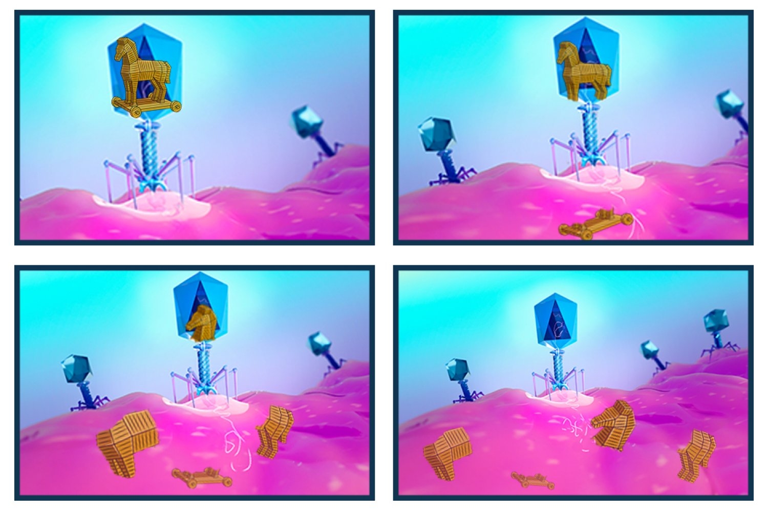
What if the Trojan horse had been pulled to pieces, revealing the ruse and fending off the invasion, just as it entered the gates of Troy?

That’s an apt description of a newly characterized bacterial defense system that chops up foreign DNA.

Bacteria and the viruses that infect them, bacteriophages — phages for short — are ceaselessly at odds, with bacteria developing methods to protect themselves against phages that are constantly striving to overcome those safeguards.

New research from the Department of Biology at MIT, recently published in Nature, describes a defense system that is integrated into the protective membrane that encapsulates bacteria. SNIPE, which stands for surface-associated nuclease inhibiting phage entry, contains a nuclease domain that cleaves genetic material, chopping the invading phage genome into harmless fragments before it can appropriate the host’s molecular machinery to make more phages.

Daniel Saxton, a postdoc in the Laub Lab and the paper’s first author, was initially drawn to studying this bacterial defense system in E. coli, in part because it is highly unusual to have a nuclease that localizes to the membrane, as most nucleases are free-floating in the cytoplasm, the gelatinous fluid that fills the space inside cells.

“The other thing that caught my attention is that this is something we call a direct defense system, meaning that when a phage infects a cell, that cell will actually survive the attack,” Saxton says. “It’s hard to fend off a phage directly in a cell and survive — but this defense system can do it.”

Light it up

For Saxton, the project came into focus during a fluorescence-based experiment in which viral genetic material would light up if it successfully penetrated the bacteria.

“SNIPE was obliterating the phage DNA so fast that we couldn’t even see a fluorescent spot,” Saxton recalls. “I don’t think I’ve ever seen such an effective defense system before — you can barrage the bacteria with hundreds of phage per cell, but SNIPE is like god-tier protection.”

When the nuclease domain of SNIPE was mutated so it couldn’t chop up DNA, fluorescent spots appeared as usual, and the bacteria succumbed to the phage infection.

Bacteria maintain tight control over all their defense systems, lest they be turned against their host. Some systems remain dormant until they flare up, for example, to halt all translation of all proteins in the cell, while others can distinguish between bacterial DNA and foreign, invading phage DNA. There were only two previously characterized mechanisms in the latter category before researchers uncovered SNIPE.

“Right now, the phage field is at a really interesting spot where people are discovering phage defense systems at a breakneck pace,” Saxton says.

Problems at the periphery

Saxton says they had to approach the work in a somewhat roundabout way because there are currently no published structures depicting all the steps of phage genome injection. Studying processes at the membrane is challenging: Membranes are dense and chaotic, and phage genome injection is a highly transient process, lasting only a few minutes.

SNIPE seems to discern viral DNA by interacting with proteins the phage uses to tunnel through the bacteria’s protective membrane. This “subcellular localization,” according to Saxton, may also prevent SNIPE from inadvertently chopping up the bacteria’s own genetic material.

The model outlined in the paper is that one region of SNIPE binds to a bacterial membrane protein called ManYZ, while another region likely binds to the tape measure protein from the phage.

The tape measure protein got its name because it determines the length of the phage tail — the part of the phage between the small, leglike protrusions and the bulbous head, which contains the phage’s genetic material. The researchers revealed that the phage’s tape measure protein enters the cytoplasm during injection, a phenomenon that had not been physically demonstrated before.

There may also be other proteins or interactions involved.

“If you shunt the phage genome injection through an alternate pathway that isn’t ManYZ, suddenly SNIPE doesn’t defend against the phage nearly as well,” Saxton says. “It’s unclear exactly how these proteins interact, but we do know that these two proteins are involved in this genome injection process.”

Future directions

Saxton hopes that future work will expand our understanding of what occurs during phage genome injection and uncover the structures of the proteins involved, especially the tunnel complex in the membrane through which phages insert their genome.

Members of the Laub Lab are already collaborating with another lab to determine the structure of SNIPE. In the meantime, Saxton has been working on a new defense system in which molecular mimicry — bacterial proteins imitating phage proteins — may play a role.

Michael T. Laub, the Salvador E. Luria Professor of Biology and a Howard Hughes Medical Institute investigator, notes that one of the breakthrough experiments for demonstrating how SNIPE works came from a brainstorming session at a lab retreat.

“Daniel and I were kind of stuck with how to directly measure the effect of SNIPE during infection, but another postdoc in the lab, Ian Roney, who is a co-author on the paper, came up with a very clever idea that ultimately worked perfectly,” Laub recalls. “It’s a great example of how powerful internal collaborations can be in pushing our science forward.”

Building the blocks of life

Computational biologist Sergei Kotelnikov is working to develop new methods in protein modeling as part of the School of Science Dean’s Postdoctoral Fellowship.

Lyn Nanticha Ocharoenchai | School of Science
March 31, 2026

Billions of years ago, simple organic molecules drifted across Earth’s primordial landscape — nothing more than basic chemical compounds. But as natural forces shaped the planet over hundreds of millions of years, these molecules began to interact and bond in increasingly complex ways. Along the way, something spectacular emerged: life.

“Life is, to some degree, magical,” says computational biologist Sergei Kotelnikov. Simple organic compounds congregate into polymers, which assemble into living cells and ultimately organisms — the whole being greater than the sum of its parts.

“You can write formulas on how a molecule behaves,” he says, referring to the world of quantum mechanics. “But yet somehow, a few orders of magnitude above, on a bigger scale, it gives rise to such a mystery.”

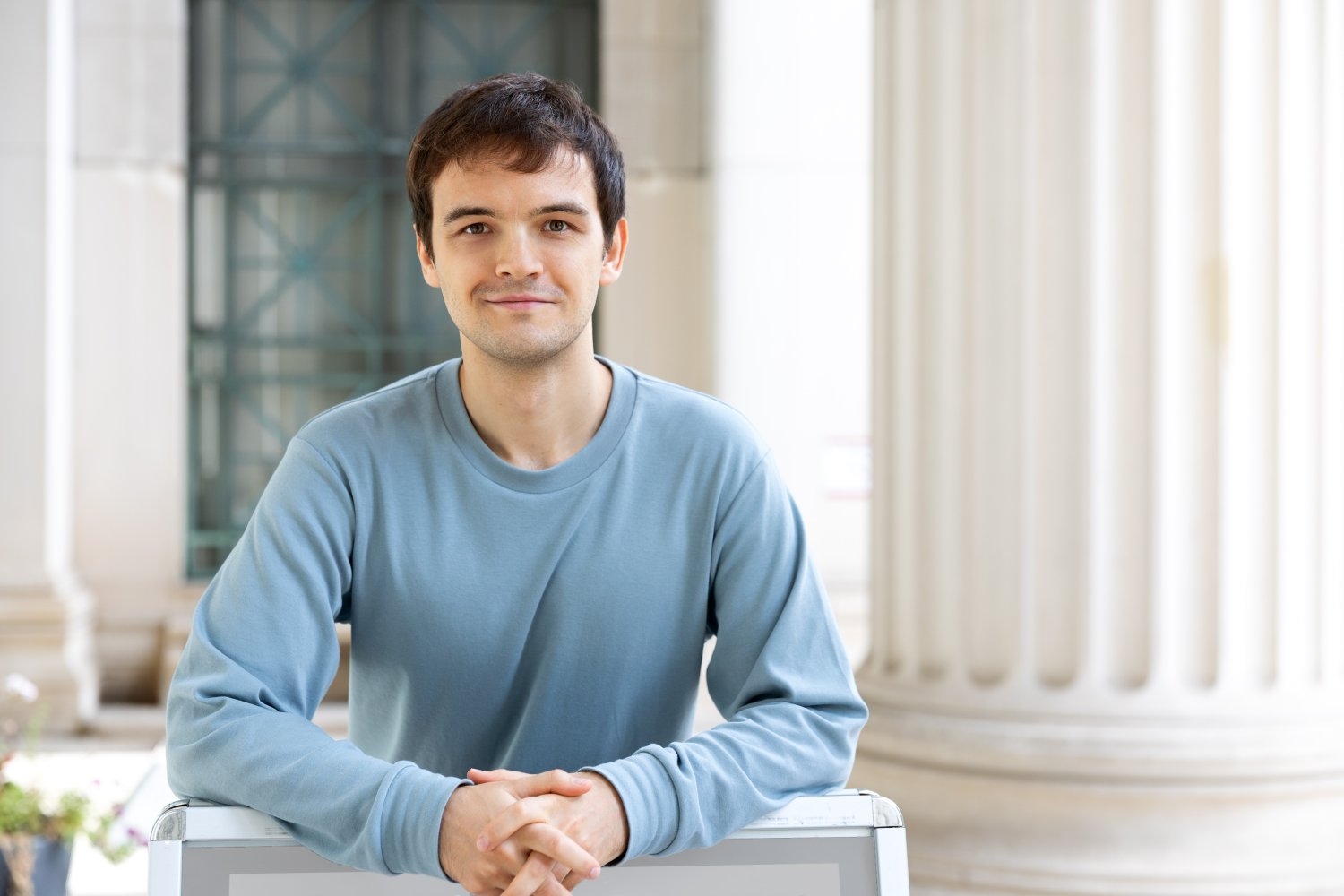
Kotelnikov builds models to analyze and predict the structure of these biomolecules, particularly proteins, the fundamental building blocks of every organism. This year, he joined MIT as part of the School of Science Dean’s Postdoctoral Fellowship to work with the Keating Lab, where researchers focus on protein structure, function, and interaction. Using machine learning, his goal is to develop new methods in protein modeling with potential applications that span from medicine to agriculture.

A hunger for problems to solve

Kotelnikov grew up in Abakan, Russia, a small city sitting right in the center of Eurasia. As a child, one of his favorite pastimes was playing with Lego bricks.

“It encouraged me to build new things, rather than just following instructions,” he says. “You can do anything.”

Kotelnikov’s father, whose background lies in engineering and economics, would often challenge him with math problems.

“Your brain — you can feel some kind of expansion of understanding how things work, and that’s a very satisfactory feeling,” Kotelnikov says.

This itch to solve problems led him to join science Olympiad competitions, and later, a science-focused public boarding school located near the Russian Academy of Sciences, from which he often encountered scientists.

“It was like a candy shop,” he recalls, describing the period as a life-changing experience.

In 2012, Kotelnikov began his bachelor of science in physics and applied mathematics at the Moscow Institute of Physics and Technology — considered one of the leading STEM universities in Russia, and globally — and continued there for his master’s degree. It was there that biology came into the picture.

During a course on statistical physics, Kotelnikov was first introduced to the idea of the “emergence of complexity.” He became fascinated by this “mysterious and attractive manifestation of biology … this evolution that sharpens the physical phenomenon” to create, drive, and shape life as we know it today. By the time he completed his master’s degree, he realized he had only scratched surface of the field of computational biology.

In 2018, he began his PhD at Stony Brook University in New York and began working with Dima Kozakov, who is recognized as one of the world’s leaders in predicting protein interactions and complex structures.

Studying the architecture of life  

Proteins act like the bricks that construct an organism, underpinning almost every cellular process from tissue repair to hormone production. Like pieces of a Lego tower, their structures and interactions determine the functions that they carry out in a body.

However, diseases arise when they’re folded, curled, twisted, or connected in unusual ways. To develop medical interventions, scientists break down the tower and examine each individual piece to find the culprit and correct their shape and pairing. With limited experimental data on protein structures and interactions currently available, simulations developed by computational biologists like Kotelnikov provide crucial insight that inform fundamental understanding and applications like drug discovery.

With the guidance of Kozakov at Stony Brook’s Laufer Center for Physical and Quantitative Biology, Kotelnikov carried over his understanding of physics to create modeling methods that are more effective, efficient, reliable, and generalizable. Among them, he developed a new way of predicting the protein complex structures mediated by proteolysis-targeting chimeras, or PROTACs, a new class of molecules that can trigger the breakdown of specific proteins previously considered undruggable, such as those found in cancer.

PROTACs have been challenging to model, in part because they are composed of proteins that don’t naturally interact with each other, and because the linker that connects them is flexible. Imagine trying to guess the overall shape of a bendy Lego piece attached to two other pieces of different irregular, unmatched shapes. To efficiently find all possible configurations, Kotelnikov’s method conceptually cuts the linker into two halves and models each separately, then reformulates the problem and calculates it using a powerful algorithm called Fast Fourier Transform.

“It’s kind of like applied math judo that you sometimes need to do in order to make certain intractable computations tractable,” he says.

Kotelnikov’s state-of-the-art methods have been instrumental to his team’s top performance in numerous international challenges including the Critical Assessment of protein Structure Prediction (CASP) competition — the same contest in which the Nobel Prize-winning AlphaFold system for protein 3D structure prediction was presented.

Physics and machine learning

At MIT, Kotelnikov is working with Amy Keating, the Jay A. Stein (1968) Professor of Biology, biology department head, and professor of biological engineering, to study protein structure, function, and interactions.

A recognized leader in the field, Keating employs both computational and experimental methods to study proteins, their interactions, as well as how this can impact disease. By infusing physics with machine learning, Kotelnikov’s goal is to advance modeling methods that can vastly inform applications such as cancer immunology and crop protection.

“Kotelnikov stands to gain a lot from working closely with wet lab researchers who are doing the experiments that will complement and test his predictions, and my lab will benefit from his experience developing and applying advanced computational analyses,” says Keating.

Kotelnikov is also planning to work with professors Tommi Jaakkola and Tess Smidt in MIT’s Department of Electrical Engineering and Computer Science to explore a field called geometric deep learning. In particular, he aims to integrate physical and geometric knowledge about biomolecules into neural network architectures and learning procedures. This approach can significantly reduce the amount of data needed for learning, and improve the generalizability of resulting models.

Beyond the two departments, Kotelnikov is also excited to see how the diversity and interdisciplinary mix of MIT’s community will help him come up with ideas.

“When you’re building a model, you’re entering this imaginary world of assumptions and simplifications and it might feel challenging because of this disconnect with reality,” Kotelnikov says. “Being able to efficiently communicate with experimentalists is of high value.”

Leading with rigor, kindness, and care

“We cannot be effective scientists if we are unhappy or unhealthy outside of the lab,” says “Committed to Caring” honoree Sara Prescott.

Leila Hudson | Office of Graduate Education
March 27, 2026

Professor Sara Prescott embodies the kind of mentorship every graduate student hopes to find: grounded in scientific rigor, guided by kindness, and defined by a deep commitment to well-being. Her approach reflects a simple but powerful belief that transformative mentorship is not only about advancing research, but about cultivating confidence, belonging, and resilience in the next generation of scholars.

A member of the 2025–27 Committed to Caring cohort, Prescott exemplifies the program’s spirit, which honors faculty who go above and beyond in nurturing both the intellectual and personal development of MIT’s graduate students.

Prescott is the Pfizer Inc. – Gerald D. Laubach Career Development Professor in the MIT departments of Biology and Brain and Cognitive Sciences, and an investigator at the Picower Institute for Learning and Memory. Her research addresses fundamental questions in body-brain communication, with a focus on lung biology, early-life adversity, women’s health, and the impacts of climate change on respiratory health.

A culture of compassion

Prescott’s mentoring philosophy begins with a focus on professional sustainability. “We cannot be effective scientists if we are unhappy or unhealthy outside of the lab,” she says.

She pushes back against what she sees as an unhelpful narrative in academia. “There’s this idea that you must choose between a successful PhD or having a personal life. This is a false dichotomy, and a problematic attitude.” Instead, she reminds her mentees that “graduate school is a marathon, not a sprint,” encouraging them to place importance not only on their research, but also on their mental and physical well-being.

This set of values shines through within her lab climate as a whole. Students describe support for flexible scheduling and mental health leave, a willingness to reimburse meals during late-night lab sessions, and encouragement during stretches of experimental failure. In addition to these more technical supports, nominators also shared stories of Prescott engaging in the smaller details: prioritizing connection for her students, celebrating their milestones, organizing lab retreats, and fostering a culture where people feel valued beyond their productivity.

Students recognize Prescott as a safe haven within the often complex and challenging world of research. Joining Prescott’s lab was a turning point for one student who was recovering from a damaging prior mentorship experience. They arrived uncertain, struggling to trust faculty and questioning whether they belonged in science at all. Prescott met them with empathy and professionalism, offering patience and trust not just in their work, but in them as a person. They describe steady support that, over time, helped them “fall back in love with science” and envision a future they had nearly abandoned.

Prescott draws inspiration from the mentorship she received early in her career. As a trainee, she had mentors who helped her believe that she could succeed. Now in a mentoring role herself, she does her best to pass this sense of confidence on to her advisees.

She is intentional about creating space where students can grow without fear. From their very first meetings, one nominator wrote, Prescott emphasized that “graduate school is a place for learning and curiosity.” They never felt judged for not knowing something; instead, they were encouraged to ask questions, share ideas, and take intellectual risks. That environment, the student explained, allowed them to grow into their scientific identity with confidence.

Prescott reinforces this message often. Success, she tells students, grows from effort, learning, and persistence, rather than from fixed traits. When working with students, she does her best to reframe failure as part of the process, emphasizing its importance within the scientific journey. Through these avenues, she cultivates a lab culture where nominators are challenged to think boldly while feeling genuinely supported, and where her students are seen not only as researchers, but as whole people.

Advocacy beyond the bench

Prescott’s commitment to caring extends well beyond day-to-day lab work. Her nominators relate that she actively supports her students’ professional development, encouraging them to pursue writing projects, certificates, internships, leadership roles, and community engagement.

Nominators also highlight Prescott’s focus on supporting underserved communities within the field as a whole. Students highlight her involvement with Graduate Women in Biology (GwiBio), where she volunteered as a speaker for the “Glass Shards” series. Her talk “Failure as the Path to Success,” in which she candidly shared pivots and setbacks in her own career, was described as one of the organization’s most impactful sessions.

Her dedication to inclusion is equally evident in her mentorship of scholars whose role in her lab is more temporary.  She welcomes international visiting scholars, temporary lab techs, and undergraduate interns in the MIT Summer Research Program. When one intern encountered barriers at their home institution, Prescott ensured they had a continued research home in her lab at MIT. These additional resources allowed them to complete their undergraduate thesis and graduate on time from their university.

Prescott says that she views mentorship as an evolving practice, regularly soliciting feedback from her students. Effective leadership, in her view, grows from mutual trust and open communication.

For many nominators, Prescott’s impact extends beyond their careers. “She has taught me what positive and supportive mentoring relationships look like,” one student reflected. “When I think about the type of mentor I want to be, I hope I can emulate the ways in which she supports and guides nominators to develop their scientific independence and confidence.”

In lifting up the people behind the science as thoughtfully as the science itself, Sara Prescott demonstrates that the most enduring legacy of a mentor is not only the discoveries from their lab, but the composure and courage their advisees carry forward.

CryoPRISM: A new tool for observing cellular machinery in a more natural environment

The method allows researchers to observe biomolecular complexes in a quick, accurate, and budget-friendly way, providing new insights into bacterial protein synthesis.

Ekaterina Khalizeva | Department of Biology
March 20, 2026

The blobfish, once considered the ugliest animal in the world, has since had quite the redemption arc. Years after it was first discovered, scientists realized that the deep-sea creature appeared so unnervingly blobby only because it went through an extreme change in pressure when it was brought up to the surface. In its natural environment, 4,000 feet underwater, the fish looks perfectly handsome.

Structural biologists, whose goal is to deduce a molecule’s structure and function within a cell, face the risk of making a similar mistake. If biomolecular complexes are extracted from the cell, better-quality images can be obtained, but the molecules may not look natural. On the other hand, studying molecules without disrupting their environment at all is technically challenging, like filming deep underwater.

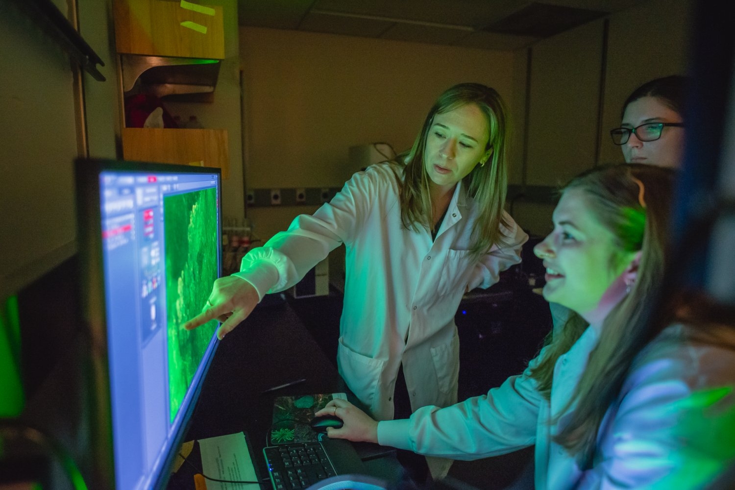
A new method, called purification-free ribosome imaging from subcellular mixtures (cryoPRISM), offers an appealing compromise. Developed by graduate students Mira May and Gabriela López-Pérez in the Davis lab in the MIT Department of Biology and recently published in PNAS, the technique allows biologists to visualize molecular complexes without taking them too far out of their natural context.

CryoPRISM captures molecular structures in cells that have just been broken open. This comes as close to preserving the natural interactions between molecules as possible, short of the extremely resource-intensive in-cell structural imaging, according to associate professor of biology Joey Davis, the faculty lead of the study.

“We think that the cryoPRISM method is a sweet spot where we preserve much of the native cellular contacts, but still have the resolution that lets us actually see molecular details,” Davis says. “Even in the extremely well-trodden system of translation in E. coli, which people have worked on for over 50 years, we are still finding new states that had just escaped people’s attention.”

A negative control that was not so negative

The development of cryoPRISM, as many discoveries in science, resulted from an unexpected observation that Mira May, the co-first author of the study, made while working on a different project.

Like all living organisms, bacteria rely on a process called translation to manufacture the proteins that carry out essential functions within the cell, from copying DNA to digesting nutrients. A key machine involved in translation is the ribosome — a biomolecular complex that assembles proteins based on instructions encoded by another molecule called mRNA. To regulate its activity, cells employ additional proteins that can change the shape of the ribosome, thus guiding its function.

May sought to identify new players in ribosomal regulation using cryoEM, by rapidly freezing lots of purified molecules and collecting thousands of 2D images to reconstruct their 3D structures. May was trying to pull ribosomes out of cells to visualize them together with their regulators. For her experiments, she designed a negative control containing unpurified bacterial lysate — a mixture of everything spilled from burst cells.

May expected to get noisy, low-quality images from this sample. To her surprise, instead, she saw intact ribosomes together with their natural interacting partners.

In just a few days, this technique experimentally validated data that would have taken months to acquire using other approaches.

“As I found more and more ribosomal states, this project became a method, not just a one-off finding,” May recalls.

Discovering new biology in a saturated field

Once May and her colleagues were confident that cryoPRISM could detect known ribosomal states, they began searching for ones that had previously escaped detection.

“It’s not just that we can recapitulate things that have been previously observed, but we can actually also discover novel ribosomal biology,” May says.

One of the novel states May identified has important implications for our understanding of the evolution of translation regulation.

During active translation, bacterial ribosomes are accompanied by a group of helper proteins called elongation factors. These factors bring in the materials for protein synthesis, like tRNAs and amino acids.

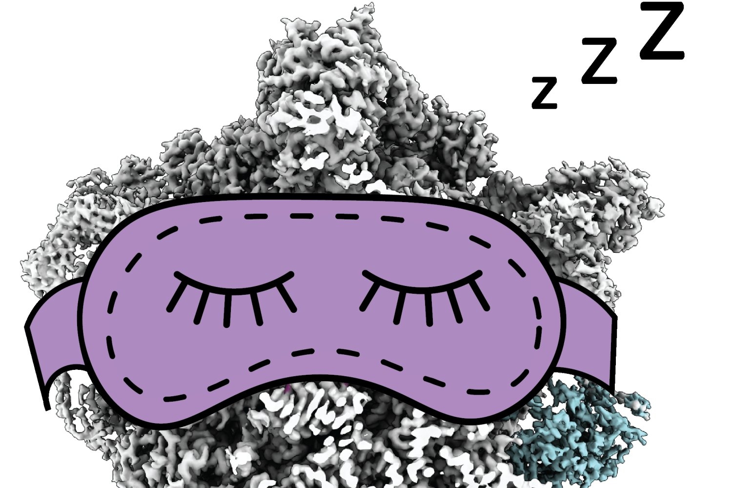
When cells encounter unfavorable conditions, such as colder temperatures, they reduce translation, which means that many ribosomes are out of work. These idle, hibernating ribosomes stop decoding mRNA, and the interface where they usually interact with helper molecules gets blocked by a hibernation factor called RaiA. This protein helps idle ribosomes avoid reactivation, like a sleeping mask that prevents a person from being woken up by light.

May observed the idle ribosomal state in her data, which on its own did not surprise her – this state had been described before. What surprised her was that some inactive ribosomes were interacting not only with RaiA, but also with an elongation factor called EF-G, which in bacteria was previously believed to only interact with active ribosomes.

A similar phenomenon has been seen before in more complex organisms, but observing it in a microbe suggests that its evolutionary origin may be older than previously thought.

“It fits an emerging model in the field, that elongation factors might bind to hibernating ribosomes to protect both the ribosome and themselves from degradation during periods of stress,” May explains. “Think of it like short-term storage.”

An unstressed cell might quickly eliminate unneeded inactive ribosomes, but because any stressor that puts ribosomes to sleep could be temporary, the cell may prefer to hold off on destroying them. That way, the ribosomes can be quickly reactivated if conditions improve.

The future of cryoPRISM

May has already teamed up with other MIT researchers to use cryoPRISM to visualize ribosomes in cells that are notoriously difficult to work with, including pathogenic organisms, which can be challenging to culture at the scale required for particle purification, and red blood cells isolated from patients, which cannot be cultured at all.

Besides its immediate application for translation research, cryoPRISM is a stepping stone toward the broader goal of structural biology: studying biomolecules in their natural environment.

To truly learn about deep-sea fish, scientists need to look at them in the deep sea; and to learn about cellular machines, scientists need to look at them in cells. According to Davis, cryoPRISM perfectly fits into the “theme of structural biology moving closer and closer to cellular context.”

Studying the genetic basis of disease to explore fundamental biological questions

Eliezer Calo’s studies of craniofacial malformations have yielded insight into protein synthesis and embryonic development.

Anne Trafton | MIT News
March 6, 2026

When Associate Professor Eliezer Calo PhD ’11 was applying for faculty positions, he was drawn to MIT not only because it’s his alma mater, but also because the Department of Biology places high value on exploring fundamental questions in biology.

In his own lab, Calo studies how craniofacial malformations arise. One motivation is to seek new treatments for those conditions, but another is to learn more about fundamental biological processes such as protein synthesis and embryonic development.

“We use genes that are mutated in disease to uncover fundamental biology,” Calo says. “Mutations that happen in disease are an experiment of nature, telling us that those are the important genes, and then we follow them up not only to understand the disease, but to fundamentally understand what the genes are doing.”

Calo’s work has led to new insights into how ribosomes form and how they control protein synthesis, as well as how the nucleolus, the birthplace of ribosomes in eukaryotic cells, has evolved over hundreds of millions of years.

In addition to earning his PhD at MIT, Calo is also an alumnus of MIT’s Summer Research Program (MSRP), which helps to prepare undergraduate students to pursue graduate education. Since starting his lab at MIT, Calo has made a point to serve as a research mentor for the program every summer.

“I feel that it’s important to pay back to the program that helped me realize what I wanted to do,” he says.

A nontraditional path

Growing up in a mountainous region of Puerto Rico, Calo was the first person from his family to finish high school. While attending the University of Puerto Rico at Rio Piedras, the largest university in Puerto Rico, he explored a few different majors before settling on chemistry.

One of Calo’s chemistry professors invited him to work in her lab, where he did a research project studying the pharmacokinetics of cell receptors found on the surface of astrocytes, a type of brain cell.

“It was a good mix of biology and chemistry,” he says. “I think that that was the catalyst to my pursuit of a career in the sciences.”

He learned about MSRP from Mandana Sassanfar, a senior lecturer in biology at MIT and director of outreach for several MIT departments, at an event hosted by the University of Puerto Rico for students interested in careers in science. He was accepted into the program, and during the summer after his junior year, he worked in the lab of Stephen Bell, an MIT professor of biology. That experience, he says, was transformative.

“Without that experience, I would have probably chosen another career,” Calo says. In Puerto Rico, “science was fun, but it was a struggle. We had to make everything from scratch, and then you spend more time making reagents than doing the experiments. When I came to MIT, I was always doing experiments.”

During that time, he realized he liked working in biology labs more than chemistry labs, so when he applied to graduate school, he decided to move into biology. He applied to five schools, including MIT. “Once MIT sent me the acceptance, I just had to say yes. There was no saying no.”

At MIT, Calo thought he might study biochemistry, but he ended up focusing on cancer biology instead, working with Jacqueline Lees, an MIT biology professor, to study the role of the tumor suppressor protein Rb.

After finishing his PhD, Calo felt burnt out and wasn’t sure if he wanted to continue along the academic track. His thesis committee advisors encouraged him to do a postdoc just to try it out, and he ended up going to Stanford University, where he fell in love with California and switched to a new research focus. Working with Joanna Wysocka, a professor of developmental biology at Stanford, he began investigating how development is affected by the regulation of proteins that make up cellular ribosomes — a topic his lab still studies today.

Returning to MIT

When searching for faculty jobs, Calo focused mainly on schools in California, but also sent an application to MIT. As he was deciding between offers from MIT and the University of California at Berkeley, a phone call from Angelika Amon, the late MIT professor of biology, convinced him to take the cross-country leap back to MIT.

“She had me on the phone for more than one hour telling me why I should come to MIT,” he recalls. “And that was so heartwarming that I could not say no.”

Since starting his lab in 2017, Calo has been studying how defects in the production of ribosomes give rise to diseases, in particular craniofacial malformations such as cleft palate.

Ribosomes, the organelles where protein synthesis occurs, consist of two subunits made of about 80 proteins. A longstanding question in biology has been why mutations that affect ribosome formation appear to primarily affect the development of the face, but not the rest of the body.

In a 2018 study, Calo discovered that this is because the mutations that affect ribosomes can have secondary effects that influence craniofacial development. In embryonic cells that form the face, a mutation in a gene called TCOF1 activates p53 at a higher level than in other embryonic cells. High levels of p53 cause some of those cells to undergo programmed cell death, leading to Treacher-Collins Syndrome, a disorder that produces underdeveloped bones in the jaw and cheek.

His lab has shown that p53 overactivation is also responsible for craniofacial disorders caused by mutations in RNA splicing factors.

Calo’s work on ribosome formation also led him to explore another cell organelle known as the nucleolus, whose role is to help build ribosomes. In 2023, he found that a gene called TCOF1, which can lead to craniofacial malformations when mutated, is critical for forming the three compartments that make up the nucleolus.

That finding, he says, could help to explain a major evolutionary shift that occurred around 300 million years ago, when the nucleolus transitioned from two to three compartments. This “tripartite” nucleolus is found in all reptiles, birds, and mammals.

“That was quite surprising,” Calo says. “Studying disease-related genes allowed us to understand a very fundamental biological process of how the nucleolus evolved, which has been a question in the field that nobody could figure out the answer for.”