When entering the office of Building 68’s manager, you will likely be greeted first with an amiable nose boop and wagging tail from Shadow, a four-year-old black lab, followed by a warm welcome from the office’s other occupant, John Fucillo. Fucillo is an animal lover, and Shadow is the gentlest of roughly nine dogs and one Siamese cat he’s taken care of throughout his life. Fortunately for MIT Biology, Shadow is not the only lab Fucillo cares for.
A Boston area local, Fucillo spent two years working at Revere Beach, then learned skills as an auto mechanic, and later completed an apprenticeship with the International Brotherhood of Electrical Workers. In 1989, Fucillo came to MIT Biology and says he couldn’t be happier.
As Building 68’s manager, Environment, Health & Safety coordinator, and Chemical Hygiene Officer, Fucillo’s goal is to make workflows “easier, less expensive, more desirable, and more comfortable.”
Fucillo was key for the Department’s successful move into its new home when Building 68 was completed in 1994, according to Mitchell Galanek, MIT Radiation Protection Officer and Fucillo’s colleague for over 30 years.
Throughout his time as a building manager, Fucillo has decreased routine spending and increased sustainability. He lowered the cost of lab coats by a whopping 92%–from $2,600 to $200–with just one phone call to North Star, the building’s uniform/linens provider. Auditing the building’s plastic waste generation inspired the institute-wide MIT Lab Plastics Recycling Program, which now serves over 200 labs across campus. More than 50,000 lbs of plastic have been recycled in the last four years alone.
“John is not a cog in the wheel, but an integral part of the whole system,” says Anthony Fuccione, Technical Instructor and Manager of the Biology Teaching lab.
Fucillo says one of his favorite parts of the job is chatting with researchers and helping them achieve their goals. He reportedly clocks about 10,000 steps per day on campus, responding to requests from labs, collaborating with colleagues, and connecting Biology to the institute’s Environment, Health, & Safety office.
“John is called upon — literally and figuratively — morning, noon, and night,” says Whitehead Professor of Molecular Genetics Monty Krieger. “He has had to become an expert in so very many areas to support staff, faculty, and students. His enormous success is due in part to his technical talents, in part to his genuine care for the welfare of his colleagues, and in part to his very special and caring personality.”
When MIT needed to comply with the EPA’s decree to improve safety standards across campus, Fucillo sat on the committees tasked with meeting those standards while avoiding undue burden on researchers, establishing the Environmental Health and Safety Management system in 2002.
“From a safety perspective, that was one of the most challenging things MIT had to go through–but it came out at the end a better, safer, place,” says John Collins, EHS Project Technician and friend and colleague to Fucillo for over 20 years.
Fucillo later co-led the initiative for a 2011 overhaul of MIT’s management of regulated medical waste (RMW), such as Petri dishes, blood, and needles. Fucillo volunteered to pilot a new approach in Building 68 — despite a lukewarm response to the proposal from other Biology EHS representatives, according to Galanek. This abundantly successful management system is now used by all MIT departments that generate RMW. It’s not only less expensive, but also does a better job at decontaminating waste than the previous management system.
“Anyone who has worked with John during his MIT career understands it is truly a privilege to partner with him,” Galanek says. “Not only does the work get done and done well, but you also gain a friend along the way.”
After consolidating a disparate group of individual lab assistants, Fucillo took on a supervisory role for the centralized staff tasked with cleaning glassware, preparing media, and ensuring consistency and sterility across Building 68 labs.
According to maintenance mechanic James “Jimmy” Carr, “you can’t find a better boss.”
“He’s just an easy-going guy,” says Karen O’Leary, who has worked with Fucillo for over 30 years. “My voice matters–I feel heard and respected by him.”
Although there are still many updates Fucillo hopes to see in Building 68, which will soon celebrate its 30th birthday, he is taking steps to cut back on his workload.
He recently began passing on his knowledge to Facilities Manager and EHS Coordinator Cesar Duarte, who joined the department in 2023.
“It’s been a pleasure working alongside John and learning about the substantial role and responsibility he’s had in the Biology department for the last three decades,” Duarte says. “Not only is John’s knowledge of Building 68 and the department’s history unparalleled, but his dedication to MIT and continued care and commitment to the health and well-being of the Biology community throughout his career are truly remarkable.”
As he winds down his time at MIT, Fucillo hopes to spend more time on music, one of his earliest passions, which began when he picked up an accordion in first grade. He still plays guitar and bass nearly every day. When he rocks out at home more often, he’ll be leaving behind the foundations of innovation, leadership, and respect in Building 68.

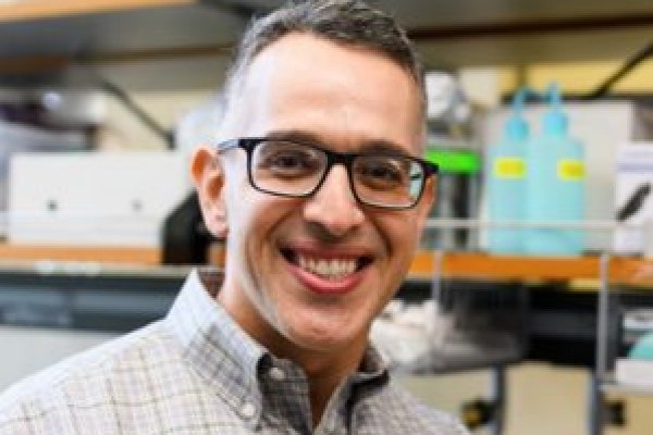
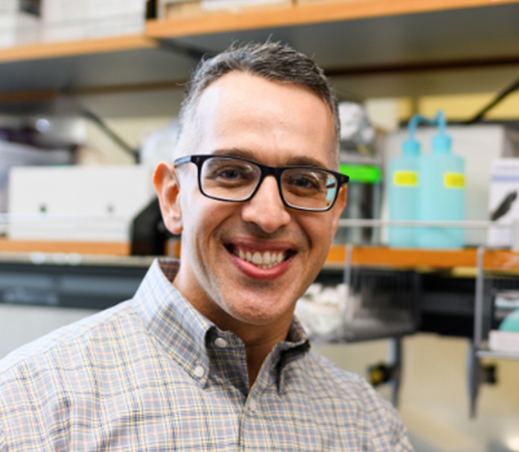
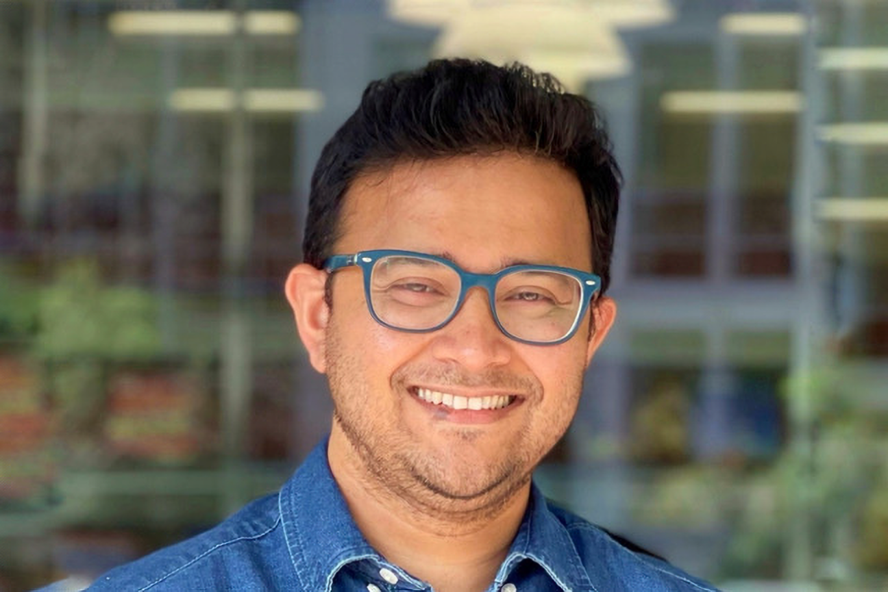
When Albert E. Almada PhD ’13 embarks on a new project, he always considers two criteria instilled in him during his time as a graduate student in the Department of Biology at MIT.
“If you want to make a big discovery, you have to approach it from a unique perspective — a unique angle,” Almada says. “You also have to be willing to dive into the unknown and go to the leading edge of your field.”
This is not without its challenges — but with an innovative spirit, Almada says, one can find ways to apply technologies and approaches to a new area of research where a roadmap doesn’t yet exist.
Now an assistant professor of orthopedic surgery and stem cell biology and regenerative medicine at the Keck School of Medicine of the University of Southern California (USC), Almada studies the mechanics of how stem cells rebuild tissues after trauma and how stem cell principles are dysregulated and drive conditions like degenerative disease and aging, exploring these topics through an evolutionary lens.
He’s also trying to solve a mystery that has intrigued scientists for centuries: Why can some vertebrate species like fish, salamanders, and lizards regenerate entire body parts, but mammals cannot? Almada’s laboratory at USC tackles these critical questions in the musculoskeletal system.
Almada’s fascination with muscle development and regeneration can be traced back to growing up in southern California. Almada’s brother had a degenerative muscle disease called Duchenne muscular dystrophy — and, while Almada grew stronger and stronger, his brother grew weaker and weaker. Last summer, Almada’s brother, unfortunately, lost his battle with his disorder at the age of 41.
“Watching his disease progress in those early years is what inspired me to become a scientist,” Almada recalls. “Sometimes science can be personal.”
Almada went to the University of California at Irvine for his undergraduate degree, majoring in biological sciences. During his summers, he participated in the Undergraduate Research Program (URP) at the Cold Spring Harbor Laboratory and the MIT Summer Research Program-Bio (now the Bernard S. and Sophie G. Gould MIT Summer Research Program in Biology, BSG-MSRP-Bio), where he saw the passion, rigor, and drive that solidified his desire to pursue a PhD.
Despite his interest in clinical applications, skeletal muscle, and regenerative biology, Almada was drawn to the Department of Biology at MIT, which is focused on basic fundamental research.
“I was willing to bet that it all came down to understanding basic cellular processes and things going wrong with the cell and how it interacts with its environment,” he says. “The MIT biology program really helped me define an identity for myself and gave me a template for how to tackle clinical problems from a molecular perspective.”
Almada’s PhD thesis work was based on a curious finding that Phillip Sharp, Institute Professor emeritus, professor emeritus of biology, and intramural faculty at the Koch Institute for Integrative Cancer Research, had made in 2007 — that transcription, the process of copying DNA into a messenger molecule called RNA, can occur in both directions at gene promoters. In one direction, it was long understood that fully formed mRNA is transcribed and can be used as a blueprint to make a protein. The transcription Sharp observed, in the opposite direction, results in a very short RNA that is not used as a gene product blueprint.
Almada’s project dug into what those short RNA molecules are — their structure and sequence, and why they’re not produced the same way that coding messenger RNA is. In two papers published in PNAS and Nature, Almada and colleagues discovered that a balance between splicing and transcription termination signals controls the length of an RNA. This finding has wider implications because toxic RNAs are produced and can build up in several degenerative diseases; being able to splice out or shorten RNAs to remove the harmful segments could be a potential therapeutic treatment.
“That experience convinced me that if I want to make big discoveries, I have to focus on basic science,” he says. “It also gave me the confidence that if I can succeed at MIT, I can succeed just about anywhere and in any field of biology.”
At the time Almada was in graduate school, there was a lot of excitement about transcription factor reprogramming. Transcription factors are the proteins responsible for turning on essential genes that tell a cell what to be and how to behave; a subset of them can even theoretically turn one cell type into another.
Almada began to wonder whether a specialized set of transcription factors instructs stem cells to rebuild tissues after trauma. After MIT, Almada moved on to a postdoctoral position in the lab of Amy Wagers, a leader in muscle stem cell biology at Harvard University, to immerse himself in this problem.
In many tissues in our bodies, a population of stem cells typically exists in an inactive, non-dividing state called quiescence. Once activated, these stem cells interact with their environment, sense damage signals, and turn on programs of proliferation and differentiation, as well as self-renewal, which is critical to maintaining a pool of stem cells in the tissue.
One of the biggest mysteries in the field of regenerative biology is how stem cells transition from dormancy into that activated, highly regenerative state. The body’s ability to turn on stem cells, including those in the skeletal muscle system, declines as we age and is often dysregulated in degenerative diseases — diseases like the one Almada’s brother suffered from.
In a study Almada published in Cell Reports several years ago, he identified a family of transcription factors that work together to turn on a critical regenerative gene program within hours of muscle trauma. This program drives muscle stem cells out of quiescence and speeds up healing.
“Now my lab is studying this regenerative program and its potential dysregulation in aging and degenerative muscle diseases using mouse and human models,” Almada says. “We’re also drawing parallels with super-healing species like salamanders and lizards.”
Recently, Almada has been working on characterizing the molecular and functional properties of stem cells in lizards, attempting to understand how the genes and pathways differ from mammalian stem cells. Lizards can regenerate massive amounts of skeletal muscle from scratch — imagine if human muscle tissue could be regrown as seamlessly as a lizard’s tail can. He is also exploring whether the tail is unique, or if stem cells in other tissues in lizards can regenerate faster and better than the tail, by comparing analogous injuries in a mouse model.
“This is a good example of approaching a problem from a new perspective: We believe we’re going to discover new biology in lizards that we can use to enhance skeletal muscle growth in vulnerable human populations, including those that suffer from deadly muscle disorders,” Almada says.
In just three years of starting his faculty position at USC, his work and approach have already received recognition in academia, with junior faculty awards from the Baxter Foundation and the Glenn Foundation/American Federation of Aging Research. He also received his first RO1 award from the National Institutes of Health with nearly $3 million in funding. Almada and his first graduate student, Alma Zuniga Munoz, were also awarded the HHMI Gilliam Fellowship last summer. Zuniga Munoz is the first to be recognized with this award at USC; fellowship recipients, student and advisor pairs, are selected with the goal of preparing students from underrepresented groups for leadership roles in science.
Almada himself is a second-generation Mexican American and has been involved in mentoring and training throughout his academic career. He was a graduate resident tutor for Spanish House at MIT and currently serves as the chair of the Diversity, Equity, and Inclusion Committee in the Department of Stem Cell Biology and Regenerative Medicine at USC; more than half of his lab members identify as members of the Hispanic community.
“The focus has to be on developing good scientists,” Almada says. “I learned from my past research mentors the importance of putting the needs of your students first and providing a supportive environment for everyone to excel, no matter where they start.”
As a mentor and researcher, Almada knows that no question and no challenge is off limits — foundations he built in Cambridge, where his graduate studies focused on teaching him to think, not just do.
“Digging deep into the science is what MIT taught me,” he says. “I’m now taking all of my knowledge in molecular biology and applying it to translationally oriented questions that I hope will benefit human health and longevity.”

MIT graduate student Juana De La O describes herself as a food-motivated organism, so it’s no surprise that she reaches for food and baking analogies when she’s discussing her thesis work in the lab of undergraduate officer and professor of biology Adam Martin.
Consider the formative stages of a croissant, she offers, occasionally providing homemade croissants to accompany the presentation: When one is forming the puff pastry, the dough is folded over the butter again and again. Tissues in a developing mouse embryo must similarly fold and bend, creating layers and structures that become the spine, head, and organs — but these tissues have no hands to induce those formative movements.
De La O is studying neural tube closure, the formation of the structure that becomes the spinal cord and the brain. Disorders like anencephaly and craniorachischisis occur when the head region fails to close in a developing fetus. It’s a heartbreaking defect, De La O says, because it’s 100 percent lethal — but the fetus fully develops otherwise.
“Your entire central nervous system hinges on this one event happening successfully,” she says. “On the fundamental level, we have a very limited understanding of the mechanisms required for neural closure to happen at all, much less an understanding of what goes wrong that leads to those defects.”
Hypothetically speaking
De La O hails from Chicago, where she received an undergraduate degree from the University of Chicago and worked in the lab of Ilaria Rebay. De La O’s sister was the first person in her family to go to and graduate from college — De La O, in turn, is the first person in her family to pursue a PhD.
From her first time visiting campus, De La O could see MIT would provide a thrilling environment in which to study.
“MIT was one of the few places where the students weren’t constantly complaining about how hard their life was,” she says. “At lunch with prospective students, they’d be talking to each other and then just organically slip into conversations about science.”
The department emails acceptance letters and sends a physical copy via snail mail. De La O’s letter included a handwritten note from department head Amy Keating, then a graduate officer, who had interviewed De La O during her campus visit.
“That’s what really sold it for me,” she recalls. “I went to my PI [principal investigator]’s office and said, ‘I have new data’” and I showed her the letter, and there was lots of unintelligible crying.”
To prepare her for graduate school, her parents, both immigrants from Mexico, spent the summer teaching De La O to make all her favorite dishes because “comfort food feels like home.”
When she reached MIT, however, the Covid-19 pandemic ground the world to a halt and severely limited what students could experience during rotations. Far from home and living alone, De La O taught herself to bake, creating the confections she craved but couldn’t leave her apartment to purchase. De La O didn’t get to work as extensively as she would have liked during her rotation in the Martin lab.
Martin had recently returned from a sabbatical that was spent learning a new research model; historically a fly lab, Martin was planning to delve into mouse research.
“My final presentation was, ‘Here’s a hypothetical project I would hypothetically do if I were hypothetically going to work with mice in a fly lab,’” De La O says.
Martin recalls being impressed. De La O is skilled at talking about science in an earnest and engaging way, and she dug deep into the literature and identified points Martin hadn’t considered.
“This is a level of independence that I look for in a student because it is important to the science to have someone who is contributing their ideas and independent reading and research to a project,” Martin says.
After agreeing to join the lab — news she shared with Martin via a meme — she got to work.
Charting mouse development
The neural tube forms from a flat sheet whose sides rise and meet to create a hollow cylinder. De La O has observed patterns of actin and myosin changing in space and time as the embryo develops. Actin and myosin are fibrous proteins that provide structure in eukaryotic cells. They are responsible for some cell movement, like muscle contraction or cell division. Fibers of actin and myosin can also connect across cells, forming vast networks that coordinate the movements of whole tissues. By looking at the structure of these networks, researchers can make predictions about how force is affecting those tissues.
De La O has found indications of a difference in the tension across the tissue during the critical stages of neural tube closure, which contributes to the tissue’s ability to fold and form a tube. They are not the first research group to propose this, she notes, but they’re suggesting that the patterns of tension are not uniform during a single stage of development.
“My project, on a really fundamental level, is an atlas for a really early stage of mouse development for actin and myosin,” De La O says. “This dataset doesn’t exist in the field yet.”
However, De La O has been performing analyses exclusively in fixed samples, so she may be quantifying phenomena that are not actually how tissues behave. To determine whether that’s the case, De La O plans to analyze live samples.
The idea is that if one could carefully cut tissue and observe how quickly it recoils, like slicing through a taught rubber band, those measurements could be used to approximate force across the tissue. However, the techniques required are still being developed, and the greater Boston area currently lacks the equipment and expertise needed to attempt those experiments.
A big part of her work in the lab has been figuring out how to collect and analyze relevant data. This research has already taken her far and wide, both literally and virtually.
“We’ve found that people have been very generous with their time and expertise,” De La O says. “One of the benefits we, as fly people, brought into this field is we don’t know anything — so we’re going to question everything.”
De La O traveled to the University of Virginia to learn live imaging techniques from associate professor of cell biology Ann Sutherland, and she’s also been in contact with Gabriel Galea at University College London, where Martin and De La O are considering a visit for further training.
“There are a lot of reasons why these experiments could go wrong, and one of them is that I’m not trained yet,” she says. “Once you know how to do things on an optimal setup, you can figure out how to make it work on a less-optimal setup.”
Collaboration and community
De La O has now expanded her cooking repertoire far beyond her family’s recipes and shares her new creations when she visits home. At MIT, she hosts dinner parties, including one where everything from the savory appetizers to the sweet desserts contained honey, thanks to an Independent Activities Period course about the producers of the sticky substance, and she made and tried apple pie for the first time with her fellow graduate students after an afternoon of apple picking.
De La O says she’s still learning how to say no to taking on additional work outside of her regular obligations as a PhD student; she’s found there’s a lot of pressure for underrepresented students to be at the forefront of diversity efforts, and although she finds that work extremely fulfilling, she can, and has, stretched herself too thin in the past.
“Every time I see an application that asks ‘How will you work to increase diversity,’ my strongest instinct is just to write ‘I’m brown and around — you’re welcome,’” she jokes. “The greatest amount of diversity work I will do is to get where I’m going. Me achieving my goals increases diversity inherently, but I also want to do well because I know if I do, I will make everything better for people coming after me.”
De La O is confident her path will be in academia, and troubleshooting, building up protocols, and setting up standards for her work in the Martin Lab has been “an excellent part of my training program.”
De La O and Martin embarked on a new project in a new model for the lab for De La O’s thesis, so much of her graduate studies will be spent laying the groundwork for future research.
“I hope her travels open Juana’s eyes to science being a larger community and to teach her about how to lead a collaboration,” Martin says. “Overall, I think this project is excellent for a student with aspirations to be a PI. I benefited from extremely open-ended projects as a student and see, in retrospect, how they prepared me for my work today.”
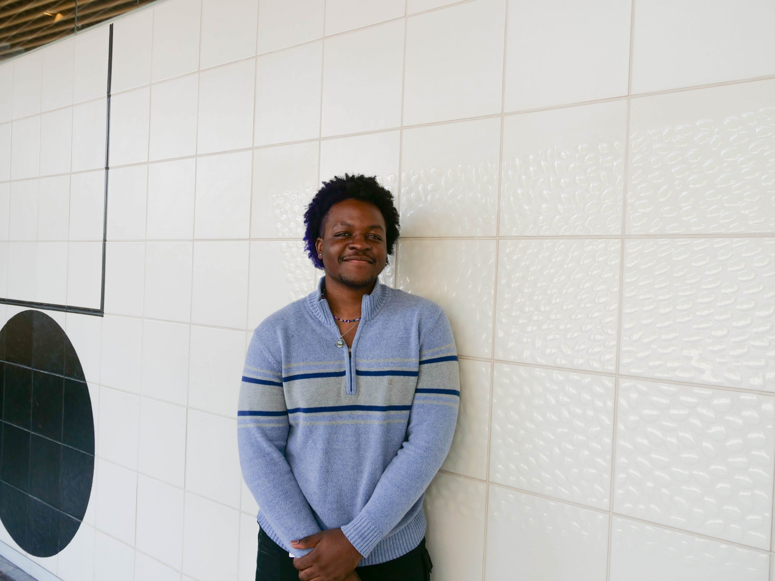
Cyrille Teforlack first stumbled across the work of MIT Professor of Biology Peter Reddien on YouTube while Teforlack was taking a cell biology class at Bethune-Cookman University, where he is now a rising senior. Teforlack became fascinated by Reddien’s work on regeneration in planarians, freshwater flatworms.
“That was how I figured out what kind of science I was interested in. I remember watching the video over and over,” Teforlack recalls. “I was like, ‘I have to figure out where this guy works.'”
The answer was, of course, at the Whitehead Institute, where Reddien is a Core Member and Associate Director. Teforlack spent the summer working in Reddien’s lab as part of the Bernard S. and Sophie G. Gould MIT Summer Research Program in Biology (BSG-MSRP-Bio). The program offers students the opportunity to work on cutting-edge research that isn’t available at their home institutions.
During the summer, Teforlack was working on eye regeneration and how different proteins and secreted factors affect the planarian’s cartoon-like crossed eyes. To understand the underlying requirements for regeneration, Teforlack used a technique called RNA interface (RNAi) to silence genes and see how it affected the planarian’s morphology when they regenerated.
The phenotypes Teforlack saw—he had a list of more than 100 candidate genes to work with—ranged from barely noticeable to strikingly defective. He studied regeneration by slicing off the head of the worm to see how the head regenerated and removing the eyes from the intact head to see how just that organ regrew.
“The eye has some connections to the brain and the rest of the body,” Teforlack explains. “But when you cut off their head, there’s no brain—but they can still regrow everything. It’s a no-brainer.”
However, some gene changes proved fatal or didn’t result in regeneration—the flatworms died and melted away. Teforlack didn’t realize that at first, however. There was one instance early in the program when he went to feed some worms—and was shocked to find they were missing.
“It was a really scary day for me in the lab,” Teforlack jokes. “I lost 12 worms, somehow. But I remember feeding them yesterday. How do you lose worms?”
Other phenotypes were more successful—showing atypical structures in the regenerated head or eyes. The optic cup of the eye regenerated in the wrong shape, for example, or separated in the middle as it regenerated.
Teforlack has been surprised by how much is still unknown about planarian regeneration. For example, a particular gene is expressed when the worm needs to regenerate wounded tissue on the half of the body facing the head, as opposed to the tail; it is unclear, however, how the planarian body detects which side the wound is on. .
“Even though it’s basic science, it’s still so intricate, and there’s so many little things that can build up to culminate in a bigger question,” Teforlack says.
Teforlack became known as “the worm guy” among his fellow students.
“Having a cool organism to talk about makes talking about it more exciting for myself, and for everyone else that’s listening,” he says. “This is something I never thought I would be a part of. It feels really great to be at a cutting-edge place doing really cool research.”
In addition to hands-on lab work, MSRP-Bio students often meet to discuss their work and do activities together. Teforlack says the program created plenty of opportunities to find community in his cohort, from arts and crafts to dodgeball.
The program also offers professional development activities like presentations from faculty including with the undergraduate and graduate officers Adam Martin and Mary Gehring to answer questions about applying for graduate school. Teforlack says he also found that faculty, despite their busy schedules, are always willing to take time out of their day for a chat.
“It’s been cool to meet all these different people and see the diversity of science that goes on and how many of them collaborate together on a variety of different projects,” Teforlack says. “This experience has helped me think like a scientist and value my own opinions. Being in an environment where your ideas are accepted, and you can learn from these scientists, has been really exciting.”
Teforlack worked closely with HHMI staff scientist Lucila Simone and graduate student Bryanna Canales. Canales herself participated in the program. As an MSRP-Bio student, Canales worked on metastasis in zebrafish in the lab of Daniel K. Ludwig Professor for Cancer Research, and Koch Institute Intramural Faculty Richard O. Hynes.
Canales says explaining her work to her peers during her time in MSRP-Bio was invaluable because it was more like teaching—she had to explain things in simple terms and found she was not the only one who sometimes struggled to do that.
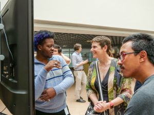
“The program made me feel more comfortable talking to people that I could learn from,” Canales recalls. “The MSRP-Bio experience humanized the institution and the people here. Everyone here is really smart, but going through the process with the students in my cohort, it feels like less of a big deal if you don’t know something.”
Canales says she’s seen Teforlack’s confidence grow this summer, taking the initiative and staying one step ahead instead of asking what he should be doing.
“It’s been nice to know that I can do science by myself—more or less—and still feel accomplished and know that everything I’m doing is the correct step, and if it’s not, I know how to troubleshoot things,” Teforlack says.
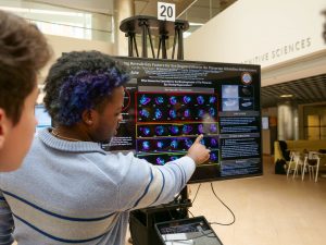
Teforlack’s work culminated in a poster session in August for MSRP-Bio students, where he showed some of the defective phenotypes he was characterizing and a short movie of the planarians eating the RNAi delivery system: liver.
“Cyrille showed a captivating movie of the small worms eating liver laced with double-stranded RNA that can downregulate specific genes,” says MIT Biology Department Head Amy Keating. “He also had beautiful images of the resulting phenotypes, which included disrupted optic cup structure. I always learn something new at the MSRP poster session!”
Long term, Teforlack plans to pursue a PhD in stem cell biology, and he says the program has reinforced that desire.
“It’s been cool to be around so many scientists,” Teforlack says. “Ten weeks isn’t enough time for anyone to learn anything perfectly. I’m excited to grow as a researcher.”
Although the MSRP-Bio program has come to a close for 2023, Teforlack’s time isn’t done in Reddien’s lab: he will return to continue his work in 2024.
“The MSRP program is a great opportunity for students to directly immerse in research here at MIT and to learn new concepts and methods,” Reddien says. “Cyrille was a terrific student and contributed a lot over the summer. I look forward to seeing his next steps with research into regeneration.”