Cambridge, MA – According to research conducted in mice by Whitehead Institute scientists, surgery in breast cancer patients, which while often curative, may trigger a systemic immunosuppressive response, allowing the outgrowth of dormant cancer cells at distant sites whose ability to generate tumors had previously been kept in check by the immune system. Taking a non-steroidal anti-inflammatory drug (NSAID) around the time of surgery may thwart such early metastatic relapse without impeding post-surgical wound healing.
The team’s work was published in the April 11 issue of the journal Science Translational Medicine.
“This represents the first causative evidence of surgery having this kind of systemic response,” says Jordan Krall, the first author of the paper and a former postdoctoral researcher in the lab of Whitehead Founding Member Robert Weinberg. “Surgery is essential for treating a lot of tumors, especially breast cancer. But there are some side effects of surgery, just as there are side effects to any treatment. We’re starting to understand what appears to be one of those potential side effects, and this could lead to supportive treatment alongside of surgery that could mitigate some of those effects.”
Although the association between surgery and metastatic relapse has been documented, a causal line between the two has never been established, leading many to consider early metastatic relapse to be the natural disease progression in some patients. Previous studies of breast cancer patients have shown a marked peak in metastatic relapse 12-18 months following surgery. Although the underlying mechanism for such a spike has not been understood, a 2010 retrospective clinical trial conducted in Belgium provides a clue: Breast cancer patients taking a non-steroidal anti-inflammatory (NSAID) for pain following tumor resection had lower rates of this early type of metastatic relapse than patients taking opioids for post-surgical pain. Anti-inflammatory drugs also have previously been shown to directly inhibit tumor growth, but Krall and Weinberg thought that the NSAIDs’ effects in these studies may be independent of the mechanism responsible for the effects noted in the retrospective clinical trial.
To investigate the causes of early metastatic relapse after surgery, the team created a mouse model that seems to mirror the immunological detente keeping in check dormant, disseminated tumor cells in breast cancer patients. In this experimental model, the mice’s T cells stall the growth of tumors that are seeded by injected cancer cells. When mice harboring dormant cancer cells underwent simulated surgeries at sites distant from the tumor cells, tumor incidence and size dramatically increased. Analysis of the blood and tumors from wounded mice showed that wound healing increases levels of cells called inflammatory monocytes, which differentiate into tumor-associated macrophages. Such macrophages, in turn, can act at distant sites to suppress the actions of T lymphocytes that previously succeeded in keeping the implanted tumors under control. Krall and Weinberg then tested the effects of the NSAID meloxicam (Mobic®), thinking that this anti-inflammatory drug might block the effects of immuno-suppressive effects of wound healing. In fact, when mice received the NSAID after or at the time of surgery, the drug prevented a systemic inflammatory response created by the wound healing and the meloxicam-treated mice developed significantly smaller tumors than wounded, untreated mice; often these tumors completely disappeared. Notably, meloxicam did not impede the mice’s wound healing
Still, Weinberg cautions that scientists are just beginning to understand the connections between post-surgical wound healing, inflammation, and metastasis.
“This is an important first step in exploring the potential importance of this mechanism in oncology,” says Weinberg, who is also a professor of biology at Massachusetts Institute of Technology (MIT) and director of the MIT/Ludwig Center for Molecular Oncology.
This work was supported by the Advanced Medical Research Foundation, the Transcend Program (a partnership between the Koch Institute and Janssen Pharmaceuticals Inc.), the Breast Cancer Research Foundation, the Ludwig Center for Molecular Oncology at MIT, and the Samuel Waxman Cancer Research Foundation, the American Cancer Society, Hope Funds for Cancer Research, the Charles A. King Trust, the National Health and Medical Research Council of Australia (NHMRC APP1071853), the National Institutes of Health (NIH/NCI 1K99CA201574-01A1), the American Cancer Society Ellison Foundation (PF-15-131-01-CSM), and the U.S. Department of Defense (W81XWH-10-1-0647).
* * *
Robert Weinberg’s primary affiliation is with Whitehead Institute for Biomedical Research, where his laboratory is located and all his research is conducted. He is also a professor of biology at Massachusetts Institute of Technology and director of the MIT/Ludwig Center for Molecular Oncology.
* * *
Full Citation:
“The systemic response to surgery triggers the outgrowth of distant immune-controlled tumors in mouse models of dormancy”
Science Translational Medicine, April 11, 2018.
Jordan A. Krall (1), Ferenc Reinhardt (1), Oblaise A. Mercury (1), Diwakar R. Pattabiraman (1), Mary W. Brooks (1), Michael Dougan (1,2), Arthur W. Lambert (1), Brian Bierie (1), Hidde L. Ploegh (1,3 *) Stephanie K. Dougan (1,4), Robert A. Weinberg (1,3,5).
1. Whitehead Institute for Biomedical Research, Cambridge, MA 02142, USA.
2. Division of Gastroenterology, Massachusetts General Hospital, Boston, MA 02114, USA.
3. Department of Biology, Massachusetts Institute of Technology, Cambridge, MA 02142, USA.
4. Department of Cancer Immunology and Virology, Dana-Farber Cancer Institute, Boston, MA 02215, USA.
5. Ludwig Center for Molecular Oncology, Massachusetts Institute of Technology, Cambridge, MA 02142, USA.
*Present address: Program in Cellular and Molecular Medicine, Boston Children’s Hospital, Boston, MA 02115, USA.
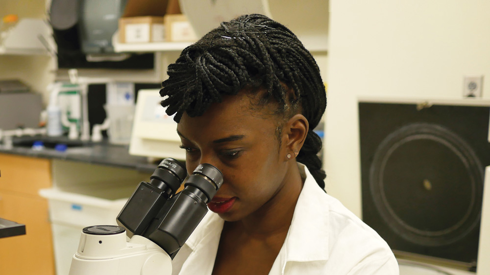
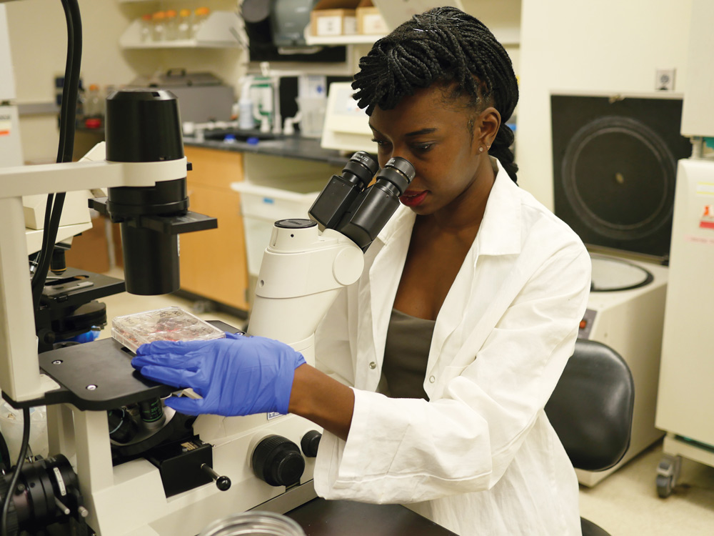
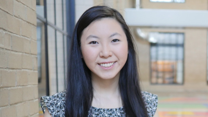
Elizabeth Li ’18 has worked in three cancer-related labs over the past six years, and one day intends to start her own.
Raleigh McElvery
March 6, 2018
Senior Elizabeth Li recreates miniature organs — lungs and intestines — outside the body. She does so in order to study cancer progression in both environments, and over the past six years has worked in three separate cancer-focused labs: two at MIT and another beginning her junior year of high school. One day, she aims to run her own.
“I’ve been into math and science ever since I was little,” she explains, “but in third grade I met a friend who was pretty important to me. She was diagnosed with a very malignant form of brain cancer and ended up dying from it. From that point on — even though I was still very young — I knew I wanted to do cancer research.”
In 9th grade, Li began at the School for Science and Math at Vanderbilt, a joint program between the university and Metro Nashville Public Schools. “I got to skip school once a week to learn research techniques, and had the opportunity to join Andries Zijlstra’s lab my junior year,” she recalls. “I’m actually still part of that group, and I’ve been working on the same project related to cancer metastasis for six years now.”
When it came time to select a university, Li was torn between Vanderbilt — where she was already performing research — and MIT, which she describes as “the place of opportunity.” She was ultimately swayed by MIT’s vast array of research areas, and fully sold after an overnight to preview the undergraduate culture.
Li opted for Course 7 in order to continue doing cancer research, and joined Omer Yilmaz’s lab in 2015 to investigate intestinal tumorigenesis. Here, she spent most of her time doing organoid work, studying the progression of colorectal cancer in miniaturized and simplified versions of the intestine. Li removed individual intestinal stem cells — or sometimes the entire “crypts” in which they reside — and grew them inside a 3D gel. This environment allowed the cells to differentiate and interact as they would in the colon, rather than growing on a flat, plastic dish.
Li and other members of the Yilmaz lab watched these cells multiply, observing their shape and the regeneration process. Li’s method of assessment varied depending on the research question: on some days, she stained the cells for proliferation markers, and on others she exposed them to different metabolites or drugs to see how the cells responded.
“On a typical day, I would come in during the morning between classes, and again in the afternoons and evenings,” she says. “The experiments differed, but we tended to do a lot of genetic manipulation. We’d make plasmids, CRISPR-Cas9 knockouts, or test for gene and protein expression using qPCR and Western blots.”
After two years, Li’s primary mentor finished her postdoctoral training, and Li transitioned to Jackie Lees’ lab at the beginning of her senior year. Li is now working with a fellow undergraduate on an independent project, centered on the enzyme protein arginine methyltransferase 5 (PRMT5).
PRMT5 catalyzes the transfer of methyl groups to the amino acid arginine in certain proteins, modifying their function. The enzyme also plays a key role in regulating gene splicing, the process by which segments of pre-mRNA are removed — changing the genetic code so that multiple genes can be encoded by the same initial transcript.
The Lees lab is interested in PRMT5 because it affects glioblastoma formation, the most common form of adult brain malignancy. As Li explains, when PRMT5 expression increases, so does tumor formation. Since there are still relatively few therapeutic options to treat glioblastomas, she’s hoping to use small molecules to inhibit PRMT5 expression and thus impede tumor initiation and progression.
“We’re considering using nanoparticles to deliver them,” she says, “and in doing so, hoping to gain a better understanding of how PRMT5 inhibition might impact cancer progression and tumorigenesis.”
Li is testing one small molecule PRMT5 inhibitor in lung organoids and several 2D cell lines — determining how sensitive the cells are and if the organoids will form, to gauge whether a tumor would still develop in the presence of the drug. “Depending on when you add the drug, you can test different aspects of tumorigenesis,” she explains.
She’s also split the past four years between the Biology Undergraduate Student Association (of which she was faculty liaison, outreach chair, and then co-president), the MIT Pre-Medical Society,MIT Lion Dance, Asian Dance Team, Wind Ensemble, and Improv-a-Do! She’s also heavily involved in DynaMIT, which organizes an annual, week-long science program for economically disadvantaged middle school students.
“There are a lot of extracurriculars to do,” Li admits. “But it’s pretty easy to get involved in the MIT community and still stay on top of your coursework, if you keep it to four or five classes per semester. It’s worked out for me so far — I’m still alive and happy and have time for eating, sleeping, and friends.”
As Li applies to MD-PhD programs, she hopes one day to practice medicine (perhaps pediatric oncology) while running her own lab.
“My advice for incoming MIT undergrads would be to remember to have fun,” she says. “You only have four years, so take advantage of your time here: hang out with your friends, take the classes you want to take, and do things that you enjoy. Hopefully most of those activities will be one and the same.”
Photo credit: Raleigh McElvery
Matthew Vander Heiden helped revive the forgotten— but critical—study of cancer metabolism.
Sam Apple | MIT Technology Review
February 21, 2018
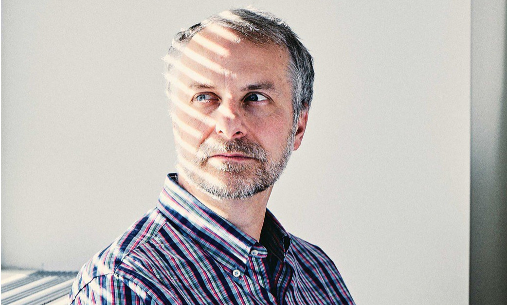
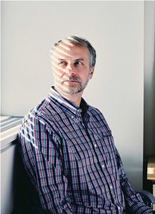
One day last October, MIT biology professor Matthew Vander Heiden showed up in one of his trademark plaid shirts to teach his undergraduate course on cancer biology. As usual, he peppered his lecture with questions, filling six sliding chalkboards with arrows mapping cellular pathways; he had to erase the boards halfway through class to make room for more notations. But what might have seemed like an ordinary lecture was far from ordinary in one respect: although Vander Heiden was explaining some of the most fundamental aspects of how tumors grow, most of what he was teaching his students would have been absent from nearly every introductory course on cancer biology a decade ago. The science Vander Heiden discussed that afternoon amounted to a once-lost but recently rediscovered chapter in the history of cancer research.
What he didn’t mention in class is that he’d played as large a role as anyone in bringing it back.
That lost chapter focuses on metabolism, and how cancers use nutrients for energy and as building blocks for new cancer cells. It began with a discovery in the early 1920s that most cancers stuff themselves with glucose and then use it in an unusual way. Whereas normal cells typically break down glucose by burning it with oxygen, cancer cells extract much of its energy through fermentation—essentially the same process microorganisms use to make yogurt, beer, and other foods. Indeed, early-20th-century researchers noticed that cancer cells seemed to behave more like yeast than the cells of an animal. But though it would briefly become a major school of cancer research, metabolism fell by the wayside in the 1960s as researchers turned their attention to how cancer-causing genes signal cells to divide.
Cancer metabolism research appeared to be dead, until Vander Heiden helped launch its revival around two decades ago. Today it’s among the hottest areas of the field, spawning conferences, journals, and promising new therapies. And it has fundamentally changed the way many researchers understand cancer and its origins.
Modest revolutionary
Metabolism’s downfall as a research area in the late 20th century was largely a reflection of the faddish nature of science. It didn’t help that Otto Warburg, the German scientist who discovered the unusual metabolism of cancer cells, was so arrogant that much of the scientific community disliked him. So it’s probably a good thing that Vander Heiden, a down-to-earth type who’s been known to downplay his own role on research papers to give his students and postdocs first-author billing, has been so central to the metabolism revival.
Vander Heiden, 45, grew up in Port Washington, Wisconsin, a small town on Lake Michigan once known for its lawnmower factories, and he lives up to every stereotype of his background. “He carries his Midwestern sensibilities with him everywhere he goes,” says his wife, Brooke Bevis, a biologist and the operations manager for Vander Heiden’s lab at MIT’s Koch Institute for Integrative Cancer Research. “I finally made him give up my old 1995 Honda Civic just a few years ago.”
When Vander Heiden enrolled at the University of Chicago in 1990, medicine was already on his mind. His younger brother had suffered from a rare blood disorder as a child, and Vander Heiden spent much of his own childhood hanging around children’s hospitals. But he had little thought of becoming an academic scientist until he began a work-study job washing out equipment in a University of Chicago biology lab. The work was not glamorous but came with a perk: the graduate students in the lab would let Vander Heiden make solutions for them and show him how they did their experiments.
After graduating, he enrolled in Chicago’s MD-PhD program and landed in the lab of Craig B. Thompson. Today Thompson is the president and CEO of the Memorial Sloan Kettering Cancer Center, but at the time he was studying immunology, looking at how the body eliminated huge numbers of immune cells once they were no longer needed.
When Vander Heiden arrived in Thompson’s lab in 1996, part of the explanation was already understood. Those cells would simply commit suicide, a process known as apoptosis. It was also known that a family of proteins named Bcl-2 could stop a cell from committing suicide—and that they appeared to do so through their impact on mitochondria, tiny organelles known as the powerhouses of the cell for their role in energy production.
Vander Heiden had just joined a cutting–edge immunology lab interested in protein signaling. Yet he had been asked to investigate how Bcl-2 proteins affect mitochondria, a relic of the old, outdated metabolism research. When it became clear that no one in the lab knew much about metabolism, Vander Heiden reread the relevant sections of his undergraduate biochemistry textbook. He also teamed up with Navdeep Chandel, a metabolism researcher at Northwestern University who was then a graduate student in a University of Chicago cellular physiology lab.
When another lab showed that proteins released from the mitochondria could trigger apoptosis, Vander Heiden and -Chandel got an important clue: the decision to commit suicide could now be traced directly to the mitochondria. And yet the deeper question of what was happening inside them remained mysterious until the two researchers arrived at an answer, thanks to a series of elegant experiments designed by Vander Heiden (whom Chandel calls “a world-class experimentalist”) to study how molecules moved through the mitochondrial membrane. They discovered that the release of the mitochondrial proteins was a sign of a failing powerhouse, a notice to the cell that a brownout was under way so it was time to abort. But brownouts weren’t inevitable; the Bcl-2 proteins, like emergency workers called to the scene of an imminent disaster, could resuscitate the metabolic function of the mitochondria and keep things from getting to that point. The suicide signal, in turn, would never be released.
Daniel Schmidt, a postdoc in Vander Heiden’s lab, prepares cells to study how metabolism affects cancer cell proliferation. Credit: BUCK SQUIBBDaniel Schmidt, a postdoc in Vander Heiden’s lab, prepares cells to study how metabolism affects cancer cell proliferation.
BUCK SQUIBB
For Vander Heiden, this was a “watershed moment.” Among other things, it meant that metabolic enzymes weren’t merely supplying energy from food. Metabolism was governing the most fundamental decision a cell has to make—whether to live or die. That meant it had to be interwoven into the signaling cascades that molecular biologists studied. His feeling at the time, he recalls, was “Oh my goodness. We don’t really understand metabolism.”
Vander Heiden might not have envisioned himself delving into research areas that had been discarded decades earlier, but what was more surprising was how little research was then being done in an area that was “as fundamental as you get in terms of how biology works,” he says. “I looked around and no one was studying it.”
Thompson, recognizing the opportunity, shifted the focus of his lab to metabolism. Vander Heiden, meanwhile, continued to pursue Thompson’s broader question of how the body eliminates unwanted immune cells. He already knew that growth factors, messages sent from one cell to the next, kept cells from committing suicide, but how the signals delivered their survival message remained unclear. What he discovered in a series of studies carried out in the late ’90s followed perfectly from his previous research. Growth factors kept cells alive by giving them permission to eat. Without that permission, a cell soon faced an energy crisis, and the mitochondria released their death signals.
The takeaway was clear: our bodies eliminate unwanted cells by starving them to death.
Solving the metabolism mystery
As Vander Heiden’s MD-PhD program was coming to an end, he hadn’t yet begun to focus on cancer, but its possible links with his research on cell suicide were intriguing. Cancer cells were the other side of the coin—cells that were resistant to suicide, that no longer cared about instructions from other cells. So in 2004, after completing a residency in oncology at Brigham and Women’s Hospital in Boston, he was anxious to investigate cancer metabolism for his postdoc research.
Finding the right lab wasn’t easy. At the time, telling leading researchers he wanted to study how cancer cells consumed glucose was like approaching a high-tech manufacturer and announcing you wanted to study the trucks that brought fuel to the factory. It sounded, Vander Heiden says, “like a really ridiculous thing.”
Vander Heiden eventually found a home in the Harvard lab of Lewis Cantley, who now directs the Meyer Cancer Center at Weil Cornell. His research in Cantley’s lab would help solve one of the central riddles of cancer metabolism: why cancer cells are so ravenous for glucose. Researchers had once assumed that cancer cells were turning to fermentation because they’d lost the ability to use oxygen properly and needed some other way to produce energy. But Vander Heiden’s research on a mutated form of the enzyme pyruvate kinase showed something else. Rather than being used for energy, much of the glucose was being shunted into pathways used to build new molecules. What a growing cancer needs most of all from its food, the research suggested, is more spare parts—raw materials for making new DNA, membranes, and proteins.
Rethinking chemotherapy
Vander Heiden’s research with Cantley would also lead to his involvement with Agios Pharmaceuticals, the company behind one of the most promising new drugs to emerge from the metabolism revival. (Cantley says he played a major role in building the company’s science in its early days.) The drug, AG-221, treats acute myelogenous leukemia, a cancer of the blood and bone marrow. It works by blocking the product of a mutated form of the mitochondrial enzyme IDH-2. Approved by the US Food and Drug Administration in August, it has been hailed as the first real advance for the disease in 30 years.
The approval of AG-221 isn’t the only thing generating excitement in the cancer world. Unlike almost all other cancer drugs, AG-221 doesn’t kill the cancer cells but, rather, allows them to develop out of their deranged state into noncancerous, mature, functioning cells. That a single metabolic enzyme could have such profound effects on which genes are expressed in a cell is now one of the many signs that changes in metabolism are not just a response to the needs of a growing cancer. Often, they may actually be causing the cancer itself. It represents a major shift in thought: many cancer-causing genes long known for their ability to signal cells to keep dividing have now been shown to have additional roles in signaling cells to keep eating. Some researchers now believe the overeating typically comes first, driving the transformations that follow.
Since his arrival at MIT and the opening of his lab at the Koch Institute in 2009, Vander Heiden has treated cancer patients and continued to search for better therapies. In recent years he has focused on improving understanding of chemotherapy. Though typically thought of as general poisons, most chemotherapy drugs work because they disrupt metabolic functions. That much has long been known, but less clear is why a particular drug works for some cancers and not for others, even when two cancers carry the same mutations.
It was while explaining to his undergrad cancer biology students how targeted drugs work that Vander Heiden first thought of an answer. As a cancer doctor, he knew that chemotherapies are often chosen on the basis of where in the body a tumor first arose, but what was it about this location that made the difference?
Vander Heiden’s research in mice now suggests that the answer may lie in which foods are available to the cancer as it forms. Melanoma and colon cancer, for example, often have the same mutations, and yet, as he explains, because the two cancers “grow in very different places in the body,” they likely “have access to different nutrients.” He adds, “It has nothing to do with the genetics.” If he turns out to be right, it could lead to a fundamental change in how oncologists think about which drugs to give their patients.
As Vander Heiden turns his attention to old chemotherapy drugs, rethinking why and how they work, he is once again looking to the past for new insights on cancer. It might be more than a coincidence. As Bevis, his wife, says, the outdated Honda Civic isn’t the only item he has struggled to let go of. “The list goes on and on,” she says. “He hates waste and will use items long after someone else would have replaced them with a newer, shinier model.”
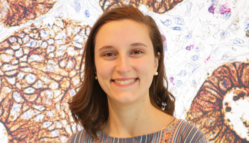
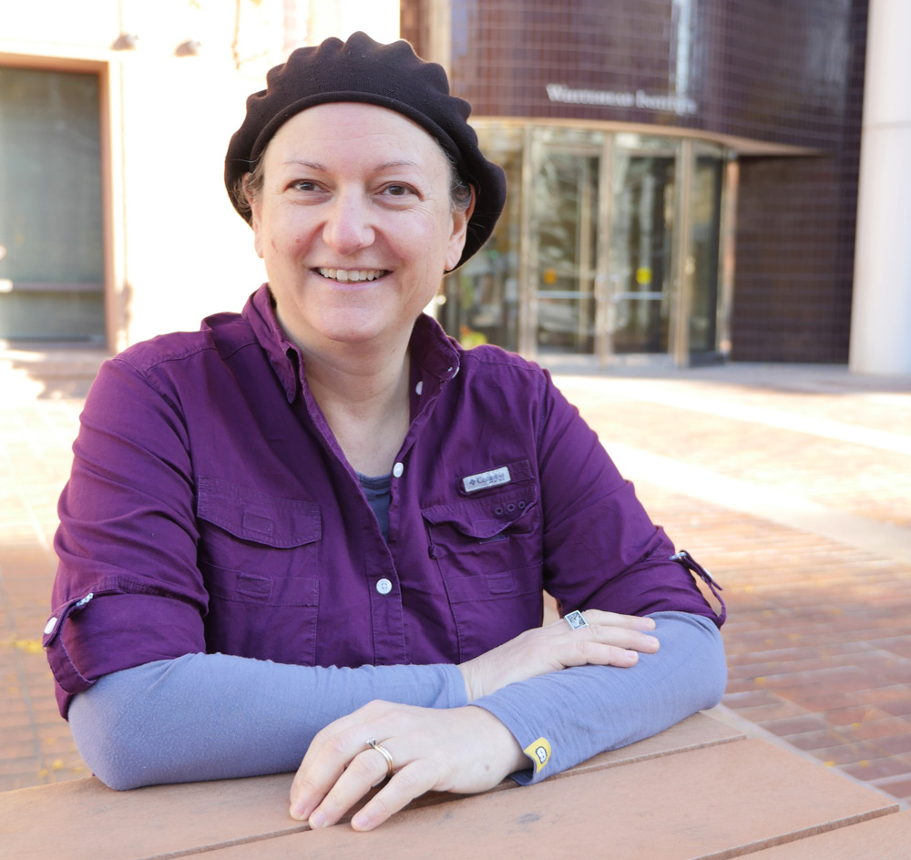
Alissandra Hillis ’18 has spent all four years at MIT in the same cancer metabolism lab, deciphering the basic science behind pancreatic cancer.
Raleigh McElvery
February 19, 2018
Senior Alissandra Hillis attributes her appetite for the basic sciences to her craving for fundamental knowledge. She’s spent her four years at MIT in the same lab, committed to unraveling the molecular mechanics of pancreatic cancer — the fourth leading cause of cancer death for both men and women, given that symptoms do not often appear until the disease is quite advanced.
“I was always very curious growing up,” she says. “I taught myself how to read at a very young age, just because I wanted to know about things and how they worked. But I didn’t become interested in biology and chemistry specifically until I came to MIT and started taking my General Institute Requirements.”
In doing so, Hillis became enthralled by the prospect of breaking down life into its most fundamental, biological units to decipher cellular function and disease. Originally a Course 7 major with a chemistry minor, she declared Course 5-7 (Chemistry and Biology) as soon as it became available in the fall of 2017 — applying her study of biochemistry and cell metabolism to cancer research.
“When I was quite young, my grandfather was diagnosed with stomach cancer, and ended up having almost three quarters of his stomach removed,” she says. “I was too little to really understand the severity of the situation, but as soon as I came to MIT I started to wonder what was going on at a cellular level. Most people today know someone who is fighting cancer, and yet we’re still lacking effective treatments for its most severe forms.”
Hillis joined Matthew Vander Heiden’s cancer metabolism lab the first semester of her freshman year, and has been there ever since.
“Professor Vander Heiden does an excellent job of tailoring the research project to the individual, and there is no hierarchy among lab members,” she says. “I really liked it from the onset, so I stayed.”
For nearly two years, Hillis has been investigating the role of one enzyme, pyruvate kinase muscle isozyme M2 (PKM2), in pancreatic cancer. PKM2 is responsible for catalyzing the final step in glycolysis, which is required to create the energy that fuels cells. Glycolysis is also important in tumor metastasis and growth, since cancer cells demand energy in order to proliferate.
Cancer cells often preferentially express PKM2 over other types of pyruvate kinases such as PKM1. This spurred William Israelsen PhD ’14, a former graduate student in the Vander Heiden lab working in breast cancer models, to delete the PKM2 gene and see what happened. Since PKM2 is critical for glycolysis, and cancer cells require energy to proliferate, he anticipated that removing PKM2 would hinder energy production and thus disrupt tumor development. To his surprise, he found the opposite: deleting PKM2 actually accelerated tumor formation and promoted liver metastasis in mice.
In his 2014 paper, Israelsen concluded that PKM2 might permit cancer cells to maintain their “plasticity,” shifting from one specialized role to another even after they’ve fully matured. In the absence of PKM2, he proposed PKM1 might take over PKM2’s influential role.
Hillis wondered if she could replicate Israelsen’s breast cancer results in a model for pancreatic cancer, especially given the conflicting findings in human data regarding PKM2 expression in the latter. Some studies suggest that high PKM2 expression correlates with accelerated disease, whiles others indicate just the opposite: that high PKM2 expression is associated with better survival rates.
“Going into the project, we were expecting similar effects in both pancreatic and breast cancer models because both cancers preferentially express PKM2, and we were using the same method of PKM2 deletion, just bred into a different cancer model,” Hillis explains. “We anticipated that PKM2 deletion would accelerate pancreatic tumor size and tumor genesis, and decrease the mouse’s lifespan. But we’ve noticed that these effects — if they exist — are very much attenuated in the pancreatic cancer model; there is only a slight decrease in lifespan and increase in tumor size without PKM2.”
Right now, her working hypothesis holds that PKM2’s influence varies depending on the tissue in question. This might explain why her own results don’t exactly parallel what Israelsen found in his breast cancer model. For instance, the method they were using to delete PKM2 is quite effective in the breast and pancreatic cells themselves, but less so in the dense scar-like tissue characteristic of pancreatic tumors in particular. It’s possible, she thinks, that this fibrous tissue may still express some PKM2 even post-knockout, perhaps hindering both a drastic decrease in lifespan and increase in tumor size.
Hillis hopes piecing together PKM2’s mechanism of action will help us better diagnose — and eventually treat — certain cancers. Her most recent results were published in the November 2017 issue Cancer & Metabolism.
Although Hillis enjoys tackling the more fundamental questions concerning cancer, she’s also interested in translating this work from bench to bedside. That’s why she decided to intern with David Ting at the Massachusetts General Hospital Cancer Center this past summer.
“I wanted to try a different type of research before applying to graduate school,” she says. “The Department of Biology frequently sends out emails about job opportunities, and there was one advertising that the Ting lab was looking for a research technician.”
Although she was still a junior at the time, she contacted Ting — an MIT alumnus with a dual degree in 7A and 10 — and together they fashioned a summer position just for her, studying the role of miniscule, fluid-filled transportation structures called exosomes in cancer development and diagnosis.
“That was the first time I’d worked with samples from actual patients,” she says. “Many of the assays were the same, but I felt closer to a clinical application than I ever had before. I really enjoy doing the foundational work to identify the basic problem, but there’s definitely something to be said for experiencing research targeted at creating a diagnostic tool. I can see the pros and cons of both approaches.”
As Hillis begins her final semester at MIT, she’s continuing her work in the Vander Heiden lab, while also finishing up the requirements for her HASS concentration in legal studies. She’s still set on pursuing a PhD in cancer biology, but the propensity to ask tough questions that drew her to science in the first place has led her to realize that the questions she raises in her own research have ramifications far beyond her lab bench. Taking policy-oriented classes in addition to her science-related ones has inspired her to pursue a law degree in conjunction with her PhD — weaving together her love for science with a newfound interest in the rules and regulations that govern how science is funded, performed, shared, applied, and monetized.
“I really enjoy doing research, and that’s something I probably will continue to do,” she says, “but I also want to influence science-related regulations, which is something I couldn’t possibly do without a law degree. I would still be heavily immersed in science, while applying the subjects I love in new and exciting ways.”
Photo credit: Raleigh McElvery
New cancer research initiative eyes individualized treatment for patients.
Koch Institute
February 1, 2018
Details matter — perhaps most noticeably in the fight against cancer. Some patients respond to a given anticancer therapy, and some do not. A new initiative at MIT takes aim at those details, and the name of the game is precision.
The recently launched MIT Center for Precision Cancer Medicine (CPCM) is housed within MIT’s Koch Institute for Integrative Cancer Research and headed by physician-scientist Michael B. Yaffe, the David H. Koch Professor of Science and professor of biology and biological engineering. The center brings together leading Institute faculty members to focus on key research themes to accelerate the clinical translation of novel cancer discoveries, treatments, and technologies.
Engineering approaches to the clinic
While other institutions have begun efforts in precision medicine as well, the MIT Center for Precision Cancer Medicine stands out for using engineering approaches to solve complex clinical challenges in cancer treatment that are rooted in biology. In particular, the CPCM combines understandings of biological circuitry — along with engineering, computational, and mathematical techniques (as well as genomic ones) — to focus on signaling networks and pathways that are aberrantly regulated in cancer cells. This strategy is supported by the fact that most state-of-the-art molecularly targeted cancer therapies are focused on these key pathways.
At its core, the CPCM is driven by both internal and external collaboration, and is devoted to translational research to help the substantial number of patients who do not respond well to traditional cancer therapies — for example, those with triple-negative breast cancer, ovarian cancer, non-small cell lung cancer, or advanced prostate cancer.
To improve outcomes for these patients, CPCM investigators are focused on four key areas of research. First among these is identifying and targeting the processes, signals, and mechanisms that determine an individual patient’s response to chemotherapy. Recent discoveries by CPCM researchers include mechanisms that cancer cells use to repair chemotherapy damage that should have killed them, to hide from drugs in protected “niches” in the body, or to grow when and where they should not.
CPCM members are also working on a second research pillar, which involves finding ways to use existing FDA-approved cancer drugs more effectively, particularly in carefully designed combinations. Combination therapies are currently used in the clinic to treat some cancers, yet the discovery process for these has been largely empirical. By contrast, CPCM investigators are integrating their knowledge of cancer biology, understandings of drugs’ mechanisms of action, and sophisticated analytical techniques, to identify or design specific combinations that work synergistically to disarm and then destroy cancer cells.
“We believe we can significantly alter cancer patients’ outcomes by determining the right combination of therapies and the right sequence of drugs for the right patients,” says Yaffe. “We’re also concentrating on innovative ways to give these drugs, like time-staggered dosages and nanoparticle delivery.” He notes that, as part of their analyses of drugs and combination regimens currently administered in the clinic, CPCM members expect to identify combinations of drugs that are not as efficacious when given simultaneously as when given sequentially, at specific intervals. Yaffe stresses that these will be important findings that could help reduce the toxicity of treatment by not exposing people to multiple drug toxicities at the same time.
In parallel with their efforts to use existing drugs more effectively, CPCM investigators are also working to identify compounds, materials, and approaches that can engage key “undruggable” genetic and molecular targets and disrupt processes driving drug resistance. The “undruggable” label often refers to the fact that a target protein or molecule lacks a site to which drugs can bind, and thus is not considered a good drug target by the pharmaceutical industry. However, using novel chemistry approaches, CPCM researchers have made early inroads against several such high-value cancer targets, including specific transcription factors and RNA-binding proteins. The center will continue and expand these efforts as the third part of its research platform, including collaborations with industry.
Finally, the fourth component of the CPCM’s efforts will be harnessing MIT’s particular expertise in big data analysis and tools to begin new and expedite existing cancer research efforts. For example, the researchers plan to use data analytics to identify selective panels of biomarkers that can be used to prioritize which of their drug combinations, treatment protocols, and formulations are best suited to a particular patient’s tumor.
Getting discoveries out the door
“Patients will be the ultimate beneficiaries of the work of the new MIT Center for Precision Cancer Medicine,” says Tyler Jacks, director of the Koch Institute and the David H. Koch Professor of Biology. “This research is, by its nature, imminently and rapidly translatable. By concentrating efforts on which patients will benefit from particular existing drugs or combinations of drugs, there is a relatively small step from laboratory to a treatment that is benefitting a cancer patient.”
While work on combinations of approved therapies, like that at the CPCM, may be more rapidly translatable than other cancer research, it can be challenging for industry to pursue, particularly when those drugs hail from multiple companies. Overcoming this disjuncture is one of the goals behind the establishment of the MIT Center for Precision Cancer Medicine, which was made possible by a generous gift from an anonymous donor.
Yaffe and his CPCM colleagues are committed to finding viable routes to move their cancer research into the clinic, particularly through collaborations between CPCM members, hospitals, and industry. Logistically, this means more work for the center’s research groups, including advanced laboratory and preclinical studies, safety and scale-up studies, and clinical-grade manufacturing, as well as staff to carry it out. Woven into these efforts, CPCM investigators will tap into MIT’s celebrated tradition of entrepreneurship and, even more so, the Institute’s expanding network of clinical collaborators. The philanthropic investment behind the center will provide stable financial support for the researchers’ endeavors.
The new hub in town
In addition to supporting the research of member investigators, the CPCM offers a robust training ground for young engineers and scientists interested in precision medicine. Moreover, it will serve as the hub of precision cancer medicine research at MIT and beyond, connecting with researchers across the MIT campus and partnering with clinical investigators in Greater Boston’s noted health care centers and around the country.
Five outstanding cancer researchers make up the center’s founding faculty:
Michael B. Yaffe, MD, PhD, director, MIT Center for Precision Cancer Medicine; David H. Koch Professor of Science, professor of biology and biological engineering
Efforts are currently underway to recruit an assistant director and a scientific advisory board.
As part of its charge, and key to spurring the new collaborations in precision cancer medicine that are its focus, the MIT Center for Precision Cancer Medicine will also convene lectures, events, and scientific exchanges and symposia, the first of which is slated for the fall.
December 14, 2017
Lab coat meets legislation
Undergraduate Courtney Diamond combines biology and policy to tackle real-world challenges
Raleigh McElvery
Undergraduate Courtney Diamond arrived at MIT determined to be an oncologist. Five years later, she’s leaving with a broader focus on human health, grappling with real-world, biomedical problems by way of public policy rather than medicine or research.
Although Diamond had completed her requirements for a degree in biology at the beginning of her senior year, she decided then to add a second major: Course 17 (Political Science), and with it a fifth year of study at MIT.
“I came into MIT wanting to be a doctor, but the more I thought about it the less it felt like medical school would be a good fit,” she says. “I spent a long time narrowing my interests within the realm of human health, and recently realized there was another dimension to that interest related to public policy, which was also this common thread among my extracurriculars.”
Diamond grew up in a small town in Massachusetts called Millbury, not too far from MIT, which she describes as special to her but “rather unremarkable” in most other ways — with the exception of one particularly zealous and articulate high school biology teacher. His infectious enthusiasm sparked Diamond’s passion for the life sciences, but over the course of her senior year this interest became far more personal. It was around that time that her mother developed breast cancer, and Diamond resolved to be an oncologist.
“My mom had been diagnosed once before with a different kind of cancer, cervical cancer,” she says. “But I was in sixth grade back then, and assumed she was just at home resting. By the time the breast cancer rolled around, I was old enough to understand that most people are lucky to survive cancer once. But twice?”
Her mother has since entered remission, and the year Diamond began at MIT her interests matured away from a career in medicine and towards biomedical research. In April 2014, she applied to the MIT Undergraduate Research Opportunities Program (UROP). “I wanted to figure out which part of biology excited me — which area I really wanted to drill down on,” she recalls.
She began working with a postdoctoral fellow in Professor Darrell Irvine’s lab at the Koch Institute for Integrative Cancer Research, tackling research questions related to cancer immunology. Diamond’s job was to analyze murine tumors as they developed over time, in order to understand how they were affected by changes to their cellular environments.
After a year, Diamond took a break from research in order to focus on her classes. But she didn’t stay away for long.
“I’ve had a life-long obsession with Australia,” she says, “and in the fall of my sophomore year, I told my advisor, Professor Bob Horvitz, that my dream was to study biochemistry in Melbourne.” One email and two hours later, she received an offer from theWalter and Eliza Hall Institute for Medical Research to spend a summer abroad in Jeff Babon’s lab. “It turns out the director of the Institute did his postdoc at MIT, and liked the UROP system so much he decided to bring it back to Australia,” she explains.
There, Diamond helped to unravel the structure of a protein complex known as JAK-STAT. This complex is involved in many diverse processes — from cell proliferation and programmed cell death to immunity — making it critical to understand how the different molecular components of the complex fit together to influence function.
When she returned to MIT, Diamond decided to maintain her focus on structural biology. She completed her thesis in Professor Thomas Schwartz’s lab, studying the Y complex, a component of the nuclear pore — a channel that allows mRNA and other molecules to pass into the cell’s nucleus. Diamond helped creat a library of fluorescing antibodies that could adhere to the Y complex, allowing her to visualize its position within the nuclear pore. After a year, she opted to broaden her interests by taking classes outside her major.
One of those classes, recommended by a friend, was in political science: 17.309 (Science, Technology, and Public Policy), taught by Professor Kenneth Oye. During one of his lectures, Oye made a quip about a small Massachusetts town called Millbury.
“I came up to him after class to ask him, ‘Did you know I’m actually from there?’ and he thought it was the funniest thing,” she says. “That initial, informal interaction led to more meaningful conversations, and I ended up working with him on a few projects.”
Today, she is pursuing a final UROP with Oye, looking at technologies and policies related to synthetic biology. At Oye’s weekly working group of graduate students and postdocs, she debates the possible repercussions of using gene editing techniques like CRISPR-Cas9 to control the transmission of certain traits throughout a given population. For example, what would happen if mosquitos in the regions where malaria is most prevalent carried a gene encoding malaria resistance — would that eradicate the illness? But might there be unintended, negative consequences?
As part of a separate project, Diamond is researching U.S. consent and privacy policies in the realm of health information technology. She’s also hard at work on her political science thesis, focusing on ways to incentivize companies and researchers to develop new and more effective antibiotics to combat antimicrobial resistance.
Diamond is now applying for public health consulting jobs, and she plans to pursue graduate training in epidemiology, followed by a master’s in public health. Long-term, she sees herself at theCenters for Disease Control and Prevention or the World Health Organization.
“I mean, that’s the current plan,” she says. “Check back in with me in two years.”
Photo credit: Raleigh McElvery
Carolyn Lanzkron discovered bench science while attending community college with her son, and followed her newfound passion to MIT
Raleigh McElvery
December 3, 2017
From DNA forensics to cancer metabolism
Carolyn Lanzkron discovered bench science while attending community college with her son, and followed her newfound passion to MIT
Raleigh McElvery
Carolyn Lanzkron spent 20 years as a stay-at-home mother raising five children before starting at MIT. Life has taught her patience, which she, in turn, has tried to pass on to her kids: “A successful person falls down many times and gets up — just pick a direction and move forward.”
Those were the same words she told her teenage son back in 2011 when she encouraged him to attend community college.
“I figured I would just take a few courses with him,” she says. “He enjoyed his chemistry classes, so I was looking at the chemistry offerings, and on the wall there was a poster for Dr. Bruce Jackson’s unique Forensic DNA Science program.” Lanzkron was intrigued, and decided to enroll.
The students aided Jackson with real cases, and were given dedicated lab space and materials to follow their curiosities, as well as design their own inquiries. The program was based on a peer-mentoring model, and Lanzkron was appointed chief of peer mentors and forensic case manager. Under Jackson’s tutelage, she worked on lineage cases tracing ancestry and criminal cases for defense and prosecution.
“I was hoping my son would join me in a chemistry class, but he wasn’t so interested in having his mom as a lab partner — go figure,” she says. “But we carpooled to school together for a year, and by that time I’d developed a love for bench science.”
After two years, Lanzkron had completed her degree, but it wasn’t enough. So she applied to several institutions within her carpool radius, including MIT. Like all transfers here, she began as a sophomore.
“I love bench science because I really appreciate the combination of being part of a team and solving a big, important question, but at the same time having tasks in my day that allow me to focus on small details — like keeping track of the labels on my tubes,” she says. “That balance works really well for me; it satisfies my need for a quest while still having control over a small environment.”
She’s turned her attention from DNA forensics to cancer metabolism, an interest which has become far more personal over the past year. Last spring, Lanzkron’s mother was diagnosed with lung cancer, and Lanzkron took a leave of absence to care for her.
“Right now, my mother is doing really well, and we are enjoying a window of stability,” Lanzkron says, “which has allowed me to come back to MIT and finish my degree.”
Although Lanzkron is not currently in a lab, lest that period of stability suddenly end, she’s worked in several over the course of her three years at MIT. She began in Jean Francois Hamel’s chemical engineering lab, adapting an adherent cell line to grow in a suspension-like culture in various bioreactors using microcarriers.
Later, Lanzkron joined David Sabatini’s lab in the Whitehead Institute for Biomedical Research, aiding two separate projects: one spearheaded by then-postdoc Yoav Shaul, and the other led by MD-PhD student Walter Chen.
Chen was hard at work developing a new method for profiling undamaged mitochondria, while Shaul had discovered a unique set of 44 metabolic genes that were upregulated in certain cancers that expressed mesenchymal markers (which he called the “Mesenchymal Metabolic Signature,” or “MMS”), indicating that those cells were acquiring cancerous characteristics. Lanzkron collaborated with Shaul as he worked to further characterize the metabolic requirements and behavior of the MMS. She also helped him refine his web-based gene analysis tool, Metabolic gEne RApid Visualizer (MERAV), which queries a database comprising ∼4,400 microarrays, representing human gene expression in normal tissues, cancer cell lines, and primary tumors.
The summer after Shaul completed his postdoctoral training, Lanzkron interned in his lab in at the Hebrew University of Jerusalem at Hadassah Ein Kerem through the MISTI/Israel program, to continue working with him on these projects.
“When I went to Israel, my husband stayed in Boston and took care of the kids,” she recalls. “Without family responsibilities, I could work in lab around the clock, and that was great. I was actually able to finish things up, prepare them for the next day, and cover for other people and really focus; I look forward to being able to do that again as the kids get older.”
Lanzkron admits these aren’t the only aspects of the MIT undergraduate experience she’s missed — not just because she lives off campus and can’t meet at odd hours of the night to collaborate on problem sets — but also because she’s a generation and a half older than her classmates.
But in some ways she considers this an advantage. For instance, she now has the tools to guide her own children through today’s college process.
“I no longer have this outdated view of what it’s like to apply to schools and navigate the SAT,” she says. “Granted, MIT is not your average school. It’s been quite the ride to be at the community college where I had to bring my own masking tape to complete the gel trays because we didn’t have any sealing rings — I didn’t even know there was such a thing as a seal back then. And to go from that to the MIT Department of Biology and the Whitehead Institute where the resources are phenomenal, it’s just mind blowing. I have learned so much from both situations — having to make do, and having an abundance of resources.”
While Lanzkron intends to graduate this spring, her future plans depend on her mother’s health.
“I picked my classes this semester so that I could take her to her cancer treatment,” Lanzkron says, “so, though I’m ultimately planning to go to graduate school, right now things are still in flux.”
While maintaining this school-family balance would be inconceivable for most, Lanzkron takes her personal and academic responsibilities in stride.
“Honestly I’m so happy here at MIT,” she says. “I tell my kids, ‘Don’t get too worked up about the college process. You’ll get where you need to go — the starting point almost doesn’t matter; what matters is what you do when you get there.’”
Photo credit: Raleigh McElvery
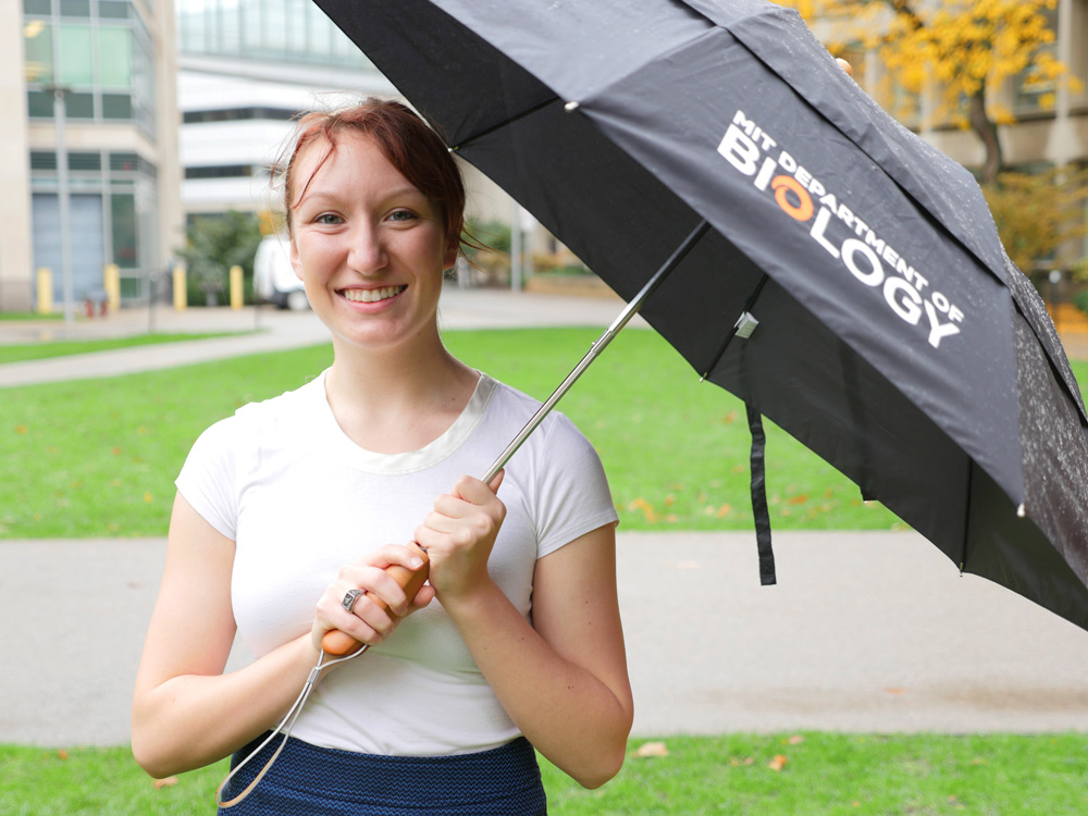
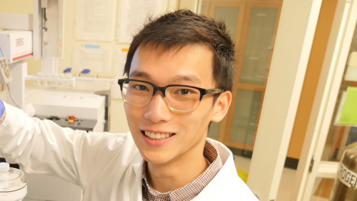
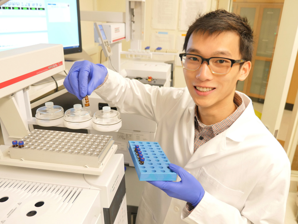
Graduate student Zhaoqi Li investigates how cancer cells grow by harnessing exceptional chemical reactions
Justin Chen
January 11, 2018
Cancer cells use extreme measures to fuel their growth. In fact, researchers like Zhaoqi Li, a third-year graduate student, witness chemical reactions in these cells that would be impossible in the context of normal cells. In a petri dish, normal cells stop dividing once they cover the bottom of the dish and fit neatly together like mosaic tiles. In contrast, cancer cells continue to proliferate and pile haphazardly into small mounds. Within the human body, this abnormal growth — when combined with the spread of cancer cells throughout the body — interferes with organ function and causes death.
Li, a member of Professor Matthew Vander Heiden’s lab located in the Koch Institute, studies cancer metabolism. His work describes the chemical reactions cancer cells use to create energy and materials to make new cells such as membranes, proteins, and DNA. By tracking the flow of nutrients through cancer cells, Li and his labmates are learning how such cells change their metabolism to stimulate growth. These insights will help scientists develop new ways to treat the disease.
Cell metabolism comprises all the chemical reactions occurring in the cell, but researchers are particularly interested in a few reactions that aren’t required by normal cells but are critical for cancer growth. Stopping these reactions with drugs would disrupt the metabolism of cancer cells and hinder tumor development.
“Even though many people may not think of metabolism as a treatment target for cancer, this strategy has been used unwittingly for a long time,” Li says. “Many chemotherapies, such as antifolates, were originally used by doctors without knowing exactly how they worked. Since then, we’ve discovered that those treatments target metabolic pathways. By understanding the details of cancer metabolism we are hoping to design drugs in a more rational way.”
– –
Li might never have joined the Vander Heiden lab or studied cancer metabolism were it not for the unique structure of graduate training at MIT.
During their first year at MIT, graduate students are required to take four classes. Unlike their counterparts at many other PhD programs, they do not work in laboratories until their second semester. This allows students to focus initially on coursework — covering biochemistry, genetics, and research methodology — designed to build a foundation of knowledge. As a result, students discover new interests and develop the confidence to move out of their comfort zones. When it comes time to select a lab, they can choose from 56 spread across six locations, spanning a wide breadth of biological research.
Li could study how the brain forms memories, interpret X-rays to deduce protein structure, or even build miniature organs for drug testing. Before making his decision, he rotated in three laboratories. During each month-long rotation, he performed a small project allowing him to experience the culture of the lab and learn more about its research.
“The first two labs I visited were studying topics I was familiar with and thought were interesting,” he says. “But when I visited the Vander Heiden lab it was so different and caught me off guard. That’s why I eventually joined, even though I had never imagined myself working in a metabolism lab before.”
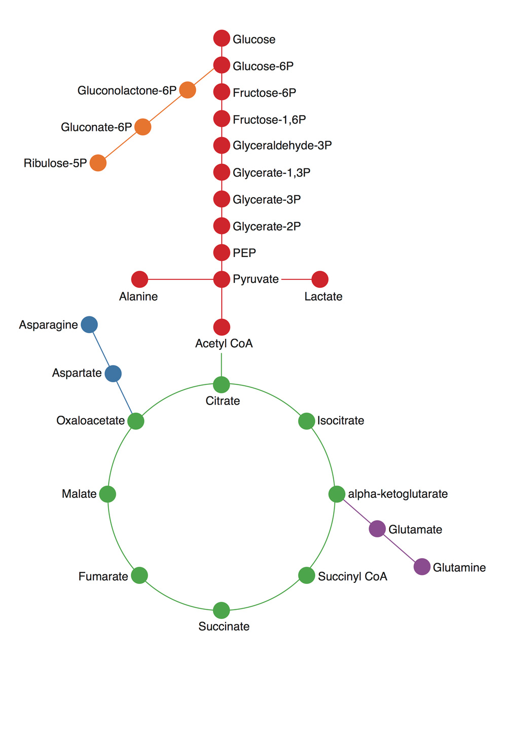
Cellular metabolism is comprised of a network of interconnected biochemical reactions resembling a subway system. Zhaoqi Li compares normal and diseased cells to determine the differences in the way nutrients travel through this network. Credit: Justin Chen
– –
Although he is new to the community of researchers specializing in metabolism, Li has long known that he wanted to interact with the world through science. As an immigrant who moved from China to southern Tennessee at the age of six, Li struggled to learn English and began to view science as a universal language that transcended culture.
“My parents were also non-native speakers and the English as a Second Language classes in my elementary school were geared towards Spanish speakers, so I had a really hard time,” Li says. “I joke that the only reason I passed the first grade was because I was good at math.”
Li’s contrasting relationship with science and English continued as an undergraduate at Columbia University. There he majored in biochemistry and also studied literature of the Western Canon to fulfill his general degree requirements.
“I took four semesters worth of classes that started with Plato and ended with Virginia Woolf,” he says, “It was an eye-opening experience, but I never really loved it. I found biology more intuitive because it doesn’t rely on being familiar with a specific cultural lens. Most every society in the world values the scientific method to some extent.”
Li began working in a lab during his sophomore year at Columbia. To his surprise, he was mentored by a professor who valued his input and encouraged creative thinking. Li’s supervisor also introduced him to basic science — a type of research driven not by the desire to find a specific answer or cure, but by curiosity and the need to better understand the natural world.
– –
During his second semester rotation at MIT, Li searched for similarly open-minded environments, and was attracted to cancer metabolism because the field was relatively young.
“In other more established areas of biology, if you have a question someone has probably answered it in some capacity,” Li says. “The Vander Heiden lab was using new techniques so there was a lot of space to explore. Many questions I asked — even during my initial rotation — didn’t have an answer, which was exciting.”
The great challenge confronting the metabolism field is translating decades’ worth of research on enzymes — proteins that manage chemical reactions — from the test tube to the cell and human body. By studying enzymes individually in the controlled setting of test tubes, researchers have documented almost all the chemical reactions that occur in the cell. When combined, these reactions look like a giant subway map where each stop, indicated by a dot, is a different molecule, and the line between stops represents a chemical reaction where atoms are added or subtracted. Some pathways are a straight line but others have nodes or intersections where a molecule can take part in several different reactions. Other pathways are circular where the molecule that starts the pathway is remade at the end so that the line circles back on itself.
Despite the ability to study chemical reactions in a test tube, scientists have struggled to understand what is actually happening in the complex environment of cells, which coordinate millions of reactions that not only affect each other, but are also influenced by outside stresses like nutrient deprivation.
To Li, using the metabolism map to figure out what chemical reactions are occurring and how atoms are moving through the cell is like using a subway map to track how people are traveling through a city.
“The map describes all the possible routes people could take,” Li says, “but you have to track the passengers to figure out where they are actually going. You could imagine people commuting into the city during the week and going to entirely different places on the weekend. There are a lot of different patterns of movement that you can’t infer just from looking at a map.”
To analyze what chemical reactions are occurring in the cell, Li utilizes cutting edge technology to track carbon atoms — an essential element that is required to build all components of the cell. By tagging carbon with an extra neutron, Li makes the experimentally altered atom heavier and distinguishable from naturally occurring carbon in the cell. Feeding cells nutrients like glucose made with heavy carbons allows Li to compare how molecules are broken down and used by normal and cancerous cells.
“Returning to the subway map analogy, this labeling technique is similar to not only being inside the subway, but also giving everyone in Downtown Boston a red shirt,” Li says. “After 12 hours, we can look at the rest of the city. If we see a lot of red shirts in Allston, we would know that this particular route is really popular.”
In the case of glucose, Li and his labmates observed that normal cells break down the sugar to release energy and heavy carbons in the form of carbon dioxide. In contrast, cancer cells alter their metabolism so that the heavy carbons originally found in glucose are used to build new parts of the cells that are required for cancer cells to grow, such as membranes, DNA, and proteins.
Li’s observations demonstrate how cancer cells sustain abnormal growth by accumulating carbon. For his thesis project, Li has chosen to investigate one of the main tricks cancer cells use to hoard carbon atoms: a process known as carbon fixation. This type of chemical reaction, originally studied in plants performing photosynthesis, attaches carbon dioxide to other molecules. Li’s initial findings suggest that a protein, Malic Enzyme 1, helps cancer cells use carbon dioxide to build components required for growing and dividing.
“This is surprising,” he says, “because the textbook version of this enzyme actually catalyzes the reverse reaction in normal cells where carbon dioxide is removed from molecules. Malic Enzyme 1 is an example of how cancer performs remarkable chemical reactions — who would have thought that cancer cells use carbon like plants do?”
Li is at the beginning stages of his research, and can’t predict where his project will take him. His current goal is to determine how cancer cells react when they are missing Malic Enzyme 1. Such loss could slow growth, but Li will have to perform experiments to be sure, since cancer is a resourceful and elusive target.
Like a detour rerouting travelers around a closed metro stop, cancer cells may further contort their metabolism, taking advantage of little-used or still unidentified chemical reactions to maintain growth. In the face of such adaptability, Li and his labmates believe the best course of action is to be as curious as possible to understand as much as they can about how cancer works. Working together, they discuss confounding results, adjust hypotheses, and design new experiments.
“It’s really encouraging to be part of Matt’s lab and the Koch Institute in general where researchers take a basic science approach,” Li says. “We try to keep an open mind because there’s probably no single thing that cancer cells depend on. Everyone’s work builds together to form a cumulative understanding.”
Photo credit: Raleigh McElvery
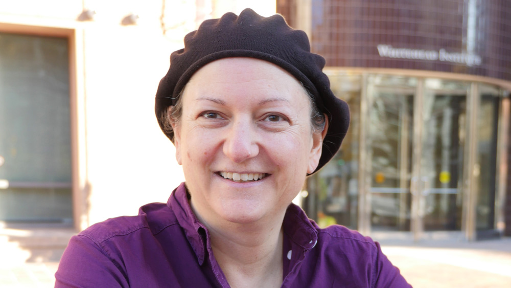
Graduate student Faye-Marie Vassel investigates a protein that helps cells tolerate DNA damage, sharing her expertise with budding scientists to further STEM education
Raleigh McElvery
December 8, 2017
Combatting chemotherapy resistance
Graduate student Faye-Marie Vassel investigates a protein that helps cells tolerate DNA damage, sharing her expertise with budding scientists to further STEM education
Raleigh McElvery
Faye-Marie Vassel has a protein. Well, as a living entity, technically she has many, but just one she affectionately refers to as her own. “My protein, REV7.” And it makes sense — if you were hard at work characterizing a single protein for all six years of your graduate career, you’d be pretty attached, too. Plus, the stakes are high. REV7, which aids in DNA damage repair, could ultimately provide insight into ways to combat chemotherapy resistance.
Although Vassel’s mother trained as an OB/GYN in Russia before moving to the U.S., serving as what Vassel describes as a “quiet” scientific role model, Vassel spent her early childhood emulating her father, a social worker, and engrossed in the social sciences. She intended to one day work in science policy — until high school when she joined an after-school program at the American Museum of Natural History in New York City, and discovered an additional interest.
Here, Vassel took a series of molecular biology classes and met her first female research mentor, a postdoctoral fellow at Rockefeller University, who encouraged her to participate in another, more advanced science program funded by the National Science Foundation.
“I initially had my doubts, but just having that support changed everything,” Vassel says. “That was my first time doing research of any kind, and I got a sense of the sheer diversity of potential research projects. That’s also when I heard there was something called biophysics.”
From that point on, Vassel was hooked. As an undergraduate at Stony Brook University, she initially declared a major in physics before switching to biochemistry. Later, when it came time to select a graduate school, she was split between MIT and the University of California, Berkeley. As she recalls, MIT’s graduate preview weekend made all the difference.
“I had the chance to stay with biology students and speak with professors,” she says. “The whole experience made the department seem personal, and demystified the graduate school process by making it more tangible.”
She proposed a joint position between two labs: Graham Walker’s lab, based in Building 68, and Michael Hemann’s lab situated in the Koch Institute for Integrative Cancer Research. Walker’s lab focuses on microbiology, DNA repair, and antibiotic resistance, while Hemann’s lab investigates chemotherapy resistance in hopes of improving cancer therapies. After stumbling upon one of their joint papers, Vassel decided she’d like to combine the two.
“It’s invaluable to have both perspectives,” she says. “Mike’s lab just celebrated its 10th anniversary, while Graham‘s just had its 35th. It’s been interesting seeing the different ways they approach their respective research questions, because they were trained in such different scientific eras.”
Although Vassel is currently the only student formally working in both labs, the collaboration between Walker and Hemann, aimed at combatting chemotherapy resistance, has been ongoing.
Frontline chemotherapies, including one anticancer agent called cisplatin, kill cancer cells by damaging their DNA and preventing them from synthesizing new genetic material. Just how sensitive cancer cells are to cisplatin — and therefore how effective the treatment is — depends on whether the cell can repair the damage and bypass DNA-damage induced cell death. In some cases, cells increase production of “translesion polymerases,” which are specialized DNA polymerases that can help cells tolerate certain kinds of DNA damage by synthesizing across from damaged DNA or DNA bound to a carcinogen.
Vassel’s protein, REV7, is a structural subunit of one key translesion polymerase, and its expression is deregulated in many different cancer cells. As Vassel suggests, if one aspect of these translesion polymerases — say, the REV7 subunit — could be altered to hinder repair, then perhaps cancer-ridden cells could regain drug sensitivity.
Thanks to recently-developed CRISPR-Cas9 gene editing techniques, Vassel has removed REV7 entirely from drug resistant lung cancer cells, and watched as cisplatin sensitivity was restored. She also conducted rescue experiments, adding REV7 back into cell lines lacking the protein to see whether those cells become resistant to the drug once again. Most recently, she has been working in murine models to see whether REV7 has similar effects in a living system.
If her hypothesis is correct, REV7 would be a powerful target for drug development. Treatments that inhibit REV7, she explains, could be used in tandem with frontline chemotherapies like cisplatin to prevent resistance.
Since her foray into biology at the American Museum of Natural History almost a decade ago, Vassel has maintained her passion for science outreach. During her time at MIT, she has served as a math tutor for middle schoolers in the Cambridge public school system. She also volunteered as a science and math mentor for high school students, as part of a dual athletic and academic program founded by MIT.
As Vassel wraps up her final year of graduate studies, she is torn between completing an academic postdoc and indulging her early interest in science education policy.
“Growing up in New York City, it was not lost on me that — despite the city’s wonderful diversity — people from historically underserved groups were still missing from many science-related positions,” Vassel says. “It got me thinking about the dire need for policymakers to improve curricula to make science more inclusive of all life experiences. There’s this idea that science is apolitical when it’s really not, and that mindset can have detrimental effects on equity and diversity in science.”
Photo credit: Raleigh McElvery
Drug that targets a key cancer protein could combat leukemia and other types of cancer.
Anne Trafton | MIT News Office
January 15, 2018
MIT biologists have designed a new peptide that can disrupt a key protein that many types of cancers, including some forms of lymphoma, leukemia, and breast cancer, need to survive.
The new peptide targets a protein called Mcl-1, which helps cancer cells avoid the cellular suicide that is usually induced by DNA damage. By blocking Mcl-1, the peptide can force cancer cells to undergo programmed cell death.
“Some cancer cells are very dependent on Mcl-1, which is the last line of defense keeping the cell from dying. It’s a very attractive target,” says Amy Keating, an MIT professor of biology and one of the senior authors of the study.
Peptides, or small protein fragments, are often too unstable to use as drugs, but in this study, the researchers also developed a way to stabilize the molecules and help them get into target cells.
Loren Walensky, a professor of pediatrics at Harvard Medical School and a physician at Dana-Farber Cancer Institute, is also a senior author of the study, which appears in the Proceedings of the National Academy of Sciences the week of Jan. 15. Researchers in the lab of Anthony Letai, an associate professor of medicine at Harvard Medical School and Dana-Farber, were also involved in the study, and the paper’s lead author is MIT postdoc Raheleh Rezaei Araghi.
A promising target
Mcl-1 belongs to a family of five proteins that play roles in controlling programmed cell death, or apoptosis. Each of these proteins has been found to be overactive in different types of cancer. These proteins form what is called an “apoptotic blockade,” meaning that cells cannot undergo apoptosis, even when they experience DNA damage that would normally trigger cell death. This allows cancer cells to survive and proliferate unchecked, and appears to be an important way that cells become resistant to chemotherapy drugs that damage DNA.
“Cancer cells have many strategies to stay alive, and Mcl-1 is an important factor for a lot of acute myeloid leukemias and lymphomas and some solid tissue cancers like breast cancers. Expression of Mcl-1 is upregulated in many cancers, and it was seen to be upregulated as a resistance factor to chemotherapies,” Keating says.
Many pharmaceutical companies have tried to develop drugs that target Mcl-1, but this has been difficult because the interaction between Mcl-1 and its target protein occurs in a long stretch of 20 to 25 amino acids, which is difficult to block with the small molecules typically used as drugs.
Peptide drugs, on the other hand, can be designed to bind tightly with Mcl-1, preventing it from interacting with its natural binding partner in the cell. Keating’s lab spent many years designing peptides that would bind to the section of Mcl-1 involved in this interaction — but not to other members of the protein family.
Once they came up with some promising candidates, they encountered another obstacle, which is the difficulty of getting peptides to enter cells.
“We were exploring ways of developing peptides that bind selectively, and we were very successful at that, but then we confronted the problem that our short, 23-residue peptides are not promising therapeutic candidates primarily because they cannot get into cells,” Keating says.
To try to overcome this, she teamed up with Walensky’s lab, which had previously shown that “stapling” these small peptides can make them more stable and help them get into cells. These staples, which consist of hydrocarbons that form crosslinks within the peptides, can induce normally floppy proteins to assume a more stable helical structure.
Keating and colleagues created about 40 variants of their Mcl-1-blocking peptides, with staples in different positions. By testing all of these, they identified one location in the peptide where putting a staple not only improves the molecule’s stability and helps it get into cells, but also makes it bind even more tightly to Mcl-1.
“The original goal of the staple was to get the peptide into the cell, but it turns out the staple can also enhance the binding and enhance the specificity,” Keating says. “We weren’t expecting that.”
Killing cancer cells
The researchers tested their top two Mcl-1 inhibitors in cancer cells that are dependent on Mcl-1 for survival. They found that the inhibitors were able to kill these cancer cells on their own, without any additional drugs. They also found that the Mcl-1 inhibitors were very selective and did not kill cells that rely on other members of the protein family.
Keating says that more testing is needed to determine how effective the drugs might be in combating specific cancers, whether the drugs would be most effective in combination with others or on their own, and whether they should be used as first-line drugs or when cancers become resistant to other drugs.
“Our goal has been to do enough proof-of-principle that people will accept that stapled peptides can get into cells and act on important targets. The question now is whether there might be any animal studies done with our peptide that would provide further validation,” she says.
Joshua Kritzer, an associate professor of chemistry at Tufts University, says the study offers evidence that the stapled peptide approach is worth pursuing and could lead to new drugs that interfere with specific protein interactions.
“There have been a lot of biologists and biochemists studying essential interactions of proteins, with the justification that with more understanding of them, we would be able to develop drugs that inhibit them. This work now shows a direct line from biochemical and biophysical understanding of protein interactions to an inhibitor,” says Kritzer, who was not involved in the research.
Keating’s lab is also designing peptides that could interfere with other relatives of Mcl-1, including one called Bfl-1, which has been less studied than the other members of the family but is also involved in blocking apoptosis.
The research was funded by the Koch Institute Dana-Farber Bridge Project and the National Institutes of Health.
 One day last October, MIT biology professor Matthew Vander Heiden showed up in one of his trademark plaid shirts to teach his undergraduate course on cancer biology. As usual, he peppered his lecture with questions, filling six sliding chalkboards with arrows mapping cellular pathways; he had to erase the boards halfway through class to make room for more notations. But what might have seemed like an ordinary lecture was far from ordinary in one respect: although Vander Heiden was explaining some of the most fundamental aspects of how tumors grow, most of what he was teaching his students would have been absent from nearly every introductory course on cancer biology a decade ago. The science Vander Heiden discussed that afternoon amounted to a once-lost but recently rediscovered chapter in the history of cancer research.
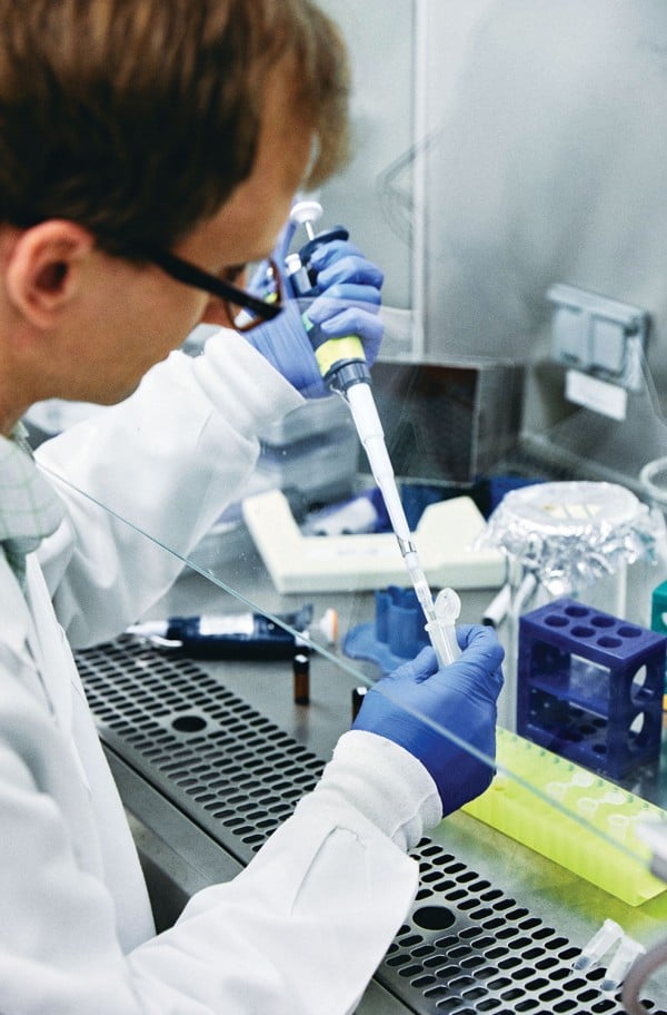
 “Returning to the subway map analogy, this labeling technique is similar to not only being inside the subway, but also giving everyone in Downtown Boston a red shirt,” Li says. “After 12 hours, we can look at the rest of the city. If we see a lot of red shirts in Allston, we would know that this particular route is really popular.”