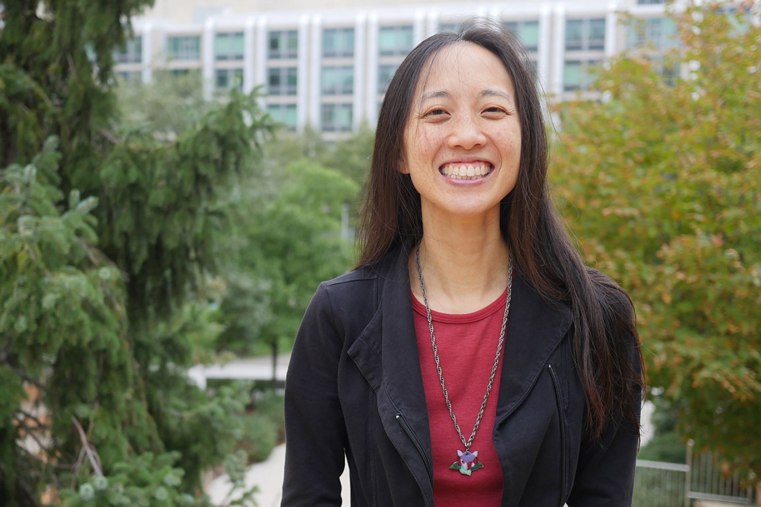
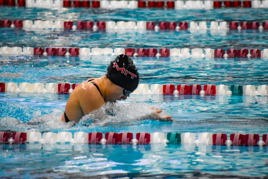
Back to the basics of gene regulation

Graduate student and Schimmel Scholar Annette Jun Diao uses a minimal system to parse the mechanisms underlying gene expression

Lillian Eden | Department of Biology
July 29, 2024

Professor Emeritus of Biology Paul Schimmel PhD ’67 and his wife Cleo Schimmel are among the biggest champions and supporters of graduate students conducting life science research in the Department of Biology at MIT, as well as in departments such as the Department of Brain and Cognitive Sciences, the Department of Biological Engineering, and the Department of Chemistry, and in cross-disciplinary degree programs including the Computational and Systems Biology Program, the Molecular and Cellular Neuroscience Program, and the Microbiology Graduate Program. In addition to the Cleo and Paul Schimmel (1967) Scholars Fund to support graduate women students in the Department of Biology, in 2021, the Schimmels established the MIT Schimmel Family Program for Life Sciences.

Their generous pledge of $50 million in matching funds called for other donors to join them in supporting the training of graduate students who will tackle some of the world’s most urgent challenges. Driven by their unwavering belief that graduate students are the driving force behind much life science research and witnessing a decline in federal funding for graduate education, the Schimmel family established their one-to-one match program. They reached the ambitious goal of $100 million in endowed support in just two years.

Annette Jun Diao’s mother loves to tell the story of Diao’s childhood aversion to the study of life — the gross and the squishy. Unlike some future biologists, Diao wasn’t the type to stomp through creeks or investigate the life of frogs. Instead, she was interested in astronomy and only ended up in a high school biology class because of a bureaucratic snafu. The physics course she’d been hoping to take was canceled due to low enrollment, and she was informed molecular biology was being offered instead.

She attended the University of Toronto and joined the molecular genetics department because of the numerous opportunities for hands-on research. She’s now a third-year graduate student in the Department of Biology at MIT.

“I’m fascinated by the mechanisms that underlie the regulation of gene expression,” Diao says. “All of our genetic information is in DNA, and that DNA is an actual molecule with chemical properties that allow it to be passed from one generation to the next.”

Every cell in our bodies contains a genome of approximately 20,000 genes, but the cells in our retinas are vastly different than the cells in our hearts — not all genes are in action simultaneously, and cell fates vary depending on how which genes are active.

“What is really awesome about the department — and what was attractive to me when I was applying to graduate school — is that I wasn’t sure exactly what methods I wanted to use to answer the questions I was interested in,” Diao says. “A huge advantage of the program was that I had a lot to choose from.”

Diao chose to pursue her thesis work with Seychelle Vos, the Robert A. Swanson (1969) Career Development Professor of Life Sciences and HHMI Freeman Hrabowski Scholar. Diao has been recognized with a Natural Sciences and Engineering Research Council of Canada Fellowship, which is similar to a National Science Foundation graduate fellowship in the United States.

Vos’s lab is generally interested in understanding how transcription is regulated, the interplay of genome organization and gene expression, and the molecular machinery involved. Diao has been working with an enzyme called RNA polymerase II (RNAP II), the molecular machine that reads DNA and creates an RNA copy called mRNA. That mRNA goes on to be read by ribosomes to create proteins.

Many questions remain about RNAP II, including what signals instruct it to begin transcription and, once engaged, whether it will transcribe and how quickly it moves.

RNAP II doesn’t work alone. Diao is working to understand how a transcription factor called negative elongation factor associates with RNAP II and whether the DNA sequence affects that interaction.

Within the broader context of the genome, DNA is packaged extremely tightly; if it were allowed to unfold, its total length could stretch from Cambridge to Connecticut. What RNAP II has access to at any given time is therefore quite restricted, which Diao is also exploring.

She has been exploring this topic in what she refers to as a “reductionist approach.” By creating a minimal system — a strand of DNA and the precise addition of certain other isolated components — she can potentially parse out what ingredients and what sequence of events are essential “in order to really get to the nitty-gritty of how genes are regulated.”

Outside of her work in the lab, Diao is part of BioREFS, a peer support group for graduate students, and gwiBio. Both organizations bring members of the department together for scientific talks and socializing activities outside of the lab, and gwiBio also participates in community outreach.

Diao is also a Schimmel Scholar, supported by Professor Emeritus of Biology Paul Schimmel PhD ’67 and his wife Cleo Schimmel.

“It was really great to learn that I was being supported by a scientist who has done a lot of awesome work that’s relevant to my world,” Diao says.

“It is awesome that they are so committed to supporting the graduate program at MIT, especially when federal resources have become more limited,” Vos says. “With their support, our lab can train basic scientists who can then use their knowledge to transform our study of disease. I hope others follow Paul and Cleo’s example.”

Whitehead Institute researchers uncover a new clue toward understanding the molecular basis of Parkinson’s disease

In Parkinson's disease, a mutation that causes protein misfolding can also turn the brain’s immune cells from friends to foes, possibly accelerating the progression of the disease. New Research from the Jaenisch Lab aims to uncover mechanisms that go awry in the brain, which may inform the development of new therapies that can halt or even reverse the progression of neurological conditions such as Parkinson's.

Shafaq Zia | Whitehead Institute
August 29, 2024

Dopamine is more than the “rush molecule”. This chemical messenger, produced by neurons in the midbrain, acts as a traffic controller that regulates the flow of electrical signals between neurons, assisting with brain functions like cognition, attention, movement, and behavior. But, in instances of Parkinson’s disease (PD), a progressive brain disorder, dopamine-producing neurons begin to die at an unprecedented rate, leading to dwindling levels of this vital chemical and impaired neural communication.

The lab of Whitehead Institute’s Founding Member Rudolf Jaenisch studies genetic and epigenetic factors — changes in gene expression that control which genes are turned on and off, and to what extent, without altering the DNA sequence itself — underlying neurological disorders like PD, Alzheimer’s disease, and Rett Syndrome. Their work aims to uncover the mechanisms that go awry in the brain, which may inform the development of new therapies that can halt or even reverse the progression of these conditions.

In their latest work, Jaenisch and former postdoctoral associate Marine Krzisch examine how a mutation in the gene that encodes for alpha-synuclein, a protein regulating the release of dopamine, affects the resident immune cells of the brain called microglia. The researchers’ detailed findings, published in the journal Biological Psychiatry on August 29, reveal that the mutation renders microglia extremely sensitive, worsening the problem of inflammation in the brain and potentially exacerbating damage to neurons in Parkinson’s disease.

“In fact, even when these mutant microglia are transplanted into a healthy, young brain, they have heightened activation upon stimulation, and low levels of the protective antioxidant catalase,” Krzisch says. “This tells us that in Familial Parkinson’s disease, which is due to genetic mutations, these microglia may be playing an important role in neuron degeneration.”

When nature’s origami falters

The human body is home to tens of thousands of unique proteins, each essential for processes sustaining life. These proteins are composed of linear chains of smaller building blocks called amino acids that are linked together in a specific sequence. For the proteins to perform their functions, the amino acid chains must crumple, rotate, and twist into stable three-dimensional structures. The stakes are high — just as precise folds and creases are crucial to the art of origami, even minor errors in the protein folding process can result in dysfunctional proteins that contribute to disease.

To date, scientists have identified over 20 causative genes in which mutations can result in Familial Parkinson’s disease, a rare, genetically inherited form of PD affecting individuals under or around the age of 50. Among them is SNCA, which encodes for alpha-synuclein, a small protein abundant in dopamine-producing neurons.

The A53T mutation in SNCA promotes the formation of dysfunctional alpha-synuclein proteins that clump together — almost like a ball of yarn — within dopamine-producing neurons. The accumulation of these protein clumps, also known as Lewy bodies, triggers inflammatory signaling in the brain, eventually killing the affected neurons. However, prior research has also shown that the A53T mutation accelerates the progression of PD, or the rate at which neurons die, although the full molecular mechanisms underlying this process are not yet fully understood.

To uncover pathways involved in this progression, researchers in the Jaenisch Lab turned their attention to star-shaped patrollers called microglia that protect the brain from foreign invaders and respond to injuries, including protein aggregates within neurons. This immune response includes activated microglia trying to clear out Lewy bodies by digesting them, recruiting additional immune cells to the site of neurons with protein aggregates, and even killing off diseased neurons to limit damage to the brain.

But these friends can quickly turn to foes. Over-activated microglia can also degrade healthy neurons in the brain, prompting Jaenisch, Krzisch, and colleagues to investigate if excessive microglia activation is one pathway that contributes to progression in PD.

Microglia go rogue

To explore how the A53T mutation in the SNCA gene affects microglia function in PD, scientists at the Jaenisch Lab began by growing human myeloid precursors — the cells that eventually develop into microglia — in lab culture and transplanting them into the brains of immune-deprived mice.

Given the complexity of the brain, it’s common for researchers to study brain cells in the Petri dish. “But in cell cultures, microglia do not have the same morphology [form] as in the brain, show signs of chronic activation, and they don’t survive for a very long time,” says Krzisch. “When we transplant them in mice, the precursors differentiate into microglia that look and function like those in the human brain, and survive for the mouse’s lifespan.”

Using this method, the researchers compared the gene expression profiles of A53T-mutant microglia with those that did not carry the mutation, revealing differences in pathways linked to inflammation, microglia activation, and DNA repair. Additionally, when A53T-mutant microglia were exposed to an immune activator called lipopolysaccharide, they exhibited a heightened inflammatory response compared to non-mutant microglia.

In fact, even in non-inflammatory conditions, A53T-mutant microglia had decreased expression of catalase, an enzyme that helps break down harmful reactive oxygen species produced in response to protein aggregates in PD.

Understanding the molecular basis of progression in PD is challenging, which explains why there are currently no drugs to alter the disease’s course. With these findings in hand, researchers at the Jaenisch Lab are now eager to explore how factors like aging also influence microglia function and contribute to an increased rate of progression in PD.

“Overactivation of microglia isn’t the only cause of neuron death in Parkinson’s,” says Jaenisch. “But if we can decrease their activation, it will help us get to the point where we can slow down or actually stop the disease.”


Pursuing the secrets of a stealthy parasite

By unraveling the genetic pathways that help Toxoplasma gondii persist in human cells, Sebastian Lourido hopes to find new ways to treat toxoplasmosis.

Anne Trafton | MIT News
August 25, 2024

Toxoplasma gondii, the parasite that causes toxoplasmosis, is believed to infect as much as one-third of the world’s population. Many of those people have no symptoms, but the parasite can remain dormant for years and later reawaken to cause disease in anyone who becomes immunocompromised.

Why this single-celled parasite is so widespread, and what triggers it to reemerge, are questions that intrigue Sebastian Lourido, an associate professor of biology at MIT and member of the Whitehead Institute for Biomedical Research. In his lab, research is unraveling the genetic pathways that help to keep the parasite in a dormant state, and the factors that lead it to burst free from that state.

“One of the missions of my lab to improve our ability to manipulate the parasite genome, and to do that at a scale that allows us to ask questions about the functions of many genes, or even the entire genome, in a variety of contexts,” Lourido says.

There are drugs that can treat the acute symptoms of Toxoplasma infection, which include headache, fever, and inflammation of the heart and lungs. However, once the parasite enters the dormant stage, those drugs don’t affect it. Lourido hopes that his lab’s work will lead to potential new treatments for this stage, as well as drugs that could combat similar parasites such as a tickborne parasite known as Babesia, which is becoming more common in New England.

“There are a lot of people who are affected by these parasites, and parasitology often doesn’t get the attention that it deserves at the highest levels of research. It’s really important to bring the latest scientific advances, the latest tools, and the latest concepts to the field of parasitology,” Lourido says.

A fascination with microbiology

As a child in Cali, Colombia, Lourido was enthralled by what he could see through the microscopes at his mother’s medical genetics lab at the University of Valle del Cauca. His father ran the family’s farm and also worked in government, at one point serving as interim governor of the state.

“From my mom, I was exposed to the ideas of gene expression and the influence of genetics on biology, and I think that really sparked an early interest in understanding biology at a fundamental level,” Lourido says. “On the other hand, my dad was in agriculture, and so there were other influences there around how the environment shapes biology.”

Lourido decided to go to college in the United States, in part because at the time, in the early 2000s, Colombia was experiencing a surge in violence. He was also drawn to the idea of attending a liberal arts college, where he could study both science and art. He ended up going to Tulane University, where he double-majored in fine arts and cell and molecular biology.

As an artist, Lourido focused on printmaking and painting. One area he especially enjoyed was stone lithography, which involves etching images on large blocks of limestone with oil-based inks, treating the images with chemicals, and then transferring the images onto paper using a large press.

“I ended up doing a lot of printmaking, which I think attracted me because it felt like a mode of expression that leveraged different techniques and technical elements,” he says.

At the same time, he worked in a biology lab that studied Daphnia, tiny crustaceans found in fresh water that have helped scientists learn about how organisms can develop new traits in response to changes to their environment. As an undergraduate, he helped develop ways to use viruses to introduce new genes into Daphnia. By the time he graduated from Tulane, Lourido had decided to go into science rather than art.

“I had really fallen in love with lab science as an undergrad. I loved the freedom and the creativity that came from it, the ability to work in teams and to build on ideas, to not have to completely reinvent the entire system, but really be able to develop it over a longer period of time,” he says.

After graduating from college, Lourido spent two years in Germany, working at the Max Planck Institute for Infection Biology. In Arturo Zychlinksy’s lab, Lourido studied two bacteria known as Shigella and Salmonella, which can cause severe illnesses, including diarrhea. His studies there helped to reveal how these bacteria get into cells and how they modify the host cells’ own pathways to help them replicate inside cells.

As a graduate student at Washington University in St. Louis, Lourido worked in several labs focusing on different aspects of microbiology, including virology and bacteriology, but eventually ended up working with David Sibley, a prominent researcher specializing in Toxoplasma.

“I had not thought much about Toxoplasma before going to graduate school,” Lourido recalls. “I was pretty unaware of parasitology in general, despite some undergrad courses, which honestly very superficially treated the subject. What I liked about it was here was a system where we knew so little — organisms that are so different from the textbook models of eukaryotic cells.”

Toxoplasma gondii belongs to a group of parasites known as apicomplexans — a type of protozoans that can cause a variety of diseases. After infecting a human host, Toxoplasma gondii can hide from the immune system for decades, usually in cysts found in the brain or muscles. Lourido found the organism especially intriguing because as a 17-year-old, he had been diagnosed with toxoplasmosis. His only symptom was swollen glands, but doctors found that his blood contained antibodies against Toxoplasma.

“It is really fascinating that in all of these people, about a quarter to a third of the world’s population, the parasite persists. Chances are I still have live parasites somewhere in my body, and if I became immunocompromised, it would become a big problem. They would start replicating in an uncontrolled fashion,” he says.

A transformative approach

One of the challenges in studying Toxoplasma is that the organism’s genetics are very different from those of either bacteria or other eukaryotes such as yeast and mammals. That makes it harder to study parasitic gene functions by mutating or knocking out the genes.

Because of that difficulty, it took Lourido his entire graduate career to study the functions of just a couple of Toxoplasma genes. After finishing his PhD, he started his own lab as a fellow at the Whitehead Institute and began working on ways to study the Toxoplasma genome at a larger scale, using the CRISPR genome-editing technique.

With CRISPR, scientists can systematically knock out every gene in the genome and then study how each missing gene affects parasite function and survival.

“Through the adaptation of CRISPR to Toxoplasma, we’ve been able to survey the entire parasite genome. That has been transformative,” says Lourido, who became a Whitehead member and MIT faculty member in 2017. “Since its original application in 2016, we’ve been able to uncover mechanisms of drug resistance and susceptibility, trace metabolic pathways, and explore many other aspects of parasite biology.”

Using CRISPR-based screens, Lourido’s lab has identified a regulatory gene called BFD1 that appears to drive the expression of genes that the parasite needs for long-term survival within a host. His lab has also revealed many of the molecular steps required for the parasite to shift between active and dormant states.

“We’re actively working to understand how environmental inputs end up guiding the parasite in one direction or another,” Lourido says. “They seem to preferentially go into those chronic stages in certain cells like neurons or muscle cells, and they proliferate more exuberantly in the acute phase when nutrient conditions are appropriate or when there are low levels of immunity in the host.”

Uphill battles: Across the country in 75 days

Amulya Aluru ’23, MEng ’24 and the MIT Spokes have spent the summer spreading science, over 3,000 miles on two wheels.

Lillian Eden | Department of Biology
August 22, 2024

Amulya Aluru ’23, MEng ’24, will head to the University of California at Berkeley for a PhD in molecular and cell biology PhD this fall. Aluru knows her undergraduate 6-7 major and MEng program, where she worked on a computational project in a biology lab, have prepared her for the next step of her academic journey.

“I’m a lot more comfortable with the unknown in terms of research — and also life,” she says. “While I’ve enjoyed what I’ve done so far, I think it’s equally valuable to try and explore new topics. I feel like there’s still a lot more for me to learn in biology.”

Unlike many of her peers, however, Aluru won’t reach the San Francisco Bay Area by car, plane, or train. She will arrive by bike — a journey she began in Washington just a few days after receiving her master’s degree.

Showing that science is accessible

Spokes is an MIT-based nonprofit that each year sends students on a transcontinental bike ride. Aluru worked for months with seven fellow MIT students on logistics and planning. Since setting out, the team has bonded over their love of memes and cycling-themed nicknames: Hank “Handlebar Hank” Stennes, Clelia “Climbing Cleo” Lacarriere, Varsha “Vroom Vroom Varsha” Sandadi, Rebecca “Railtrail Rebecca” Lizarde, JD “JDerailleur Hanger” Hagood, Sophia “Speedy Sophia” Wang, Amulya “Aero Amulya” Aluru, and Jessica “Joyride Jess” Xu. The support minivan, carrying food, luggage, and occasionally injured or sick cyclists, even earned its own nickname: “Chrissy”, short for Chrysler Pacifica.

“I really wanted to do something to challenge myself, but not in a strictly academic sense,” Aluru says of her decision to join the team and bike more than 3,000 miles this summer.

The Spokes team is not biking across the country solely to accomplish such a feat. Throughout their journey, they’ll be offering a variety of science demonstrations, including making concrete with Rice Krispies, demonstrating the physics of sound, using 3D printers, and, in Aluru’s case, extracting DNA from strawberries.

“We’re going to be in a lot of really different learning environments,” she says. “I hope to demonstrate that science can be accessible, even if you don’t have a lab at your disposal.”

These demonstrations have been held in venues such as a D.C. jaila space camp, and libraries and youth centers across the country; their learning festivals were even featured on a local news channel in Kentucky.

Some derailments

The team was beset with challenges from the first day they started their journey. Aluru’s first day on the road involved driving to every bike shop and REI store in the D.C. metro area to purchase bike computers for navigation because the ones the team had already purchased would only display maps of Europe.

Four days in and four Chrysler Pacificas later — the first was unsafe due to bald tires, the second made a weird sound as they pulled out of the rental lot, and the third’s gas pedal stopped working over 50 miles away from the nearest rental agency — the team was back together again in Waynesboro, Virginia, for the first time since they’d set out.

Since then, they’ve had run-ins with local fauna — including mean dogs and a meaner turtle — attempted to repair a tubeless bike that was not, in fact, tubeless, and slept in Chrissy the minivan after their tents got soaked and blew away.

Although it hasn’t all been smooth riding, the team has made time for fun. They’ve perfected the art of eating a Clif bar while on two wheelsplayed around on monkey bars in Colorado, met up with Stanford Spokes, enjoyed pounds of ice cream, and downed gallons of lattes.

The team prioritized routes on bike trails, rather than highways, as much as possible. Their teaching activities are scheduled between visits to National Parks like Tahoe, Zion, Bryce Canyon, Arches, and touring and hiking places like Breaks Interstate ParkMammoth Cave, and the Collegiate Peaks.

Aluru says she’s excited to see parts of the country she’s never visited before, and experience the terrain under her own power — except for breaks when it’s her turn to drive Chrissy.

Rolling with the ups and downs

Aluru was only a few weeks into her first Undergraduate Research Opportunities Program project in the late professor Angelika Amon’s lab when the Covid-19 pandemic hit, quickly transforming her wet lab project into a computational one. David Waterman, her postdoc mentor in the Amon Lab, was trained as a biologist, not a computational scientist. Luckily, Aluru had just taken two computer science classes.

“I was able to have a big hand in formulating my project and bouncing ideas off of him,” she recalls. “That helped me think about scientific questions, which I was able to apply when I came back to campus and started doing wet lab research again.”

When Aluru returned to campus, she began work in the Page Lab at the Whitehead Institute for Biomedical Research. She continued working there for the rest of her time at MIT, first as an undergraduate student and then as an MEng student.

The Page Lab’s work primarily concerns sex differences and how those differences play out in genetics, development, and disease — and the Department of Electronic Engineering and Computer Science, which oversees the MEng program, allows students to pursue computational projects across disciplines, no matter the department.

For her MEng work, Aluru looked at sex differences in human height, a continuation of a paper that the Page Lab published in 2019. Height is an easily observable human trait and, from previous research, is known to be sex-biased across at least five species. Genes that have sex-biased expression patterns, or expression patterns that are higher or lower in males compared to females, may play a role in establishing or maintaining these sex differences. Through statistical genetics, Aluru replicated the findings of the earlier paper and expanded them using newly published datasets.

“Amulya has had an amazing journey in our department,” says David Page, professor of biology and core member of the Whitehead Institute. “There is simply no stopping her insatiable curiosity and zest for life.”

Working with the lab as a graduate student came with more day-to-day responsibility and independence than when she was an undergrad.

“It was a shift I quite appreciated,” Aluru says. “At times it was challenging, but I think it was a good challenge: learning how to structure my research on my own, while still getting a lot of support from lab members and my PI [principal investigator].”

Gearing up for the future

Since departing MIT, Aluru and the rest of the Spokes team have spent their nights camping, sleeping in churches, and staying with hosts. They enjoyed the longest day of the year in a surprisingly “Brooklyn chic” house, spent a lazy afternoon on a river, and pinky-promised to be in each other’s weddings. The team has also been hosted by, met up with, and run into MIT alums as they’ve crossed the country.

As Aluru looks to the future, she admits she’s not exactly sure what she’ll study — but when she reaches the West Coast, she knows she’s not leaving what she’s built through MIT far behind.

“There’s going to be a small MIT community even there — a lot of my friends are in San Francisco, and a few people I know are also going to be at Berkeley,” she says. “I have formed a community at MIT that I know will support me in all my future endeavors.”

Study reveals the benefits and downside of fasting

Fasting helps intestinal stem cells regenerate and heal injuries but also leads to a higher risk of cancer in mice, MIT researchers report.

Anne Trafton | MIT News
August 21, 2024

Low-calorie diets and intermittent fasting have been shown to have numerous health benefits: They can delay the onset of some age-related diseases and lengthen lifespan, not only in humans but many other organisms.

Many complex mechanisms underlie this phenomenon. Previous work from MIT has shown that one way fasting exerts its beneficial effects is by boosting the regenerative abilities of intestinal stem cells, which helps the intestine recover from injuries or inflammation.

In a study of mice, MIT researchers have now identified the pathway that enables this enhanced regeneration, which is activated once the mice begin “refeeding” after the fast. They also found a downside to this regeneration: When cancerous mutations occurred during the regenerative period, the mice were more likely to develop early-stage intestinal tumors.

“Having more stem cell activity is good for regeneration, but too much of a good thing over time can have less favorable consequences,” says Omer Yilmaz, an MIT associate professor of biology, a member of MIT’s Koch Institute for Integrative Cancer Research, and the senior author of the new study.

Yilmaz adds that further studies are needed before forming any conclusion as to whether fasting has a similar effect in humans.

“We still have a lot to learn, but it is interesting that being in either the state of fasting or refeeding when exposure to mutagen occurs can have a profound impact on the likelihood of developing a cancer in these well-defined mouse models,” he says.

MIT postdocs Shinya Imada and Saleh Khawaled are the lead authors of the paper, which appears today in Nature.

Driving regeneration

For several years, Yilmaz’s lab has been investigating how fasting and low-calorie diets affect intestinal health. In a 2018 study, his team reported that during a fast, intestinal stem cells begin to use lipids as an energy source, instead of carbohydrates. They also showed that fasting led to a significant boost in stem cells’ regenerative ability.

However, unanswered questions remained: How does fasting trigger this boost in regenerative ability, and when does the regeneration begin?

“Since that paper, we’ve really been focused on understanding what is it about fasting that drives regeneration,” Yilmaz says. “Is it fasting itself that’s driving regeneration, or eating after the fast?”

In their new study, the researchers found that stem cell regeneration is suppressed during fasting but then surges during the refeeding period. The researchers followed three groups of mice — one that fasted for 24 hours, another one that fasted for 24 hours and then was allowed to eat whatever they wanted during a 24-hour refeeding period, and a control group that ate whatever they wanted throughout the experiment.

The researchers analyzed intestinal stem cells’ ability to proliferate at different time points and found that the stem cells showed the highest levels of proliferation at the end of the 24-hour refeeding period. These cells were also more proliferative than intestinal stem cells from mice that had not fasted at all.

“We think that fasting and refeeding represent two distinct states,” Imada says. “In the fasted state, the ability of cells to use lipids and fatty acids as an energy source enables them to survive when nutrients are low. And then it’s the postfast refeeding state that really drives the regeneration. When nutrients become available, these stem cells and progenitor cells activate programs that enable them to build cellular mass and repopulate the intestinal lining.”

Further studies revealed that these cells activate a cellular signaling pathway known as mTOR, which is involved in cell growth and metabolism. One of mTOR’s roles is to regulate the translation of messenger RNA into protein, so when it’s activated, cells produce more protein. This protein synthesis is essential for stem cells to proliferate.

The researchers showed that mTOR activation in these stem cells also led to production of large quantities of polyamines — small molecules that help cells to grow and divide.

“In the refed state, you’ve got more proliferation, and you need to build cellular mass. That requires more protein, to build new cells, and those stem cells go on to build more differentiated cells or specialized intestinal cell types that line the intestine,” Khawaled says.

Too much of a good thing

The researchers also found that when stem cells are in this highly regenerative state, they are more prone to become cancerous. Intestinal stem cells are among the most actively dividing cells in the body, as they help the lining of the intestine completely turn over every five to 10 days. Because they divide so frequently, these stem cells are the most common source of precancerous cells in the intestine.

In this study, the researchers discovered that if they turned on a cancer-causing gene in the mice during the refeeding stage, they were much more likely to develop precancerous polyps than if the gene was turned on during the fasting state. Cancer-linked mutations that occurred during the refeeding state were also much more likely to produce polyps than mutations that occurred in mice that did not undergo the cycle of fasting and refeeding.

“I want to emphasize that this was all done in mice, using very well-defined cancer mutations. In humans it’s going to be a much more complex state,” Yilmaz says. “But it does lead us to the following notion: Fasting is very healthy, but if you’re unlucky and you’re refeeding after a fasting, and you get exposed to a mutagen, like a charred steak or something, you might actually be increasing your chances of developing a lesion that can go on to give rise to cancer.”

Yilmaz also noted that the regenerative benefits of fasting could be significant for people who undergo radiation treatment, which can damage the intestinal lining, or other types of intestinal injury. His lab is now studying whether polyamine supplements could help to stimulate this kind of regeneration, without the need to fast.

“This fascinating study provides insights into the complex interplay between food consumption, stem cell biology, and cancer risk,” says Ophir Klein, a professor of medicine at the University of California at San Francisco and Cedars-Sinai Medical Center, who was not involved in the study. “Their work lays a foundation for testing polyamines as compounds that may augment intestinal repair after injuries, and it suggests that careful consideration is needed when planning diet-based strategies for regeneration to avoid increasing cancer risk.”

The research was funded, in part, by a Pew-Stewart Trust Scholar award, the Marble Center for Cancer Nanomedicine, the Koch Institute-Dana Farber/Harvard Cancer Center Bridge Project, and the MIT Stem Cell Initiative.

New approach enables a closer look at brain cell organelle

Microglia are involved in brain development, as well as neurodegeneration and brain cancer. A new approach from the Jaenisch Lab allows researchers to isolate and analyze microglia phagosomes.

Greta Friar | Whitehead Institute
August 14, 2024

Microglia are the immune system’s front-line enforcers in the brain. They are cells that patrol the brain and destroy anything harmful that they encounter, from invading bacteria to cellular debris. They also remove plaques and prune dysfunctional synapses between neurons. Microglia eliminate their targets by eating them: they envelope material and seal it in bubble-like organelles called phagosomes. A phagosome can then fuse with other organelles that break down its contents.

Microglial phagosomes play important roles in brain development, brain function and a plethora of brain diseases, including neurodegeneration and brain cancer. Therefore, understanding microglial phagosome biology could help to develop new therapies for currently untreatable brain diseases. However, microglia and their organelles have been difficult to study because existing stem cell and animal models insufficiently resemble microglia in the human brain, and because microglia, as vigilant immune patrollers, react to even subtle stimuli and so experimental conditions can trigger changes in the cells that confound analyses.

To overcome those issues, Whitehead Institute Founding Member Rudolf Jaenisch, also a professor of biology at the Massachusetts Institute of Technology; University of Freiburg Professor of Neuropathology Marco Prinz; and University of Freiburg neuropathologist Emile Wogram, who began this project as a postdoctoral researcher in Jaenisch’s lab, have developed a method to isolate and analyze microglia phagosomes in a rapid, gentle, and unbiased fashion.

In research shared in the journal Immunity on August 15, the researchers describe how they can isolate and profile phagosomes from stem cell-derived microglia and fresh human brain tissue. They also share new insights into phagosome biology in the human brain, regarding synaptic pruning and generation of NAD+, a broadly used molecule in the brain, by microglia.

The method that the researchers developed to isolate phagosomes from cells uses immunoprecipitation, in which antibodies latch on to a specific target protein on an organelle’s surface. When the antibodies are collected, they pull the organelles with them. This technique avoids many chemical perturbations that might alter the microglial profile. Sometimes researchers genetically engineer a target for the antibodies, but in order to isolate phagosomes from human brain tissue, Wogram had to find a naturally expressed target. Eventually, he and colleagues found one: the protein CD68.

The researchers first isolated phagosomes from stem cell-derived microglia. They co-cultured the microglia with other brain cell types to create a more brain-like environment, which led to a better match between brain and stem cell-derived microglia gene expression. They triggered some of the microglia to enter an inflammatory or disease-like state to see how that affected the phagosomes. Additionally, Wogram collaborated with the neurosurgery department at the University of Freiburg to get access to brain tissues immediately after their removal during surgery. He isolated phagosomes from brain tissue within a half hour of its removal, allowing him to profile the organelles before their contents could change much.

The profiles that the researchers built included what proteins and metabolites the phagosomes contained, and the whole-cell gene expression profile. The profiles differed significantly between sets of phagosomes, but the researchers identified a core of consistent proteins, including many known and also some unknown phagosome proteins. The results showed that phagosomes contain sensitive signaling molecules that allow them to react quickly to even subtle environmental stimuli.

Additionally, the protein contents of the co-cultured microglia provided strong evidence that when microglia prune synapses, they predominantly prune the side that sends a signal and not the side that receives one. This insight could be useful for understanding how microglia interact with synapses in health and disease.

The researchers also gained insights into a key metabolic pathway that occurs inside of microglia. In excess, the molecule quinolinic acid can be toxic to neurons; it is implicated as involved in many neurodegenerative diseases. However, cells can use quinolinic acid to make NAD+, a molecule broadly used to carry out essential cellular functions. Microglia are the only brain cells that generate NAD+. Wogram and colleagues found that key steps in this process occur in phagosomes. Phagosomes are therefore necessary both for removing excess quinolinic acid to prevent toxicity and for helping to generate NAD+ in the brain.

Finally, Wogram used brain tissues to compare phagosomes from within a tumor to those in the surrounding healthy tissue. The phagosomes in the tumor contained excess quinolinic acid. Although follow-up studies would be needed to confirm the results, these findings are consistent with research that suggests cancer cells use quinolinic acid to fuel their growth.

Collectively, these findings illuminate aspects of phagosome biology and the roles that phagosomes may play in normal brain development and maintenance, as well as in cancer and neurodegeneration. The researchers also anticipate that their method could prove useful for profiling other organelles, especially when the organelles need to be rapidly isolated from human tissue.

Talented high schoolers excel while they explore the brain

Over six years of operation, pre-college outreach programs administered by Mandana Sassanfar, Senior Lecturer and Director of Diversity and Outreach, have placed seven exceptional pre-college students, often from underserved or underrepresented backgrounds, with research groups in The Picower Institute.

David Orenstein | The Picower Institute for Learning and Memory
August 14, 2024

During the pandemic, when many classes delivered online could barely hold students’ attention, Presley Simelus became captivated by the subject of biology thanks to their boundless curiosity and their uncommonly engaging teacher at Prospect Hill Academy Charter School in Cambridge. Meanwhile for Eli Hanechak, the science bug must have bit her very early. She’s wanted to be a doctor for as long as she can remember and in fifth grade built a model of a space station the size of a car out of duct tape, cardboard and broomsticks.

Not every teenager is expected to want to spend their summer breaks exploring science at a bench in an MIT lab, but each year students like Simelus and Hanechak, who have a distinct passion for research, can bring that to The Picower Institute and other research entities around MIT. Over six years of operation, pre-college outreach programs administered by Mandana Sassanfar, Director of Diversity and Outreach, have placed seven exceptional pre-college students, often from underserved or underrepresented backgrounds, with research groups in The Picower Institute. Despite their relative lack of experience compared to the technicians, graduate students, postdocs and professors around them, the students typically thrive.

“Eli has been a wonderful addition to our lab for the summer,” said Kendyll Burnell, the graduate student in the lab of Professor Elly Nedivi who has been working closely with Hanechak. “She is a hard worker, has caught on to techniques quickly, and is constantly asking excellent questions about science and doing research.”

Simelus, too, has been not only learning but also contributing, said their summer host, Yire Jeong, a postdoc in the lab of Associate Professor Gloria Choi.

“Presley has been amazing in our lab, and I was impressed by Presley’s eagerness to learn so much about neuroscience,” Jeong said. “Even when facing technical difficulties, Presley diligently worked to overcome them and achieved meaningful results.”

‘Dive into it’

Simelus, who hails from Everett, Mass., and will be enrolling in Swarthmore College this fall to study biochemistry, first came to MIT through the Leah Knox Scholars Program. Friends who’d been in the program before encouraged them to apply and they got in. During five weeks last summer Simelus and their cohort of fellow Leah Knox high-schoolers had the geeky pleasure of extracting bacteria out of the Charles River and performing a battery of tests to genetically characterize the novel organisms they found. Sassanfar noted that Simelus did the lab work exceptionally well, which is something she looks for when determining whom she might invite back the next summer to do research in an MIT Brain and Cognitive Sciences or Biology lab.

This spring when it came time for Simelus to decide where they might like to take that opportunity, they chose the Choi lab, which studies how the central nervous systems and immune systems interact, sometimes with consequences relevant to disorders including autism. Those keywords intrigued Simelus but really they made the choice because of the potential to learn something entirely new.

It was all this stuff I just simply wasn’t familiar with and I wanted to learn more about it,” Simelus said. “With Gloria’s lab I was truly mystified and I wanted to dive into it. That’s the reason I chose it.”

This summer Simelus has been working with Jeong on a study of how brain cell activity differs when mice are sick vs. when they are well. The project has involved imaging neurons in the brain to detect telltale signs of recent activation, expression of a protein called c-fos. Learning about neuroscience and gaining skills like preparing, staining and imaging tissue have been a very fulfilling outcome of the internship, Simelus said.

“I truly have learned so much about neuroscience,” they said. “I feel like the field, anything related to the brain or neuroscience, is always under this sort of veil and nobody really knows what’s going on. But I feel like my time at the Choi lab has really allowed me to see what neuroscience is about. It’s taught be more about the brain itself and also more about different biology techniques and skills I might need.”

Now the only problem, Simelus said, is that there are even more things to be deeply curious about. Simelus feels committed to harnessing the life sciences in some way in the future to sustain human life and experience. And as someone who not only plays the viola but also composes, they’ve begun thinking more about how the brain responds to music.

There will no doubt be many chances to continue exploring these interests at Swarthmore, but during the summer at MIT, Simelus said they’ve expanded their horizons while still hanging out with friends, some of whom have been working in other nearby labs.

“I don’t think I would have changed my summer,” Simelus said.

‘The perfect opportunity’

Hanechak lives in the tiny Western Massachusetts town of Russell (population: 1,643) and commutes 45 minutes to Pope Francis Preparatory School in Springfield, where she is a rising senior.

In her freshman year at a different school, she yearned for an extra challenge so she got involved in science fair. Interested in medicine, but eager for a project in which she could make a difference without having clinical credentials, she chose to work on reducing pollution by developing a microbe-derived enzyme that could biodegrade plastics. She had read about such enzymes in the research literature and learned that they don’t work as well as engineers have hoped. In successive years she has scrounged lab space and general supervision in labs at Westfield State University and UMass Amherst to create and screen beneficial mutations in the enzyme and to synthesize structures that might help the enzyme work better. The enzyme she presented at the International Science and Engineering Fair last year can degrade plastics in 24 hours.

Sasssanfar, who also directs the Massachusetts Junior Academy of Science (MassJAS), learned of Hanechak’s award-winning science fair presentation and invited her to present at the MassJAS symposium, held at MIT last October. Hanechak did so well, Sassanfar said, she earned a spot present at the American Junior Academy of Science meeting (adjacent to the American Association for the Advancement of Science Annual Meeting) in Denver in February. She also earned Sassanfar’s invitation to join a lab this summer at MIT.

Hanechak has long had an MIT pennant on her wall at home and has admired MIT as a place where regardless of one’s background, if one has a passion for science and technology, that’s what matters.

“No one in my family has gone to college and no one has been involved in a science-related career of any kind,” she said. “One of the reasons MIT has always stood out to me is that there are especially great minds here, but they didn’t all come from established families or super prestigious backgrounds or anything like that. They kind of just were able to make their own way.”

Moreover, the chance to come to MIT to learn about the brain in the Nedivi lab seemed like a great step to take toward that longer-term goal of medicine.

“It seemed like the perfect opportunity to start transitioning into what I want my career to look like and to get some experience doing neuroscience research,” Hanechak said. “I’m very glad I’m able to have this summer experience, like learning the techniques. When I go into my college major of neuroscience, I will have a good background of what I’m doing, besides just my environmental research.”

With Burnell, Hanechak is working on finding a DNA promoter specific for a rare but interesting kind of neuron in the visual cortex, where the brain processes what the eyes see. Finding this genetic signature would allow the lab to label these cells and image them under the microscope, so that they could see how the cells contribute to visual processing.

Hanechak acknowledged she was anxious at first about joining a bigger lab with scientists who have much more experience.

“But my entire summer has been incredibly gratifying and exciting—just being able to work in Cambridge, and live in this area, and experience city life, and then also be in a lab environment where it’s so collaborative and everyone’s very friendly,” she said.

For many teens, summer provides a chance to do what they want to do. Simelus and Hanechak chose the opportunity to explore the brain at The Picower Institute and have made the most of it.

Two Whitehead Institute graduate researchers awarded the 2024 Regeneron Prize for Creative Innovation

Whitehead Institute graduate student researchers Christopher Giuliano (Lourido Lab) and Julian Roessler (Hrvatin Lab) have been awarded the 2024 Regeneron Prize for Creative Innovation.

Merrill Meadow | Whitehead Institute
July 30, 2024

Whitehead Institute graduate student researchers Christopher Giuliano and Julian Roessler have been awarded the 2024 Regeneron Prize for Creative Innovation. In addition, postdoctoral researcher Chen Weng was selected as a finalist in the postdoctoral fellows competition.

The Regeneron Prize, sponsored by global biotechnology company Regeneron Pharmaceuticals, Inc., is a competitive award designed to recognize and honor exceptional talent and originality in biomedical research. Individual graduate students and postdoctoral fellows in the biomedical sciences are nominated by the nation’s top research universities. Then, nominees outline their “Dream Projects” — potentially groundbreaking research projects that they would pursue given unrestricted access to resources and state-of-the-art technology.

The “Dream Project” proposals, presented by the nominees to a selection committee comprised of Regeneron’s leading scientists, are used to evaluate a trainee’s scientific merit, elegance, precision, and creativity. Novel research ideas and out-of-the-box thinking is encouraged — although the proposal must include a strong rationale, basic methodology and design for the project, and a discussion of how its results could advance the field. Both Giuliano and Roessler have been awarded $50,000 for their proposals, which can be used in any way the winners choose. In addition, Weng was awarded $5,000 as a finalist, and Regeneron has made a $10,000 grant to the Whitehead Institute as the home institute of the winners to support its seminar series.

This year’s awards are distinctive in that the two winners are from the same institution: Both Giuliano and Roessler are pursuing their PhDs at Massachusetts Institute of Technology (MIT) and conducting their doctoral research at Whitehead Institute.

Giuliano is a researcher in the lab of Whitehead Institute Member Sebastian Lourido, who is also an associate professor of biology at MIT and holds the Landon Clay Career Development Chair at Whitehead Institute. Giuliano’s Dream Project seeks to address the unique challenges posed by genetically based muscle disorders. “An obstacle in using current gene therapies to treat these conditions,” he explains, “is that muscle tissue comprises large syncytial cells, which contain hundreds of nuclei in a shared cytoplasm. Even when a gene therapy is able to reach an individual muscle cell, it often isn’t able to spread to every nucleus within that cell.” However, certain parasites, like Toxoplasma gondii, thrive because they have the capacity to successfully gain access to and manipulate muscle cells. T. gondii, the primary focus of the Lourido lab’s work, may infect nearly one third of all humans. “My project,” Giuliano says, “would identify the specific biological mechanisms used by the parasites to spread their virulence factor proteins throughout the cell. Using genetic screens for protein spread, we would work toward applying these protein features to improve the efficiency of muscle-directed gene therapies, and ultimately test our system in a mouse model of Duchenne muscular dystrophy.”

Roessler is a researcher in the lab of Whitehead Institute Member Siniša Hrvatin, who is also an assistant professor of biology at MIT. While Roessler’s doctoral research focuses on the neuronal circuitry underlying torpor and hibernation in small mammals, his Dream Project seeks to identify the sensory circuitry regulating the “diving reflex” displayed in land- and sea-dwelling mammals, including humans. The diving reflex occurs when an animal’s face is immersed in cold water, prompting an array of organs to reduce their function in ways that, scientists believe, privileges the flow of oxygen to the brain and muscles. “That this reflex has been conserved across millions of years of mammalian evolution suggests an extraordinary genetic advantage,” Roessler says. “Yet, researchers have given comparatively little attention to the neuronal circuits underlying this reflex, and we don’t understand even the fundamental mechanisms by which the nervous system coincidently detects both cold temperature and the presence of water.” Beyond elucidating a foundational aspect of mammalian biology, Roessler’s projects could, if pursued, underpin new interventions for conditions ranging from migraine headaches to cardiac arrhythmia that might be ameliorated by artificial stimulation or inhibition of the diving response.

Weng is a postdoctoral researcher in the lab of Whitehead Institute Member Jonathan Weissman, who is also a professor of biology at MIT, the Landon T. Clay Professor of Biology at Whitehead Institute, and an Investigator of the Howard Hughes Medical Institute. His Dream Project — which proposes a new approach to using single-cell genealogy to understand factors driving cell line evolution — is an extension of his current work. Indeed, this past year he co-developed a technology that details the family trees of human blood cells and provides new insights into the differences between lineages of hematopoietic stem cells. The technology gives researchers unprecedented access to any human cells’ histories — and a path to resolving previously unanswerable questions.

In immune cells, X marks the spot(s)

By researching the effects of sex chromosomes on two types of immune cells, researchers in the Page Lab explore the biological underpinnings of sex biases in immunity and autoimmune disease

Greta Friar | Whitehead Institute
August 6, 2024

There are many known sex differences in health and disease: cases in which either men or women are more likely to get a disease, experience a symptom, or have a certain drug side effect. Some of these sex differences are caused by social and environmental factors: for example, when men smoked more than women, men were more likely to develop lung cancer. However, some have biological underpinnings. For example, men are more likely to be red-green colorblind because the relevant gene is on the X chromosome, of which men with XY chromosomes have no backup copy for a dysfunctional version.

Often, the specific factors contributing to a sex difference are hard to tease apart; there may not be a simple way to tell what is caused by sex chromosomes versus sex hormones versus environment. To address this question, researchers in Whitehead Institute Member David Page’s lab previously developed an approach to identify the contributions of the sex chromosomes to sex differences. Now, Page and former postdoc in his lab Laura Blanton have built on that work by measuring the effects of the sex chromosomes on two types of immune cells. The work, published in the journal Cell Genomics on August 6, shows that sex chromosome gene expression is consistent across cell types, but that its effects are cell type specific.

Sex differences are common in the function and dysfunction of our immune system. Examples include the typically weaker male immune response to pathogens and vaccines, and the female-biased frequency of autoimmune diseases. Page and Blanton’s work in immune cells examines several genes that have been implicated in such sex differences.

Developing a method to measure sex chromosome influence

The approach that the researchers used is based on several facts about sex chromosomes. Firstly, although females typically have two X chromosomes and males typically have one X and one Y, there are people with rare combinations of sex chromosomes, who have anywhere from 1-5 X chromosomes and 0-4 Y chromosomes. Secondly, there are two types of X chromosome: The active X chromosome (Xa) and the inactive X chromosome (Xi). They are genetically identical, but many of the genes on Xi are either switched off or have their expression level dialed way down.

Xa does not really function as a sex chromosome since everyone in the world has exactly one Xa regardless of their sex. In people with more than one X chromosome, any additional X chromosomes are always Xi. Furthermore, Page and Blanton’s research demonstrates that Xa responds to gene expression by Xi and Y—the sex chromosomes—in the same manner as do the other 22 pairs of non-sex chromosomes—the autosomes.

With these facts in mind, the researchers collected cells from donors with different combinations of sex chromosomes. Then they measured the expression of every gene in these cells, across the donor population, and observed how the expression of each gene changed with the addition of each Xi or Y chromosome.

This approach was first shared in a Cell Genomics paper by Page and former postdoc Adrianna San Roman in 2023. They had cultured two types of cells, fibroblasts and lymphoblastoid cell lines, from donor tissue samples. They found that the effects of Xi and Y were modular—each additional chromosome changed gene expression by about the same amount. This approach allowed the researchers to identify which genes are sensitive to regulation by the sex chromosomes, and to measure the strength of the effect for each responsive gene.

In that and a following paper, Page and San Roman looked at how Xi and Y affect gene expression from Xa and the autosomes. Blanton expanded the study of Xi and Y by using the same approach in two types of immune cells, monocytes and CD4+ T cells, taken directly from donors’ blood. Studying cells taken directly from the body, rather than cells cultured in the lab, enabled the researchers to confirm that their observations applied in both conditions.

In all three papers, the researchers found that the sex chromosomes have significant effects on the expression levels of many genes that are active throughout the body. They also identified a particular pair of genes as driving much of this effect in all four cell types. The genes, ZFX and ZFY, found on the X and Y chromosomes respectively, are transcription factors that can dial up the expression of other genes. The pair originates from the same ancestral gene, and although they have grown slightly apart since the X and Y chromosomes diverged, they still perform the same gene regulatory function. The researchers found that they tended to affect expression of the same gene targets by similar though not identical amounts.

In other words, the presence of either sex chromosome causes roughly the same effect on expression of autosomal and Xa genes. This similarity makes sense: carefully calibrated gene regulation is necessary in every body, and so each sex chromosome must maintain that function. It does, however, make it harder to spot the cases in which sex chromosomes contribute to sex differences in health and disease.

“Sex differences in health and disease could stem from the rare instances in which one gene responds very differently to Xi versus Y—we found cases where that occurs,” Blanton says. “They could also stem from subtle differences in the gene expression changes caused by Xi and Y that build up into larger effects downstream.”

Blanton then combined her and San Roman’s data in order to look at how the effects of sex chromosome dosage—how many Xs or Ys are in a cell—compared across all four cell types.

The effects of sex chromosomes on immune cells

 Blanton found that gene expression from the sex chromosomes was consistent across all four cell types. The exceptions to this rule were always X chromosome genes that are only expressed on Xa, and so could be regulated by Xi and Y in the way that autosomal genes are. This contrasts with speculation that different genes on Xi might be silenced in different cells.

However, each cell type had a distinct response to this identical sex chromosome gene expression. Different biological pathways were affected, or the same biological pathway could be affected in the opposite direction. Key immune cell processes affected by sex chromosome dosage in either monocytes or T cells included production of immune system proteins, signaling, and inflammatory response.

The cell type specific responses were due to different genes responding to the sex chromosomes in each cell type. The researchers do not yet know the mechanism causing the same gene to respond to sex chromosome dosage in one cell type but not another. One possibility is that access to the genes is blocked in some of the cell types. Regions of DNA can become tightly packed so that a gene, or a DNA region that regulates the gene, becomes inaccessible to transcription factors such as ZFX and ZFY, and so they cannot affect the gene’s expression. Another possibility is that the genes might require specific partner molecules in order for their expression level to increase, and that these partners may be present in one cell type but not the other.

Blanton also measured how X chromosome dosage affected T cells in their inactive state, when there is no perceived immune threat, versus their activated state, when they begin to produce an immune response and replicate themselves. Increases in X chromosome dosage led to heightened activation, with increased expression of genes related to proliferation. This finding highlights the importance of looking at how sex chromosomes affect not just different cell types, but cells in different states or scenarios.

“As we learn what pathways the sex chromosomes influence in each cell type, we can begin to make sense of the contributions of the sex chromosomes to each cell type’s functions and its roles in disease,” Blanton says.

Although Page and Blanton found that the presence of an Xi or Y chromosome had very similar effects on most genes, the researchers did identify one interesting case in which response to X and Y differed. FCG2RB is a gene involved in immunity that has been implicated in and thought to contribute to the female bias in developing systemic lupus erythematosus (SLE). Blanton found that unlike most genes, FCGR2B is sensitive to X and not Y chromosome dosage. This strengthens the case that higher expression of FCGR2B could be driving the SLE female bias.

FCGR2B provides a promising opportunity to study the contributions of the sex chromosomes to a sex bias in disease, and to learn more about the biology of a chronic disease that affects many people around the world,” Page says.

In other cases, the researchers found that genes which have been suspected to contribute to female bias in disease did not have a strong response to X chromosome dosage. For example, TLR7 is thought to contribute to female bias in developing autoimmunity, and CD40LG is thought to contribute to female bias in developing lupus. Neither of the genes showed increased expression as X chromosome dosage increased. This suggests that other mechanisms may be driving the sex bias in these cases.

Because of the limited pool of donors, the researchers were not able to identify every gene that responds to sex chromosome dosage, and future research may uncover more sex-chromosome-sensitive genes of interest. Meanwhile, the Page lab continues to investigate the sex chromosomes’ shared role as regulators of gene expression throughout the body.

“We’ve got to recalibrate our thinking from the view that X and Y are mainly involved in differentiating males and females, to understanding that they also have largely shared functions that are important throughout the body,” Page says. “At the same time, I think that uncovering the biology of Xi is going to be incredibly important for understanding women’s health and sex differences in health and disease.”

News Brief: Lamason Lab uncovers seven novel effectors in Rickettsia parkeri infection

The enemy within: new research reveals insights into the arsenal Rickettsia parkeri uses against its host

Lillian Eden | Department of Biology
July 29, 2024

Identifying secreted proteins is critical to understanding how obligately intracellular pathogens hijack host machinery during infection, but identifying them is akin to finding a needle in a haystack.

For then-graduate student Allen Sanderlin, PhD ’24, the first indication that a risky, unlikely project might work was cyan, tic tac-shaped structures seen through a microscope — proof that his bacterial pathogen of interest was labeling its own proteins.  

Sanderlin, a member of the Lamason Lab in the Department of Biology at MIT, studies Rickettsia parkeri, a less virulent relative of the bacterial pathogen that causes Rocky Mountain Spotted Fever, a sometimes severe tickborne illness. No vaccine exists and definitive tests to diagnose an infection by Rickettsia are limited.

Rickettsia species are tricky to work with because they are obligately intracellular pathogens whose entire life cycles occur exclusively inside cells. Many approaches that have advanced our understanding of other bacterial infections and how those pathogens interact with their host aren’t applicable to Rickettsia because they can’t be grown on a plate in a lab setting. 

In a paper recently published in Nature Communications, the Lamason Lab outlines an approach for labeling and isolating R. parkeri proteins released during infection. This research reveals seven previously unknown secreted factors, known as effectors, more than doubling the number of known effectors in R. parkeri. 

Better-studied bacteria are known to hijack the host’s machinery via dozens or hundreds of secreted effectors, whose roles include manipulating the host cell to make it more susceptible to infection. However, finding those effectors in the soup of all other materials within the host cell is akin to looking for a needle in a haystack, with an added twist that researchers aren’t even sure what those needles look like for Rickettsia.  

Approaches that worked to identify the six previously known secreted effectors are limited in their scope. For example, some were found by comparing pathogenic Rickettsia to nonpathogenic strains of the bacteria, or by searching for proteins with domains that overlap with effectors from better-studied bacteria. Predictive modeling, however, relies on proteins being evolutionarily conserved. 

“Time and time again, we keep finding that Rickettsia are just weird — or, at least, weird compared to our understanding of other bacteria,” says Sanderlin, the paper’s first author. “This labeling tool allows us to answer some really exciting questions about rickettsial biology that weren’t possible before.”

The cyan tic tacs

To selectively label R. parkeri proteins, Sanderlin used a method called cell-selective bioorthogonal non-canonical amino acid tagging. BONCAT was first described in research from the Tirrell Lab at Caltech. The Lamason Lab, however, is the first group to use the tool successfully in an obligate intracellular bacterial pathogen; the thrilling moment when Sanderlin saw cyan tic-tac shapes indicated successfully labeling only the pathogen, not the host. 

Sanderlin next used an approach called selective lysis, carefully breaking open the host cell while leaving the pathogen, filled with labeled proteins, intact. This allowed him to extract proteins that R. parkeri had released into its host because the only labeled proteins amid other host cell material were effectors the pathogen had secreted. 

Sanderlin had successfully isolated and identified seven needles in the haystack, effectors never before identified in Rickettsia biology. The novel secreted rickettsial factors are dubbed SrfA, SrfB, SrfC, SrfD, SrfE, SrfF, and SrfG. 

“Every grad student wants to be able to name something,” Sanderlin says. “The most exciting — but frustrating — thing was that these proteins don’t look like anything we’ve seen before.”

Special delivery

Theoretically, Sanderlin says, once the effectors are secreted, they work independently from the bacteria — a driver delivering a pizza does not need to check back in with the store at every merge or turn.

Since SrfA-G didn’t resemble other known effectors or host proteins the pathogen could be mimicking during infection, Sanderlin then tried to answer some basic questions about their behavior. Where the effectors localize, meaning where in the cell they go, could hint at their purpose and what further experiments could be used to investigate it. 

To determine where the effectors were going, Sanderlin added the effectors he’d found to uninfected cells by introducing DNA that caused human cell lines to express those proteins. The experiment succeeded: he discovered that different Srfs went to different places throughout the host cells.  

SrfF and SrfG are found throughout the cytoplasm, whereas SrfB localizes to the mitochondria. That was especially intriguing because its structure is not predicted to interact with or find its way to the mitochondria, and the organelle appears unchanged despite the presence of the effector. 

Further, SrfC and SrfD found their way to the endoplasmic reticulum. The ER would be especially useful for a pathogen to appropriate, given that it is a dynamic organelle present throughout the cell and has many essential roles, including synthesizing proteins and metabolizing lipids. 

Aside from where effectors localize, knowing what they may interact with is critical. Sanderlin showed that SrfD interacts with Sec61, a protein complex that delivers proteins across the ER membrane. In keeping with the theme of the novelty of Sanderlin’s findings, SrfD does not resemble any proteins known to interact with the ER or Sec61. 

With this tool, Sanderlin identified novel proteins whose binding partners and role during infection can now be studied further. 

“These results are exciting but tantalizing,” Sanderlin says. “What Rickettsia secrete — the effectors, what they are, and what they do is, by and large, still a black box.” 

There are very likely other effectors in the proverbial cellular haystack. Sanderlin found that SrfA-G are not found in every species of Rickettsia, and his experiments were solely conducted with Rickettsia at late stages of infection — earlier windows of time may make use of different effectors. This research was also carried out in human cell lines, so there may be an entirely separate repertoire of effectors in ticks, which are responsible for spreading the pathogen.

Expanding Tool Development

Becky Lamason, the senior author of the Nature Communications paper, noted that this tool is one of a few avenues the lab is exploring to investigate R. parkeri, including a paper in the Journal of Bacteriology on conditional genetic manipulation. Characterizing how the pathogen behaves with or without a particular effector is leaps and bounds ahead of where the field was just a few years ago when Sanderlin was Lamason’s first graduate student to join the lab.

“What I always hoped for in the lab is to push the technology, but also get to the biology. These are two of what will hopefully be a suite of ways to attack this problem of understanding how these bacteria rewire and manipulate the host cell,” Lamason says. “We’re excited, but we’ve only scratched the surface.”