Alumni Feature: Carrie Muh, SB ’96, ’97, SM ’97

Muh came to MIT planning to pursue health policy, but ended up majoring in biology and political science, and earned a master's degree in political science before heading to Columbia University for medical school. Now she serves as the chief of pediatric neurosurgery and surgical director of the Pediatric Epilepsy Program at Maria Fareri Children’s Hospital and Westchester Medical Center in Valhalla, New York.

Kara Baskin | MIT Technology Review
December 8, 2025

Carrie Muh ’96, ’97, SM ’97 works in an office surrounded by letters from grateful parents. As the chief of pediatric neurosurgery and surgical director of the Pediatric Epilepsy Program at Maria Fareri Children’s Hospital and Westchester Medical Center in Valhalla, New York, Muh performs life-changing surgeries.

“I see parents who come into my office on their post­operative visit in tears because, for the first time, their child is able to talk or walk. Having a mom come in and say their child said ‘Mama’ for the first time is huge,” she says. Other patients can finally play sports after a lifetime of falls.

About 2% of kids have epilepsy, a neurological condition that can cause seizures, falls, and language issues. About 30% of pediatric epilepsy patients are resistant to the drugs available to treat the condition, but in some cases surgery can help. “Surgery can be such a huge game-changer. Even if it can’t cure them, it can significantly improve quality of life,” she says.

Muh came to MIT planning to pursue health policy. She majored in both biology and political science and then earned a master’s degree in political science. But after a summer interning at the White House, she saw a stronger opportunity for influence as a physician.

As a medical student at Columbia University, Muh got to observe the transplant of a heart from a child who had passed away to another child in need. That sparked her interest in pediatric surgery. “I was able to watch a surgical team save a child’s life,” she remembers.

She took a gap year during medical school to conduct brain tumor research at Columbia, shadowing neurosurgical residents and observing the precise poetry of their surgery. “I absolutely knew that was for me,” she says, adding that the need was also compelling. “There aren’t enough pediatric epilepsy surgery specialists in the country.”

Now patients often travel to Muh for laser ablation, which destroys the part of the brain responsible for seizures without damaging nearby healthy tissue. In other cases, she installs a vagal-nerve stimulator in a child’s chest, which can make seizures less frequent and intense. An additional option is to outfit a child’s brain with EEG electrodes to pinpoint areas of seizure activity; then she can treat those precise areas. For some children, a responsive neurostimulator—“a pacemaker for the brain,” she calls it—can stop a seizure in its tracks.

“Most of my research for the last five years has been on new ways to use technology to help more patients,” she says—younger people and those who have not traditionally been considered candidates for these devices.

Despite her workload, Muh finds time for Yankees games and Broadway plays with her three children. She also travels internationally to care for vulnerable patients. In April 2024, she performed some of the first pediatric epilepsy surgeries with deep brain stimulation in Ukraine. She was also scheduled to head to Kenya for similar work in September of this year.

But wherever she travels, she maintains strong ties to MIT as class secretary and as a former Undergraduate Association president. This reflects her outgoing nature, though she once doubted if she would fit in with the Institute’s intensely engineering-focused culture.

“My dad had gone to MIT and always told me how amazing it was. I loved engineering and science from a young age, so he thought I would obviously love MIT. But I didn’t know if I was ‘techy’ enough to go,” she jokes, even though in high school she did research at NASA’s Student Space and Biology program while juggling sports and theater commitments.

When she toured campus, though, she was hooked.

“I made lifelong friends at MIT and actually met my husband at the wedding of one of my sorority sisters,” she says. “I discovered MIT was a welcoming, open place. I tell my kids now: ‘I’m proud to be a nerd!’ Cool, passionate people are proud of the work they do and the things they love.”

Alumni Spotlight: Michael Franklin ’88

Franklin describes himself as an overachiever, so perhaps it’s not surprising that when he set out to become an educational counselor, one of the MIT alums who volunteers to interview applicants for undergraduate admission, he quickly started racking up record numbers.

Kathryn M. O'Neill | Slice of MIT
December 4, 2025

Michael Franklin ’88 describes himself as an overachiever. So perhaps it’s not surprising that when he set out to become an educational counselor (EC)—an MIT alum who volunteers to interview applicants for undergraduate admission—he quickly started racking up record numbers.

In his first year as an EC, Franklin did 96 interviews—a lot but not quite the most anyone conducted for the 2023–’24 admission cycle. The following year, he redoubled his efforts and earned the top spot. He did it again for students hoping to enter in 2025–’26, interviewing a whopping 160 candidates—nearly twice as many as the No. 2 interviewer.

Interviewing for MIT is a passion he shares with his wife, Debbie Birnby ’91, who conducted 44 interviews herself for students applying for this year. “We started doing this, and it turned out to be just amazing talking to people,” Franklin says. “There’s this glow about students when they talk about what they really like to do, and I enjoy seeing that.”

Birnby agrees. “You hear bad stuff on the news, and then you see young people and you have hope for the future,” she says. “They have so much energy and enthusiasm.”

A Huge Volunteer Corps

Educational counselors form one of the largest groups of MIT volunteers, with more than 7,500 people signed up during the 2024–’25 interview cycle alone. Many—like Franklin and Birnby—love it enough to come back year after year. Currently, MIT has more than 3,500 ECs who have volunteered for over five years and more than 2,000 who have been interviewing for over 10 years. Five ECs have been interviewing for over 50 years.

All play a vital role by helping MIT Admissions get a more holistic view of the candidates, according to Yi Tso ’85, the staff member who runs the EC program as director of the Educational Council. The average EC completes just about six interviews each year. So Franklin and Birnby—who also produce very informative reports on candidates, Tso emphasizes—really stand out: “They are clearly among our super-superstar volunteers.”

The couple’s large interview numbers are, in part, an accident of geography. ECs typically interview candidates who live near them, but when Franklin and Birnby decided to start interviewing in 2022, they were living in an area of Maryland without many MIT applicants. As a result, they took on interviews with “overflow” candidates—those without access to a local EC. They could conduct these interviews easily online, so the pair—who were both newly retired (Franklin was a software developer; Birnby was in lab technical service)—quickly got into a groove and just kept going.

Two years later, they moved back to the Boston area, “partly because we kept telling people how great Boston was, so we started believing it,” Franklin jokes. Since the area has a robust group of ECs, the couple—who by then had been named regional coordinators for the EC program in Boston—continued to interview students from the overflow list.

The Personal Touch

ECs start their work with very little information—just the student’s name, high school, and contact information—and EC guidelines recommend that they spend 30 to 60 minutes with each student. Birnby says she typically spends about an hour and a half. Franklin often takes even more time; he admits he happily spoke for four hours with one enthusiastic candidate. “You meet all these interesting people,” he explains, noting that he and his wife have heard students discuss a full range of interests and ambitions, including everything from competing in the sailing Junior Olympics to launching a national-scale desalination project.

ECs also answer questions from applicants, and both Franklin and Birnby say most students are eager to learn more about campus culture. “A lot of people don’t have a good idea about how weird and wonderful MIT is. It’s a really weird place in a totally good way,” Franklin says. He likes to tell students about the Banana Lounge, the Pirate Certificate, the Baker House piano drop, and other quirky traditions.

Both Franklin and Birnby hope they can help students find out if MIT will be a good fit for them—because that’s at the heart of why they care enough to give back to the Institute themselves. “At MIT I felt I had found my people. I fit there,” says Birnby, who was a biology major while Franklin studied political science. (She says they knew each other when they were both at the Institute but didn’t become a couple until decades later.)

Of course, most candidates ECs interview do not ultimately gain admission. Consider that for the 2025–’26 year, MIT admitted 1,334 undergraduates out of a competitive field of 29,282 applicants. Still, Franklin and Birnby have been able to congratulate several students each year. Today there are MIT students from all over the world—from North Carolina to Kyrgyzstan—who can say they were interviewed by one of them.

Mentors and Friends

Franklin and Birnby have made a point of keeping in touch with many of these students, who now count them as mentors and friends. The pair begin by congratulating students as soon as they can see who has been accepted, which is posted online. “We can’t see results until they see. So we’re like, check already!” Birnby says.

In the fall, they welcome the new students. Then they invite their admitted interviewees from all classes—a group that now numbers 55—to various gatherings throughout the year. In 2024, for example, the pair hosted 10 students for Thanksgiving at their house in Somerville.

“When I came to MIT, it felt so reassuring to know I always had someone to talk to and ask questions of during my MIT journey,” says Yumn Elameer ’28, whom Franklin interviewed. “I’m so grateful to have gotten Mike as an interviewer, to have gained him as a friend and as someone I know will always be there for help, a good laugh, or advice.”

RNA editing study finds many ways for neurons to diversify

When MIT neurobiologists including Troy Littleton tracked how more than 200 motor neurons in fruit flies each edited their RNA, they cataloged hundreds of target sites and widely varying editing rates. Scores of edits altered proteins involved in neural communication and function.

David Orenstein | The Picower Institute for Learning and Memory
November 20, 2025

All starting from the same DNA, neurons ultimately take on individual characteristics in the brain and body. Differences in which genes they transcribe into RNA help determine which type of neuron they become, and from there, a new MIT study shows, individual cells edit a selection of sites in those RNA transcripts, each at their own widely varying rates.

The new study surveyed the whole landscape of RNA editing in more than 200 individual cells commonly used as models of fundamental neural biology: tonic and phasic motor neurons of the fruit fly. One of the main findings is that most sites were edited at rates between the “all or nothing” extremes many scientists have assumed based on more limited studies in mammals, said senior author Troy Littleton, Menicon Professor in the Departments of Biology and Brain and Cognitive Sciences. The resulting dataset and analyses published in eLife set the table for discoveries about how RNA editing affects neural function and what enzymes implement those edits.

“We have this ‘alphabet’ now for RNA editing in these neurons,” Littleton said. “We know which genes are edited in these neurons so we can go in and begin to ask questions as to what is that editing doing to the neuron at the most interesting targets.”

Andres Crane, who earned his PhD in Littleton’s lab based on this work, is the study’s lead author.

From a genome of about 15,000 genes, Littleton and Crane’s team found, the neurons made hundreds of edits in transcripts from hundreds of genes. For example, the team documented “canonical” edits of 316 sites in 210 genes. Canonical means that the edits were made by the well-studied enzyme ADAR, which is also found in mammals including humans. Of the 316 edits, 175 occurred in regions that encode the contents of proteins. Analysis indeed suggested 60 are likely to significantly alter amino acids. But they also found 141 more editing sites in areas that don’t code for proteins but instead affect their production, which means they could affect protein levels, rather than their contents.

The team also found many “non-canonical” edits that ADAR didn’t make. That’s important, Littleton said, because that information could aid in discovering more enzymes involved in RNA editing, potentially across species. That, in turn, could expand the possibilities for future genetic therapies.

“In the future, if we can begin to understand in flies what the enzymes are that make these other non-canonical edits, it would give us broader coverage for thinking about doing things like repairing human genomes where a mutation has broken a protein of interest,” Littleton said.

Moreover, by looking specifically at fly larvae, the team found many edits that were specific to juveniles vs. adults, suggesting potential significance during development. And because they looked at full gene transcripts of individual neurons, the team was also able to find editing targets that had not been cataloged before.

Widely varying rates

Some of the most heavily edited RNAs were from genes that make critical contributions to neural circuit communication such as neurotransmitter release, and the channels that cells form to regulate the flow of chemical ions that vary their electrical properties. The study identified 27 sites in 18 genes that were edited more than 90 percent of the time.

Yet neurons sometimes varied quite widely in whether they would edit a site, which suggests that even neurons of the same type can still take on significant degrees of individuality.

“Some neurons displayed ~100 percent editing at certain sites, while others displayed no editing for the same target,” the team wrote in eLife. “Such dramatic differences in editing rate at specific target sites is likely to contribute to the heterogeneous features observed within the same neuronal population.”

On average, any given site was edited about two-thirds of the time, and most sites were edited within a range well between all or nothing extremes.

“The vast majority of editing events we found were somewhere between 20% and 70%,” Littleton said. “We were seeing mixed ratios of edited and unedited transcripts within a single cell.”

Also, the more a gene was expressed, the less editing it experienced, suggesting that ADAR could only keep up so much with its editing opportunities.

Potential impacts on function

One of the key questions the data enables scientists to ask is what impact RNA edits have on the function of the cells. In a 2023 study, Littleton’s lab began to tackle this question by looking at just two edits they found in the most heavily edited gene: Complexin. Complexin’s protein product restrains release of the neurotransmitter glutamate, making it a key regulator of neural circuit communication. They found that by mixing and matching edits, neurons produced up to eight different versions of the protein with significant effects on their glutamate release and synaptic electrical current. But in the new study, the team reports 13 more edits in Complexin that are yet to be studied.

Littleton said he’s intrigued by another key protein, called Arc1, that the study shows experienced a non-canonical edit. Arc is a vitally important gene in “synaptic plasticity,” which is the property neurons have of adjusting the strength or presence of their “synapse” circuit connections in response to nervous system activity. Such neural nimbleness is hypothesized to be the basis of how the brain can responsively encode new information in learning and memory. Notably, Arc1 editing fails to occur in fruit flies that model Alzheimer’s disease.

Littleton said the lab is now working hard to understand how the RNA edits they’ve documented affect function in the fly motor neurons.

In addition to Crane and Littleton, the study’s other authors are Michiko Inouye and Suresh Jetti.

The National Institutes of Health, The Freedom Together Foundation and The Picower Institute for Learning and Memory provided support for the study.

Research:

Andrés B CraneMichiko O InouyeSuresh K JettiJ Troy Littleton (2025) A stochastic RNA editing process targets a select number of sites in individual Drosophila glutamatergic motoneurons eLife 14:RP108282.
https://doi.org/10.7554/eLife.108282.2

Alternate proteins from the same gene contribute differently to health and rare disease

Whitehead Institute Member Iain Cheeseman, graduate student Jimmy Ly, and colleagues propose that researchers and clinicians may be able to get more information from patients’ genomes by looking at them in a different way.

Greta Friar | Whitehead Institute
November 7, 2025

In a paper published in Molecular Cell on November 7, Whitehead Institute Member Iain Cheeseman, graduate student Jimmy Ly, and colleagues propose that researchers and clinicians may be able to get more information from patients’ genomes by looking at them in a different way.

The common wisdom is that each gene codes for one protein. Someone studying whether a patient has a mutation or version of a gene that contributes to their disease will therefore look for mutations that affect the “known” protein product of that gene. However, Cheeseman and others are finding that the majority of genes code for more than one protein. That means that a mutation that may seem insignificant because it does not appear to affect the known protein could nonetheless alter a different protein made by the same gene. Now, Cheeseman and Ly have shown that mutations affecting one or multiple proteins from the same gene can contribute differently to disease.

In their paper, the researchers first share what they have learned about how cells make use of the ability to generate different versions of proteins from the same gene. Then, they examine how mutations that affect these proteins contribute to disease. Through a collaboration with co-author Mark Fleming, the pathologist-in-chief at Boston Children’s Hospital, they provide two case studies of patients with atypical presentations of a rare anemia linked to mutations that selectively affect only one of two proteins produced by the gene implicated in the disease.

“We hope this work demonstrates the importance of considering whether a gene of interest makes multiple versions of a protein, and what the role of each version is in health and disease,” Ly says. “This information could lead to better understanding of the biology of disease, better diagnostics, and perhaps one day to tailored therapies to treat these diseases.”

Rethinking how cells use genes

Cells have several ways to make different versions of a protein, but the variation that Cheeseman and Ly study happens during protein production from genetic code. Cellular machines build each protein according to the instructions within a genetic sequence that begins at a “start codon” and ends at a “stop codon.” However, some genetic sequences contain more than one start codon, many that are hiding in plain sight. If the cellular machinery skips the first start codon and detects a second one, it may build a shorter version of the protein. In other cases, the machinery may detect a section that closely resembles a start codon at a point earlier in the sequence than its typical starting place, and build a longer version of the protein.

These events may sound like mistakes: the cell’s machinery accidentally creating the wrong version of the correct protein. To the contrary, protein production from these alternate starting places is an important feature of cell biology that exists across species. When Ly traced when certain genes evolved to produce multiple proteins, he found that this is a common, robust process that has been preserved throughout evolutionary history for millions of years.

Ly shows that one function this serves is to send versions of a protein to different parts of the cell. Many proteins contain zip code-like sequences that tell the cell’s machinery where to deliver them so the proteins can do their jobs. Ly found many examples in which longer and shorter versions of the same protein contained different zip codes and ended up in different places within the cell.

In particular, Ly found many cases in which one version of a protein ended up in mitochondria, structures that provide energy to cells, while another version ended up elsewhere. Because of the mitochondria’s role in the essential process of energy production, mutations to mitochondrial genes are often implicated in disease.

Ly wondered what would happen when a disease-causing mutation eliminates one version of a protein but leaves the other intact, causing the protein to only reach one of its two intended destinations. He looked through a database containing genetic information from people with rare diseases to see if such cases existed, and found that they did. In fact, there may be tens of thousands of such cases. However, without access to the people, Ly had no way of knowing what the consequences of this were in terms of symptoms and severity of disease.

Meanwhile, Cheeseman had begun working with Boston Children’s Hospital to foster collaborations between Whitehead Institute and the hospital’s researchers and clinicians to accelerate the pathway from research discovery to clinical application. Through these efforts, Cheeseman and Ly met Fleming.

One group of Fleming’s patients have a type of anemia called SIFD—Sideroblastic Anemia with B-Cell Immunodeficiency, Periodic Fevers, and Developmental Delay—that is caused by mutations to the TRNT1 gene. TRNT1 is one of the genes Ly had identified as producing a mitochondrial version of its protein and another version that ends up elsewhere: in the nucleus.

Fleming shared anonymized patient data with Ly, and Ly found two cases of interest in the genetic data. Most of the patients had mutations that impaired both versions of the protein, but one patient had a mutation that eliminated only the mitochondrial version of the protein, while another patient had a mutation that eliminated only the nuclear version.

When Ly shared his results, Fleming revealed that both of those patients had very atypical presentations of SIFD, supporting Ly’s hypothesis that mutations affecting different versions of a protein would have different consequences. The patient who only had the mitochondrial version was anemic but developmentally normal. The patient missing the mitochondrial version of the protein did not have developmental delays or chronic anemia but did have other immune symptoms, and was not correctly diagnosed until his fifties. There are likely other factors contributing to each patient’s exact presentation of the disease, but Ly’s work begins to unravel the mystery of their atypical symptoms.

Cheeseman and Ly want to make more clinicians aware of the prevalence of genes coding for more than one protein, so they know to check for mutations affecting any of the protein versions that could contribute to disease. For example, several TRNT1 mutations that only eliminate the shorter version of the protein are not flagged as disease-causing by current assessment tools. Cheeseman lab researchers including Ly and graduate student Matteo Di Bernardo are now developing a new assessment tool for clinicians, called SwissIsoform, that will identify relevant mutations that affect specific protein versions, including mutations that would otherwise be missed.

“Jimmy and Iain’s work will globally support genetic disease variant interpretation and help with connecting genetic differences to variation in disease symptoms,” Fleming says. “In fact, we have recently identified two other patients with mutations affecting only the mitochondrial versions of two other proteins, who similarly have milder symptoms than patients with mutations that affect both versions.”

Long term, the researchers hope that their discoveries could aid in understanding the molecular basis of disease and in developing new gene therapies: once researchers understand what has gone wrong within a cell to cause disease, they are better equipped to devise a solution. More immediately, the researchers hope that their work will make a difference by providing better information to clinicians and people with rare diseases.

“As a basic researcher who doesn’t typically interact with patients, there’s something very satisfying about knowing that the work you are doing is helping specific people,” Cheeseman says. “As my lab transitions to this new focus, I’ve heard many stories from people trying to navigate a rare disease and just get answers, and that has been really motivating to us, as we work to provide new insights into the disease biology.”

Jimmy Ly, Matteo Di Bernardo, Yi Fei Tao, Ekaterina Khalizeva, Christopher J. Giuliano, Sebastian Lourido, Mark D. Fleming, Iain M. Cheeseman. “Alternative start codon selection shapes mitochondrial function and rare human diseases.” Molecular Cell, November 7, 2025. DOI: https://10.0.3.248/j.molcel.2025.10.013

Q&A: Picower researchers including MIT Biology faculty Sara Prescott join effort to investigate the ‘Biology of Adversity’

Assistant Professor Sara Prescott and Research Affiliate Ravikiran Raju are key collaborators in a new Broad Institute research project to better understand physiological and medical effects of acute and chronic life stressors.

David Orenstein | The Picower Institute for Learning and Memory
November 3, 2025

Adverse experiences such as abuse and violence or poverty and deprivation have always been understood to be harmful, but the tools to understand how they may cause specific medical conditions and outcomes have only emerged recently. Technologies such as RNA or chromatin sequencing, for instance, can help scientists observe how stressors change gene expression, which can help establish mechanistic biological explanations for why people who’ve suffered adversity also experience higher risks of conditions such as stroke or Alzheimer’s disease.

Advancing scientific understanding of the physiological connections between adversity and disease can help pharmaceutical developers, physicians and public officials to develop meaningful interventions. Led by researcher Jason Buenrostro, the Broad Institute has launched a new research program, the “Biology of Adversity” project.. As leading collaborators in the effort, Picower Institute investigator Sara Prescott, assistant professor of biology, and Tsai Lab research affiliate Ravikiran Raju, a pediatrician at Boston Children’s Hospital, plan research projects in their Picower Institute labs to better elucidate how life stress leads to medical distress.

How can biology and neuroscience studies help people who’ve experienced adversity?

Prescott: Adversity comes in many flavors. But across different types of adversity, there is a common theme that it leads to psychological and emotional distress. If you were to ask a random person on the street, they’d probably tell you that distress is simply a feeling that exists only in the mind, rather than a biological process. But this is not true. We now appreciate that stress has predictable effects on the body, and there are severe long-term health consequences of experiencing chronic stress. Unfortunately, it’s been difficult to argue based on epidemiological data that stress itself (rather than other lifestyle factors like diet, smoking or access to health care services) is causally linked to poor health outcomes. This is confounded by the fact that we haven’t had good ways to empirically measure people’s levels of adversity and stress. This is part of what we want to address at the Biology of Adversity Project.

From a scientific perspective, there is still much to be understood about stress and the biological processes that lead to stress-associated diseases. And so that’s hopefully where efforts like the Biology of Adversity Project are going to come in. We can use scientific practices to come up with better guidelines for ways to track levels of stress, develop diagnostics, and then, hopefully, one day this will turn into actionable interventions. It’s not a random process of things going awry. There are going to be biological programs that are engaged in predictable ways. And we’re trying to understand, what exactly are these neural or biological programs? How many different types of programs are there? And how do each of those programs actually work down to the cellular and molecular level?

Raju: Efforts to combat adversity and stress have largely remained in the social space to date. But what we know from a growing body of epidemiological literature is that social stressors can have profound biological impact. They cause increases in mental health disorders, physical disorders like cancer, stroke, and heart disease. Individuals who experience chronic and high levels of stress are dying sooner. I think there is an imperative to understand what these forces are doing to our biology and how they’re dysregulating our physiology. Armed with that information, we can start to be more mechanistic and evidence-based in our promotion of resilience. What are the pathways that are made vulnerable when individuals are stressed? How do we rescue those deficiencies, whether it be through existing practices or novel interventions? A lot of the research we’re doing here at Picower is focusing on pathways that could be targeted and leveraged using specific micronutrients or specific small molecules that help promote resilience and prevent the onset of premature illness in individuals who are stress exposed.

What is the Biology of Adversity Project and how are each of you involved?

Prescott: My lab studies the autonomic nervous system, and we’re involved in the project’s animal studies. We think of stress as an adaptive response to prepare the body for an impending threat. When people experience stress, what happens? You engage a fight or flight response—you sweat, start to breathe harder, your heart rate goes up, your pupils dilate. This is protective in acute settings, but can become very maladaptive when these systems are activated for too long or in inappropriate settings, like when someone is having a panic attack. We predict that a lot of the long-term health consequences associated with adversity could relate to dysregulated autonomic stress responses.

And so that’s where our lab’s tools come in. We have good ways in animals to measure their heart rate and breathing in response to stress. We also have a wide range of genetic tools to specifically target different neural pathways in the periphery, possibly blocking stress pathways at the source. With these tools, we can explore what role those circuits have in long-term changes in these animals with greater precision than what was possible in the past.

Raju: My involvement came through my work on the Environmental and Social Determinants of Child Mental Health Conference in 2023, which I co-hosted with Li-Huei Tsai. I think this conference made the scientific community in Boston more aware that this was something of deep interest to researchers at Picower and MIT. In the creation of the Biology of Adversity Project, the center director, Jason Buenrostro, was doing a survey of the landscape of folks who were studying stress and adversity, and who were passionate about it and connected with us because of that symposium. Since then, I’ve been engaged in really exciting conversations with him and a exciting group of collaborators, including Sara Prescott. And so I’m really excited that a few of our projects are being showcased as flagship projects. We are currently using animal models of early life stress to try and build preclinical models to deepen our understanding of how stress dysregulates physiology. We’re developing pipelines for trying to think about promoting resilience through targeted interventions, using those preclinical models.

What research questions do you each plan to tackle?

Prescott: Broadly, we’re interested in the body-brain connection and how this relates to stress. How do different cues from within the body—like diet, or taking a deep breath–promote or regulate stress levels? These are interesting questions about how sensory inputs from the body feed into stress circuits in the brain. We’re also interested in the other direction—understanding how stress causes changes to peripheral organs, for example, by engaging the sympathetic nervous system. It’s well understood that sympathetic neurons are responsible for making you sweat and your heart race, but do they do other things as well? For example, the field is starting to appreciate that these same neurons regulate the immune system, and can signal to stem cells to promote or suppress tissue repair. These are important pathways to understand, as they could explain some of the links between chronic stress (where sympathetic neurons are over-activated) and increased rates of diseases like cancer. It also may have therapeutic applications down the road. I’m incredibly excited for the opportunity to work with people like Ravi, and others in the project, to apply our expertise in physiology and autonomic signaling towards this immensely important problem. I’m hoping that through this work we can move to an era where we can, from a societal perspective, understand how much our stress levels are damaging our body, be able to track that, and then find better ways to prevent the damage that’s happening.

Raju:  We are leveraging three key mouse models of environmental perturbations in this work: environmental enrichment, social isolation and resource deprivation. In studying enrichment, we are trying to better study the factors that promote resilience to stress. In our previous work on resilience, for example, we identified a transcription factor that’s specifically recruited to help ensure that neurons are resilient to the onset of Alzheimer’s pathology. So we’ve leveraged these enrichment models to study that mechanism and are able to then think of how that pathway might be leveraged in stress-exposed individuals. We are also using models of stress, specifically social isolation and resource deprivation. The idea here is that because mice are social mammals and rely on resources and social interactions and social networks in order to thrive, we can modulate these in a species-relevant way, and then study the pathways that are dysregulated. This will allow us to define vulnerable pathways in these preclinical models, and then assess if those same pathways are dysregulated in humans that are experiencing analagous environmental conditions. Armed with the right model, we can then determine how to reverse the physiological derangements induced by environmental stressors.

Research Threads: One lab’s detective work reveals secrets of immortal cells

Most cells in our body live and die. But the germline, the cells that produce eggs and sperm, must carry on forever. How the germline successfully produces the next generation is a long-studied question. Through a string of discoveries made over years, the Yamashita lab is teasing apart how the germline remains immortal.

Madeleine Turner | Whitehead Institute
October 7, 2025

Most cells in our body live and die. But the germline, the cells that produce eggs and sperm, must carry on forever. How the germline successfully produces the next generation is a long-studied question. Research Threads examines how answering one question uncovers more questions to be solved. In our first installment of Research Threads, we follow the research of Whitehead Institute Member Yukiko Yamashita. Through a string of discoveries made over years, the Yamashita lab is teasing apart how the germline remains immortal.

“The germline is the only cell type responsible for transmitting the genome from generation to generation,” Whitehead Institute Member Yukiko Yamashita says. “We’ve done that for 1.5 billion years.”

The germline is the population of cells in an organism that give rise to gametes, either egg or sperm cells. These gametes contain genetic information, encoded in DNA, needed to produce the next generation. The act of transmitting this information — the instructions that a new individual needs to develop and function — is like a relay race that never ends. While a skin or gut cell may be prone to genetic errors and is generally replaceable, germline stem cells (GSCs) must maintain their genomes with precision. Otherwise, any mistakes or imbalances would be inherited by offspring and accumulated over generations, potentially driving a species to extinction. In other words, to keep passing the baton in this relay, the germline must be faithfully preserved.

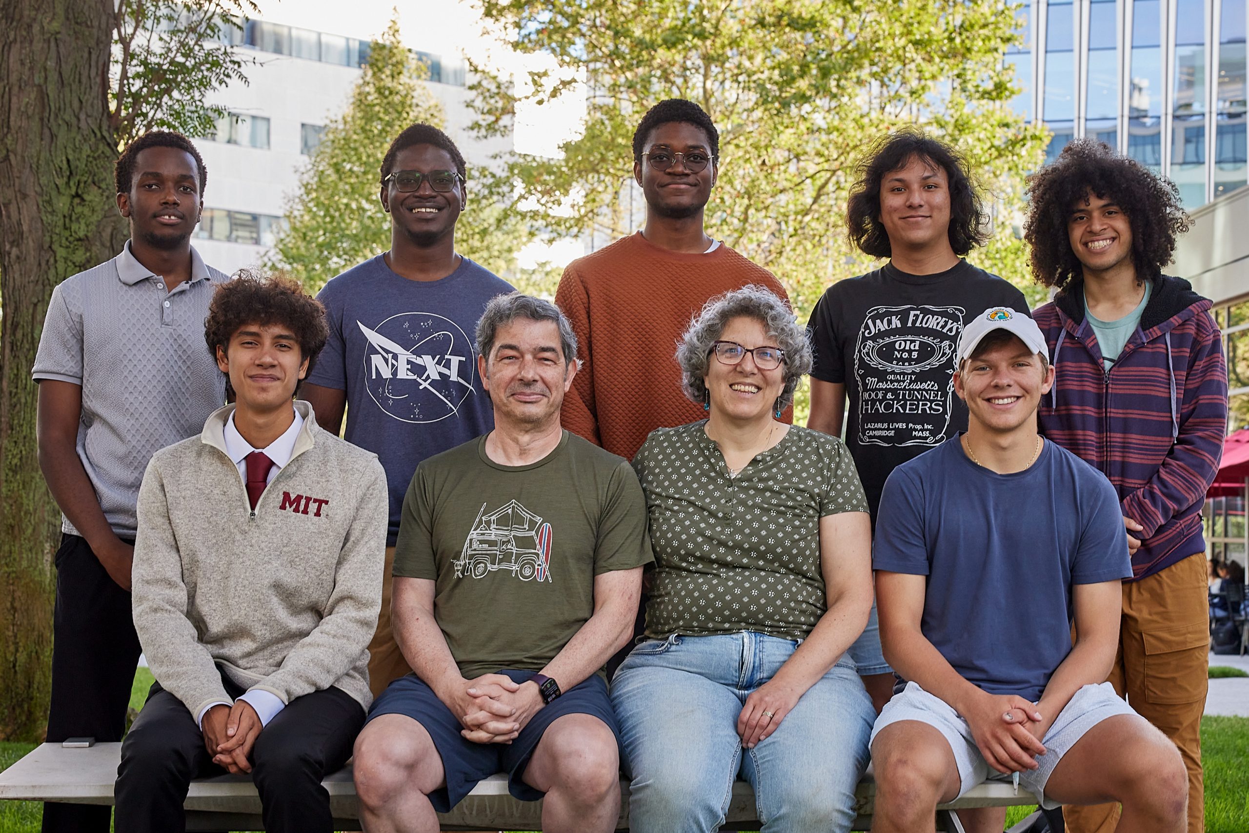
Although germline preservation is paramount to the existence and survival of a species, some fundamental parts of its biology have remained a mystery. Yamashita, who is also a professor of biology at the Massachusetts Institute of Technology and a Howard Hughes Medical Institute Investigator, has focused her research on unraveling the secrets of the germline. To study these cells’ immortality, her lab utilizes the model organism Drosophila melanogaster, or the fruit fly. Along the way, research in the Yamashita lab has highlighted how the process of scientific discovery can be circuitous, often pulling scientists in surprising directions, revealing new questions and avenues to explore.

For decades, scientists had observed an aspect of germline behavior that was hard to explain. Most cells in the body divide to produce two identical copies, or daughter cells. GSCs in male fruit flies, however, divide “asymmetrically,” meaning they yield two daughter cells that are not identical. Instead, one daughter cell becomes a new GSC, while the other goes on to become a gamete, in this case a sperm cell. It might be easy to assume that asymmetric cell division is about producing gametes while maintaining a pool of stem cells. But an additional detail complicates this theory: when a daughter cell is on the path to becoming sperm, it can loop back around to become another stem cell, instead of continuing differentiation to become a sperm cell.

“If it can do that, why divide asymmetrically in the first place?” Yamashita says.

To shed light on why GSCs divide asymmetrically, researchers wanted to know how genetic information was actually divvied up between daughter cells. “After I started my own lab, there was this question hanging in the field,” Yamashita says. It made sense to find some difference in inheritance, DNA-based or otherwise: something to distinguish between the daughter fated to become a gamete, and the other that would remain in the GSC pool.

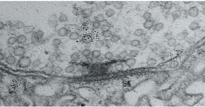
Preparing for division, a cell duplicates its DNA. Chromosomes happen to be shaped like the letter “X” as a result of this duplication: the left and right sides of the “X” are called matching sister chromatids, each a copy of the other. Later in cell division, these two sister chromatids part ways, winding up in separate daughter cells.

In 2013, Yamashita and her former graduate student, Swathi Yadlapalli, made a strange but important discovery. In fruit flies, for the X and Y chromosomes (the sex chromosomes), sister chromatids were not being sorted randomly. Instead, one was more likely to go to the daughter cell that would become a gamete; the other to the daughter on the GSC track. There had to be a reason for this preference, but no one had an explanation.

Initial experiments did not reveal obvious differences between these sister chromatid pairs. “Everyone would say, ‘oh, there’s probably some sort of epigenetic information in there,” Yamashita says, referring to heritable changes not carried in DNA. With few promising leads, the lab decided to take a systematic approach. George Watase, then a postdoc in the lab, began the painstaking work of removing different a parts of flies’ X chromosomes, seeing if the absence of any particular element would disrupt the pattern of preferential segregation.

“We thought it was going to be satellite DNA,” Yamashita says, referring to large swathes of DNA in the genome that are highly repetitive but don’t code for any genes. (While this initial guess was wrong, it kickstarted a separate project in the lab — one which led to discovering the unexpected role that satellite DNA plays when one species forks into two.)

Eventually the team narrowed in on the true culprit: ribosomal DNA (rDNA). The primary role of rDNA is to produce the components that make up ribosomes. Ribosomes, in turn, perform the crucial task of synthesizing proteins.

“We didn’t like this finding in the beginning. I always say that ribosomal DNA is ‘too important to be interesting.’ You don’t get excited about something you really need, like toilet paper,” Yamashita says. “In the case of ribosomal DNA, bacteria needs it, humans need it, everybody needs it.”

But what did rDNA have to do with asymmetric cell division in the germline?

“Around that time, we started reading lots of papers,” Yamashita says. “Then we discovered a phenomenon called rDNA magnification. These were studies from the 1960s and ’80s — most of the people in my lab were not even born yet.”

These studies from decades ago described mutant flies that lacked a sufficient amount of rDNA and, as a result, had a “bobbed” phenotype, or appearance. Since these flies possessed fewer ribosomes to produce proteins, the bristles on their back were shorter; the protective cuticle covering their bodies weakened. But when they reproduced, many of their offspring possessed a normal amount of rDNA. These observations pointed towards a mechanism that allowed flies to replenish their supply of rDNA.

At the time rDNA magnification was first observed, the phenomenon was regarded as an oddity, something that only happened in mutant flies and did not have physiological relevance. But Yamashita realized there was a connection between rDNA magnification and asymmetric division in the germline.

To make enough protein, a cell must produce ample ribosomes. To do that, the fruit fly genome contains hundreds of copies of rDNA in a row. But the DNA replication process can struggle to handle so many rDNA copies strung together, and can sometimes experience a hiccup, resulting in the loss of rDNA copies with each new division. It’s an outcome that might be okay on occasion, but would wreak havoc over many generations.

“All of a sudden, [rDNA magnification] was not about a mutant chromosome,” Yamashita says. “We were like, holy moly. This is about germline immortality.”

Soon many different pieces became part of a cohesive story: asymmetric cell division is not about producing a balance of gametes and stem cells; it’s about maintaining rDNA in the germline. Sister chromatids are almost identical — but one contains more copies of rDNA than the other, and that copy is fated to stay in the GSC pool. Through this asymmetry, the germline is replenished of rDNA; it can pass the baton to the next generation.

“For quite some time, for so many observations that everyone knew in the field, we felt we had an explanation. But from that ‘aha!’ moment, we took multiple years to test everything,” Yamashita says.

In subsequent years, the Yamashita lab pinned down additional details about how rDNA is diverted back to the germline. First, in 2022, the team identified a specific protein, which they named Indra, which binds to rDNA. The presence of Indra helps assign the sister chromatid containing more rDNA copies to the GSC daughter cell.

Their next discovery was another plot twist. If one sister chromatid contained more rDNA than the other, and those rDNA copies weren’t appearing out of thin air, it meant that one chromatid needed to be stealing rDNA from its sister. The lab discovered a genetic element that facilitated this transfer: a retrotransposon.

Retrotransposons are usually considered “genetic parasites,” copying and pasting themselves into the genome. In an attempt to reinsert itself, this particular retrotransposon, called R2, slices open sections containing rDNA on both chromatids. As the cell repairs these breaks, it may inadvertently stitch copies of rDNA from one chromosome to the other, creating an unequal number of copies between the two.

“Not many people thought retrotransposons could be beneficial to the host. They’re seen as parasites,” Yamashita says. “But it turns out that they are essential for germline immortality.”

Sometimes, one research project is a spin off of a spin off. That was true for Xuefeng Meng, a graduate student in the lab who was working on satellite DNA, the genetic element that turned out to be unrelated to asymmetric cell division, but was interesting in its own right.

While studying satellite DNA, Meng noticed that a particular stock of flies had a problem producing normal sperm, that their cells’ nuclei were abnormally packaged. The problem had to do with a gene called Stellate on the flies’ X chromosome. While most flies have few copies of Stellate, these flies had a higher number of copies.

Stellate was already known in the field as a meiotic driver, or “selfish-gene”: a genetic element that has evolved ways to preferentially transmit itself to the next generation. Some meiotic drivers, including Stellate, are on the sex chromosomes and, when not suppressed, cause an excess of either male or female progeny. In this case, Stellate produces a protein, Ste, which is found to concentrate in Y-carrying cells during meiosis, the specialized type of cell division that produces gametes (meiosis follows the earlier round of asymmetric cell division described above). High concentrations of Ste impede the proper packaging of nuclei in cells, leading to their eventual death. When Stellate is widely expressed, fewer male flies emerge in the next generation.

But here lies an inherent tension: if a selfish gene is too good at propagating itself, and produces only males or females, its host species would go extinct — and so would the gene. Meng and Yamashita were interested in this paradox. Through this work, they identified a novel mechanism that keeps Stellate in check. To balance selfish propagation with the host species’ survival, Stellate has a built-in system for pumping the brakes. After Ste concentrates in Y-carrying cells during the first meiotic division, the protein is unevenly distributed a second time. This second step spares a portion of Y-carrying cells that go on to create males.

How the germline is able to counter disruptive forces, thereby renewing itself, continues to be a ripe research area. Researchers still don’t know all the secrets to how a line of cells can achieve perpetuity — but the Yamashita lab continues to investigate the question.

“What I really like about people in my lab is that they don’t jump to the easiest conclusion,” Yamashita says. “If you start embracing surprise, then good things happen. Because you are surprised, you start testing your finding in multiple ways. Then you can build confidence about the result.”

Notes

Xuefeng Meng and Yukiko Yamashita (2025). “Intrinsically weak sex chromosome drive through sequential asymmetric meiosis.” Science Advanceshttps://doi.org/10.1126/sciadv.adv7089

Jonathan O. Nelson, Tomohiro Kumon, Yukiko M. Yamashita. (2023) “rDNA magnification is a unique feature of germline stem cells.” PNAShttps://doi.org/10.1073/pnas.2314440120

Jonathan O. Nelson, Alyssa Slicko, Yukiko M. Yamashita. (2023) “The retrotransposon R2 maintains Drosophila ribosomal DNA repeats.” PNAShttps://doi.org/10.1073/pnas.2221613120

George J. Watase, Jonathan O. Nelson, Yukiko M. Yamashita. (2022) “Nonrandom sister chromatid segregation mediates rDNA copy number maintenance in Drosophila.” Science Advanceshttps://www.science.org/doi/10.1126/sciadv.abo4443

Madhav Jagannathan and Yukiko Yamashita. (2021) “Defective satellite DNA clustering into chromocenters underlies hybrid incompatibility in Drosophila.” Molecular Biology and Evolutionhttps://doi.org/10.1093/molbev/msab221

Swathi Yadlapalli and Yukiko Yamashita (2013) “Chromosome-specific nonrandom sister chromatid segregation during stem-cell division.” Nature10.1038/nature12106

Neural activity helps circuit connections mature into optimal signal transmitters

By carefully tracking the formation and maturation of synaptic active zones in fruit flies, MIT scientists have discovered how neural activity helps circuit connections become tuned to the right size and degree of signal transmission capability over a period of days.

David Orenstein | The Picower Institute for Learning and Memory
October 14, 2025

Nervous system functions, from motion to perception to cognition, depend on the active zones of neural circuit connections, or “synapses,” sending out the right amount of their chemical signals at the right times. By tracking how synaptic active zones form and mature in fruit flies, researchers at The Picower Institute for Learning and Memory at MIT have revealed a fundamental model for how neural activity during development builds properly working connections.

Understanding how that happens is important, not only for advancing fundamental knowledge about how nervous systems develop, but also because many disorders such as epilepsy, autism, or intellectual disability can arise from aberrations of synaptic transmission, said senior author Troy Littleton, Menicon Professor in The Picower Institute and MIT’s Department of Biology. The new findings, funded in part by a 2021 grant from the National Institutes of Health, provide insights into how active zones develop the ability to send neurotransmitters across synapses to their circuit targets. It’s not instant or predestined, the study shows. It can take days to fully mature and that is regulated by neural activity.

If scientists can fully understand the process, Littleton said, then they can develop molecular strategies to intervene to tweak synaptic transmission when it’s happening too much or too little in disease.

“We’d like to have the levers to push to make synapses stronger or weaker, that’s for sure,” Littleton said. “And so knowing the full range of levers we can tug on to potentially change output would be exciting.”

Littleton Lab research scientist Yuliya Akbergenova led the study published Oct. 14 in the Journal of Neuroscience.

How newborn synapses grow up 

In the study, the researchers examined neurons that send the neurotransmitter glutamate across synapses to control muscles in the fly larvae. To study how the active zones in the animals matured, the scientists needed to keep track of their age. That hasn’t been possible before, but Akbergenova overcame the barrier by cleverly engineering the fluorescent protein mMaple, which changes its glow from green to red when zapped with 15 seconds of ultraviolet light, into a component of the glutamate receptors on the receiving side of the synapse. Then, whenever she wanted, she could shine light and all the synapses already formed before that time would glow red and any new once that formed subsequently would glow green.

With the ability to track each active zone’s birthday, the authors could then document how active zones developed their ability to increase output over the course of days after birth. The researchers actually watched as synapses were built over many hours by tagging each of eight kinds of proteins that make up an active zone. At first, the active zones couldn’t transmit anything. Then, as some essential early proteins accumulated, they could send out glutamate spontaneously, but not if evoked by electrical stimulation of their host neuron (simulating how that neuron might be signaled naturally in a circuit). Only after several more proteins arrived did active zones possess the mature structure for calcium ions to trigger the fusion of glutamate vesicles to the cell membrane for evoked release across the synapse.

Activity matters

Of course, construction does not go on forever. At some point, the fly larva stops building one synapse and then builds new ones further down the line as the neuronal axon expands to keep up with growing muscles. The researchers wondered whether neural activity had a role in driving that process of finishing up one active zone and moving on to build the next.

To find out, they employed two different interventions to block active zones from being able to release glutamate, thereby preventing synaptic activity. Notably, one of the methods they chose was blocking the action of a protein called Synaptotagmin 1. That’s important because mutations that disrupt the protein in humans are associated with severe intellectual disability and autism. Moreover, the researchers tailored the activity-blocking interventions to just one neuron in each larva because blocking activity in all their neurons would have proved lethal.

In neurons where the researchers blocked activity, they observed two consequences: the neurons stopped building new active zones and instead kept making existing active zones larger and larger. It was as if the neuron could tell the active zone wasn’t releasing glutamate and tried to make it work by giving it more protein material to work with. That effort came at the expense of starting construction on new active zones.

“I think that what it’s trying to do is compensate for the loss of activity,” Littleton said.

Testing indicated that the enlarged active zones the neurons built in hopes of restarting activity were functional (or would have been if the researchers weren’t artificially blocking them). This suggested that the way the neuron sensed that glutamate wasn’t being released was therefore likely to be a feedback signal from the muscle side of the synapse. To test that, the scientists knocked out a glutamate receptor component in the muscle and when they did, they found that the neurons no longer made their active zones larger.

Littleton said the lab is already looking into the new questions the discoveries raise. In particular, what are the molecular pathways that initiate synapse formation in the first place, and what are the signals that tell an active zone it has finished growing? Finding those answers will bring researchers closer to understanding how to intervene when synaptic active zones aren’t developing properly.

In addition to Littleton and Akbergenova, the paper’s other authors are Jessica Matthias and Sofya Makeyeva.

In addition to the National Institutes of Health, The Freedom Together Foundation provided funding for the study.

W.M. Keck Foundation to support research on healthy aging at MIT

Assistant Professor of Biology Alison Ringel will investigate the intersection of immunology and aging biology, aiming to define the mechanisms that underlie aging-related decline, thanks to grant from prestigious foundation.

Lillian Eden | Department of Biology
October 9, 2025

A prestigious grant from the W.M. Keck Foundation to Assistant Professor of Biology Alison Ringel will support groundbreaking healthy aging research at MIT.

Ringel, also a Core Member of the Ragon Institute, will draw on her background in cancer immunology to create a more comprehensive biomedical understanding of the cause and possible treatments for aging-related decline.

“It is such an honor to receive this grant,” Ringel says. “This support will enable us to draw new connections between immunology and aging biology. As the U.S. population grows older, advancing this research is increasingly important, and this line of inquiry is only possible because of the W.M. Keck Foundation.”

Understanding how to extend healthy years of life is a fundamental question of biomedical research with wide-ranging societal implications. Although modern science and medicine have greatly expanded global life expectancy, it remains unclear why everyone ages differently; some maintain physical and cognitive fitness well into old age, while others become debilitatingly frail later in life.

Our immune systems are adaptable, but they do naturally decline as we get older. One critical component of our immune system is CD8+ T cells, which are known to target and destroy cancerous or damaged cells. As we age, our tissues accumulate cells that can no longer divide. These senescent cells are present throughout our lives, but reach seemingly harmful levels as a normal part of aging, causing tissue damage and diminished resilience under stress.

There is now compelling evidence that the immune system plays a more active role in aging than previously thought.

“Decades of research have revealed that T cells can eliminate cancer cells, and studies of how they do so have led directly to the development of cancer immunotherapy,” Ringel says. “Building on these discoveries, we can now ask what roles T cells play in normal aging, where the accumulation of senescent cells, which are remarkably similar to cancer cells in some respects, may cause health problems later in life.”

In animal models, reconstituting elements of a young immune system has been shown to improve age-related decline, potentially due to CD8+ T cells selectively eliminating senescent cells. CD8+ T cells progressively losing the ability to cull senescent cells could explain some age-related pathology.

Ringel aims to build models for the express purpose of tracking and manipulating T cells in the context of aging and to evaluate how T cell behavior changes over a lifespan.

“By defining the protective processes that slow aging when we are young and healthy, and defining how these go awry in older adults, our goal is to generate knowledge that can be applied to extend healthy years of life,” Ringel says. “I’m really excited about where this research can take us.”

The W. M. Keck Foundation was established in 1954 in Los Angeles by William Myron Keck, founder of The Superior Oil Company. One of the nation’s largest philanthropic organizations, the W. M. Keck Foundation supports outstanding science, engineering and medical research. The Foundation also supports undergraduate education and maintains a program within Southern California to support arts and culture, education, health and community service projects.

Alnylam Pharmaceuticals establishes named fund in honor of co-founder

The Phil Sharp-Alnylam Fund for Emerging Scientists will support graduate students and faculty in MIT Biology.

Lillian Eden | Department of Biology
November 13, 2025

It’s no question that graduate school in fundamental research was never for the faint of heart, but academia’s nationwide funding disruptions threaten not just research happening now, but the critical pipeline for the next generation of scientists.

“What’s keeping me up at night is the uncertainty,” says Nobel Laureate Phillip A. Sharp, Institute Professor and Professor of Biology Emeritus, and Intramural Faculty at the Koch Institute.

In the short term, Sharp foresees challenges in sustaining students so they can complete their degrees, postdoctoral scholars to finish their professional preparation, and faculty to set up and sustain their labs. In the long term, the impact becomes potentially existential — fewer people pursuing academia now means fewer advancements in the decades to come.

So, when Sharp was looped into discussions about a gift in his honor, he knew exactly where it should be directed. Established this year thanks to a generous donation from Alnylam Pharmaceuticals, the Phil Sharp-Alnylam Fund for Emerging Scientists will support graduate students and faculty within life sciences.

“This generosity by Alnylam provides an opportunity to bridge the uncertainty and ideally create the environment where students and others will feel that it’s possible to do science and have a career,” Sharp says. 

The fund is set up to be flexible, so the expendable gift can be used to address the evolving needs of the Department of Biology, including financial support, research grants, and seed funding. 

“This fund will help us fortify the department’s capacity to train new generations of life science innovators and leaders,” says Amy E. Keating, Department Head and Jay A. Stein (1968) Professor of Biology and Professor of Biological Engineering. “It is a great privilege for the department to be part of this recognition of Phil’s key role at Alnylam.”

Alnylam Pharmaceuticals, a company Sharp cofounded in 2002, is, in fact, a case study for the type of long-term investment in fundamental discovery that leads to paradigm-shifting strides in biomedical science, such as: what if the genetic drivers of diseases could be silenced by harnessing a naturally occurring gene regulation process? 

Good things take time

In 1998, Andrew Fire, PhD ’83, who was trained as a graduate student in the Sharp Lab at MIT, and Craig Mello published a paper showing that double-stranded RNA suppresses the expression of the protein from the gene that encodes its sequence. The process, known as RNA interference, was such a groundbreaking revelation that Fire and Mello shared a Nobel Prize in Medicine and Physiology less than a decade later. 

Four of the five cofounders of Alnylam Pharmaceuticals: (from left to right) Tom Tuschl, Phil Sharp, David Bartel, and Phil Zamore. Not pictured: Paul Schimmel. Photo credit: Christoph Westphal

RNAi is an innate cellular gene regulation process that can, for example, assist cells in defending against viruses by degrading viral RNA, thereby interfering with the production of viral proteins. Taking advantage of this natural process to fine-tune the expression of genes that encode specific proteins was a promising option for disease treatment, as many diseases are caused by the creation or buildup of mutated or faulty proteins. This approach would address the root cause of the disease, rather than its downstream symptoms.

The details of the biochemistry of RNAi were characterized and patented, and in 2002, Alnylam was founded by Sharp, David Bartel, Paul Schimmel, Thomas Tuschl, and Phillip Zamore. 

“16 years later, we got our first approval for a totally novel therapeutic agent to treat disease,” Sharp says. “Something in a research laboratory, translated in about as short a time as you can do, gave rise to this whole new way of treating critical diseases.” 

This timeline isn’t atypical. Particularly in healthcare, Sharp notes, investments often occur five or ten years before they come to fruition. 

“Phil Sharp’s visionary idea of harnessing RNAi to treat disease brought brilliant people together to pioneer this new class of medicines. RNAi therapeutics would not exist without the bridge Phil built between academia and industry. Now there are six approved Alnylam-discovered RNAi therapeutics, and we are exploring potential treatments for a range of rare and prevalent diseases to improve the lives of many more patients in need,” says Kevin Fitzgerald, Chief Scientific Officer of Alnylam Pharmaceuticals

Today, the company has grown to over 2,500 employees, markets its six approved treatments worldwide, and has a long list of research programs that are likely to yield new therapeutic agents in the years to come. 

Change is always on the horizon

Sharp foresees potential benefits for companies investing in academia, in the way Alnylam Pharmaceuticals has through the Phil Sharp-Alnylam Fund for Emerging Scientists

“We are proud to support the MIT Department of Biology because investments in both early-stage and high-risk research have the potential to unlock the next wave of medical breakthroughs to help so many patients waiting for hope throughout the world,” says Yvonne Greenstreet, Chief Executive Officer of Alnylam Pharmaceuticals

It is prudent for industry to keep its finger on the pulse — for becoming aware of new talent and for anticipating landscape-shifting advancements, such as Artificial Intelligence. Sharp notes that academia, in its pursuit of fundamental knowledge, “creates new ideas, new opportunities, and new ways of doing things.” 

“All of society, including biotech, is anticipating that AI is going to be a great accelerator,” Sharp says. “Being associated with institutions that have great biology, chemistry, neuroscience, engineering, and computational innovation is how you sort through this anticipation of what the future is going to be.” 

But, Sharp says, it’s a two-way street: academia should also be asking how it can best support the future workplaces for their students who will go on to have careers in industry. To that end, the Department of Biology recently launched a career connections initiative for current trainees to draw on the guidance and experience of alums, and to learn how to hone their knowledge so that they are a value-add to industry’s needs.  

“The symbiotic nature of these relationships is healthy for the country, and for society, all the way from basic research through innovative companies of all sizes, healthcare delivery, hospitals, and right down to primary care physicians meeting one-on-one with patients,” Sharp says. “We’re all part of that, and unless all parts of it remain healthy and appreciated, it will bode poorly for the future of the country’s economy and well-being.”

Pathology and the Allure of Analytical Thinking

Susan Fuhrman ’75 pursued pathology because she liked providing clear answers to diagnostic questions, and has spent her retirement making complex beaded jewelry, a hobby she started more than 30 years ago as a foil for the stresses of work.

Kathryn M. O'Neill | Slice of MIT
October 7, 2025

Susan Fuhrman ’75 became a pathologist because she likes providing clear answers to diagnostic questions. “As opposed to guessing what people have, you’ve got the lab results, you have reviewed the pathology slides,” she says. “It’s pretty analytical. Your answer is the answer.”

That clarity of focus was never more valuable than in 2020, when Fuhrman was charged with answering the question everyone was asking: Is it Covid?

As the system director for pathology and laboratory services at OhioHealth, a major hospital system based in Columbus, Ohio, Fuhrman led efforts to address the epidemic—through hospital protocols and, of course, testing—all while fielding seemingly endless requests for her expertise in identifying disease.

“Everybody—from hospital vice presidents to the chief medical officer for the system— was calling me late at night and multiple times on weekends. It was incredible,” she says.

Within a year, the system’s labs had performed over half a million Covid tests and Fuhrman had been featured several times in CAP Today, a publication of the College of American Pathologists. She discussed general testing challenges as well as whom to test when and on which testing platform.

As it happened, however, Fuhrman was already famous thanks to work dating back to the 1980s.

Understanding Renal Cancer

The daughter of two chemists, Fuhrman majored in biology at MIT and earned her medical degree from the University of Michigan in 1978. She then went to the University of Minnesota Medical Center for her residency in pathology and laboratory medicine and found herself in need of a research topic. “I remember asking the head of our surgical pathology department, Dr. Juan Rosai, ‘What is a question in pathology that hasn’t been answered?’” she says. “He said, ‘Well, we don’t have a good way of determining which renal cell cancers have a bad prognosis. Currently we go by size, but there must be more than that. No one’s cracked the code. Why don’t you try that?’”

So, Fuhrman teamed up with another doctor at the Minneapolis veterans hospital, Dr. Catherine Limas, and together they developed and proposed a set of parameters to grade kidney cancers that might predict cancer outcomes. Then, Fuhrman did the painstaking work of reviewing and analyzing thousands of tumor slides, as well as cancer registry clinical data and medical charts. Her husband, Larry Lasky ’72—whom she had met at MIT and who also became a pathologist—programmed the analysis and helped her run the data she found through an early computer. “I input everything with computer cards and a teletype, super primitive stuff,” she says.

The data produced clear patterns in the predictive value of the appearance of cell nuclei, and the three published a paper proposing a grading system classifying which renal tumors are most aggressive and likely to spread based on their findings. The system, which is still the standard, is known as the Fuhrman Nuclear Grade for Clear Cell Renal Carcinoma.

American Board of Pathology President

After her residency, Fuhrman taught laboratory medicine to senior medical students as an assistant professor at the University of Minnesota for 12 years before moving to Ohio in 1994. In addition to working at OhioHealth, Fuhrman served for several years as president and CEO of CORPath, a private pathology practice. In 2022, she served a term as president of the American Board of Pathology, which later named her a life trustee in honor of her many years of service.

Fuhrman retired at the end of 2020 and has since spent much of her time making beaded jewelry—a hobby she started 35 years ago as a foil to work. “The job was stressful, and beading uses a totally different part of your brain. The left side can rest,” she says. “I can sit and sort beads by size and color for hours. That’s really weird and mindless, but I love it. I also love bead weaving; it’s like physics and architecture, building beautiful, structurally sound pieces from tiny beads.”

She creates elaborate bracelets and necklaces, often giving them away to friends or donating them to charity. “Jewelry making doesn’t pay very well, but I’m very lucky I don’t need to support myself on my hobby,” she says. “I do this for me.”