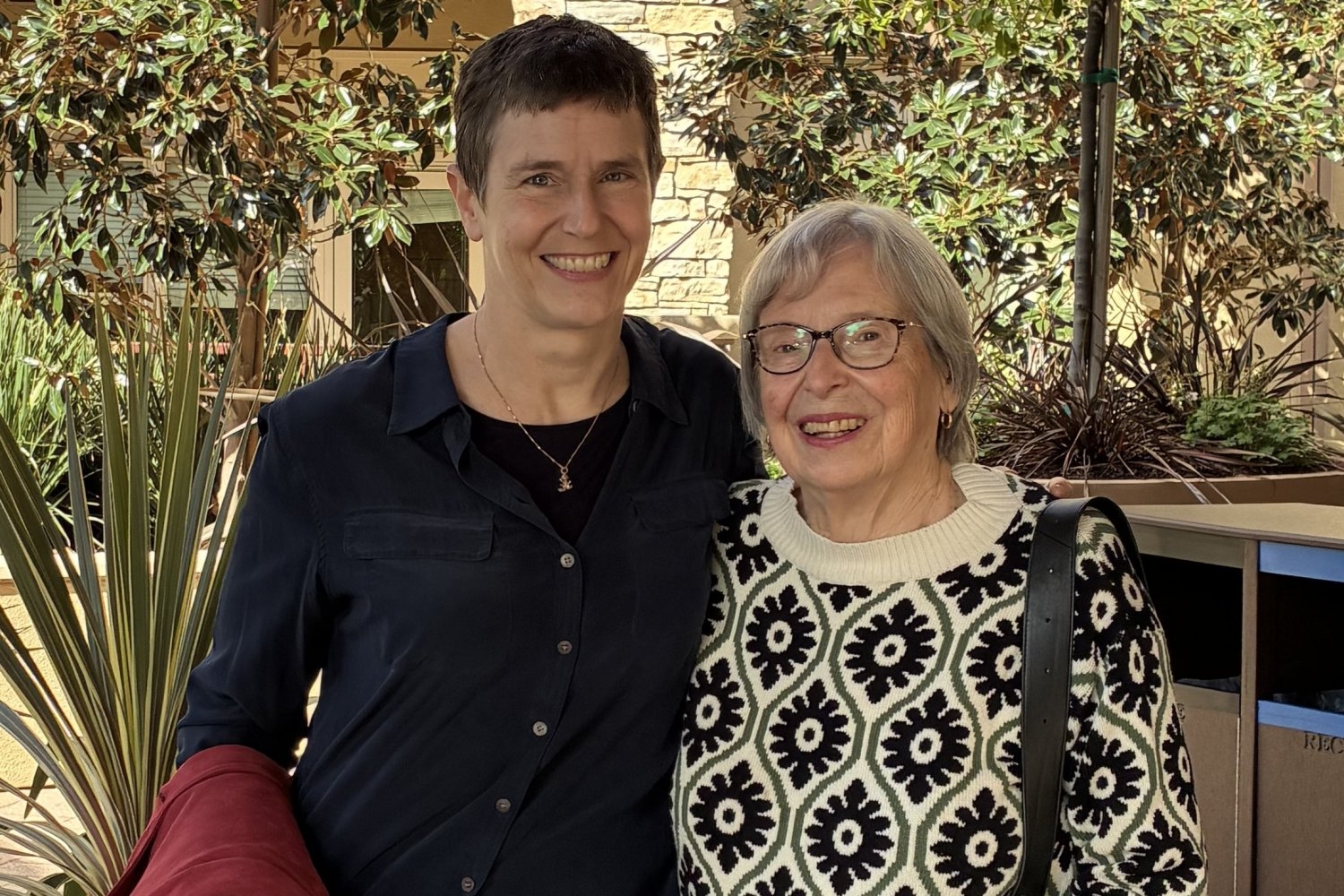
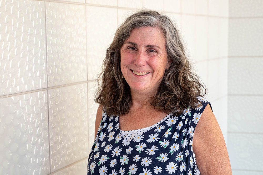
Research Threads: One lab’s detective work reveals secrets of immortal cells

Most cells in our body live and die. But the germline, the cells that produce eggs and sperm, must carry on forever. How the germline successfully produces the next generation is a long-studied question. Through a string of discoveries made over years, the Yamashita lab is teasing apart how the germline remains immortal.

Madeleine Turner | Whitehead Institute
October 7, 2025

Most cells in our body live and die. But the germline, the cells that produce eggs and sperm, must carry on forever. How the germline successfully produces the next generation is a long-studied question. Research Threads examines how answering one question uncovers more questions to be solved. In our first installment of Research Threads, we follow the research of Whitehead Institute Member Yukiko Yamashita. Through a string of discoveries made over years, the Yamashita lab is teasing apart how the germline remains immortal.

“The germline is the only cell type responsible for transmitting the genome from generation to generation,” Whitehead Institute Member Yukiko Yamashita says. “We’ve done that for 1.5 billion years.”

The germline is the population of cells in an organism that give rise to gametes, either egg or sperm cells. These gametes contain genetic information, encoded in DNA, needed to produce the next generation. The act of transmitting this information — the instructions that a new individual needs to develop and function — is like a relay race that never ends. While a skin or gut cell may be prone to genetic errors and is generally replaceable, germline stem cells (GSCs) must maintain their genomes with precision. Otherwise, any mistakes or imbalances would be inherited by offspring and accumulated over generations, potentially driving a species to extinction. In other words, to keep passing the baton in this relay, the germline must be faithfully preserved.

Although germline preservation is paramount to the existence and survival of a species, some fundamental parts of its biology have remained a mystery. Yamashita, who is also a professor of biology at the Massachusetts Institute of Technology and a Howard Hughes Medical Institute Investigator, has focused her research on unraveling the secrets of the germline. To study these cells’ immortality, her lab utilizes the model organism Drosophila melanogaster, or the fruit fly. Along the way, research in the Yamashita lab has highlighted how the process of scientific discovery can be circuitous, often pulling scientists in surprising directions, revealing new questions and avenues to explore.

For decades, scientists had observed an aspect of germline behavior that was hard to explain. Most cells in the body divide to produce two identical copies, or daughter cells. GSCs in male fruit flies, however, divide “asymmetrically,” meaning they yield two daughter cells that are not identical. Instead, one daughter cell becomes a new GSC, while the other goes on to become a gamete, in this case a sperm cell. It might be easy to assume that asymmetric cell division is about producing gametes while maintaining a pool of stem cells. But an additional detail complicates this theory: when a daughter cell is on the path to becoming sperm, it can loop back around to become another stem cell, instead of continuing differentiation to become a sperm cell.

“If it can do that, why divide asymmetrically in the first place?” Yamashita says.

To shed light on why GSCs divide asymmetrically, researchers wanted to know how genetic information was actually divvied up between daughter cells. “After I started my own lab, there was this question hanging in the field,” Yamashita says. It made sense to find some difference in inheritance, DNA-based or otherwise: something to distinguish between the daughter fated to become a gamete, and the other that would remain in the GSC pool.

Preparing for division, a cell duplicates its DNA. Chromosomes happen to be shaped like the letter “X” as a result of this duplication: the left and right sides of the “X” are called matching sister chromatids, each a copy of the other. Later in cell division, these two sister chromatids part ways, winding up in separate daughter cells.

In 2013, Yamashita and her former graduate student, Swathi Yadlapalli, made a strange but important discovery. In fruit flies, for the X and Y chromosomes (the sex chromosomes), sister chromatids were not being sorted randomly. Instead, one was more likely to go to the daughter cell that would become a gamete; the other to the daughter on the GSC track. There had to be a reason for this preference, but no one had an explanation.

Initial experiments did not reveal obvious differences between these sister chromatid pairs. “Everyone would say, ‘oh, there’s probably some sort of epigenetic information in there,” Yamashita says, referring to heritable changes not carried in DNA. With few promising leads, the lab decided to take a systematic approach. George Watase, then a postdoc in the lab, began the painstaking work of removing different a parts of flies’ X chromosomes, seeing if the absence of any particular element would disrupt the pattern of preferential segregation.

“We thought it was going to be satellite DNA,” Yamashita says, referring to large swathes of DNA in the genome that are highly repetitive but don’t code for any genes. (While this initial guess was wrong, it kickstarted a separate project in the lab — one which led to discovering the unexpected role that satellite DNA plays when one species forks into two.)

Eventually the team narrowed in on the true culprit: ribosomal DNA (rDNA). The primary role of rDNA is to produce the components that make up ribosomes. Ribosomes, in turn, perform the crucial task of synthesizing proteins.

“We didn’t like this finding in the beginning. I always say that ribosomal DNA is ‘too important to be interesting.’ You don’t get excited about something you really need, like toilet paper,” Yamashita says. “In the case of ribosomal DNA, bacteria needs it, humans need it, everybody needs it.”

But what did rDNA have to do with asymmetric cell division in the germline?

“Around that time, we started reading lots of papers,” Yamashita says. “Then we discovered a phenomenon called rDNA magnification. These were studies from the 1960s and ’80s — most of the people in my lab were not even born yet.”

These studies from decades ago described mutant flies that lacked a sufficient amount of rDNA and, as a result, had a “bobbed” phenotype, or appearance. Since these flies possessed fewer ribosomes to produce proteins, the bristles on their back were shorter; the protective cuticle covering their bodies weakened. But when they reproduced, many of their offspring possessed a normal amount of rDNA. These observations pointed towards a mechanism that allowed flies to replenish their supply of rDNA.

At the time rDNA magnification was first observed, the phenomenon was regarded as an oddity, something that only happened in mutant flies and did not have physiological relevance. But Yamashita realized there was a connection between rDNA magnification and asymmetric division in the germline.

To make enough protein, a cell must produce ample ribosomes. To do that, the fruit fly genome contains hundreds of copies of rDNA in a row. But the DNA replication process can struggle to handle so many rDNA copies strung together, and can sometimes experience a hiccup, resulting in the loss of rDNA copies with each new division. It’s an outcome that might be okay on occasion, but would wreak havoc over many generations.

“All of a sudden, [rDNA magnification] was not about a mutant chromosome,” Yamashita says. “We were like, holy moly. This is about germline immortality.”

Soon many different pieces became part of a cohesive story: asymmetric cell division is not about producing a balance of gametes and stem cells; it’s about maintaining rDNA in the germline. Sister chromatids are almost identical — but one contains more copies of rDNA than the other, and that copy is fated to stay in the GSC pool. Through this asymmetry, the germline is replenished of rDNA; it can pass the baton to the next generation.

“For quite some time, for so many observations that everyone knew in the field, we felt we had an explanation. But from that ‘aha!’ moment, we took multiple years to test everything,” Yamashita says.

In subsequent years, the Yamashita lab pinned down additional details about how rDNA is diverted back to the germline. First, in 2022, the team identified a specific protein, which they named Indra, which binds to rDNA. The presence of Indra helps assign the sister chromatid containing more rDNA copies to the GSC daughter cell.

Their next discovery was another plot twist. If one sister chromatid contained more rDNA than the other, and those rDNA copies weren’t appearing out of thin air, it meant that one chromatid needed to be stealing rDNA from its sister. The lab discovered a genetic element that facilitated this transfer: a retrotransposon.

Retrotransposons are usually considered “genetic parasites,” copying and pasting themselves into the genome. In an attempt to reinsert itself, this particular retrotransposon, called R2, slices open sections containing rDNA on both chromatids. As the cell repairs these breaks, it may inadvertently stitch copies of rDNA from one chromosome to the other, creating an unequal number of copies between the two.

“Not many people thought retrotransposons could be beneficial to the host. They’re seen as parasites,” Yamashita says. “But it turns out that they are essential for germline immortality.”

Sometimes, one research project is a spin off of a spin off. That was true for Xuefeng Meng, a graduate student in the lab who was working on satellite DNA, the genetic element that turned out to be unrelated to asymmetric cell division, but was interesting in its own right.

While studying satellite DNA, Meng noticed that a particular stock of flies had a problem producing normal sperm, that their cells’ nuclei were abnormally packaged. The problem had to do with a gene called Stellate on the flies’ X chromosome. While most flies have few copies of Stellate, these flies had a higher number of copies.

Stellate was already known in the field as a meiotic driver, or “selfish-gene”: a genetic element that has evolved ways to preferentially transmit itself to the next generation. Some meiotic drivers, including Stellate, are on the sex chromosomes and, when not suppressed, cause an excess of either male or female progeny. In this case, Stellate produces a protein, Ste, which is found to concentrate in Y-carrying cells during meiosis, the specialized type of cell division that produces gametes (meiosis follows the earlier round of asymmetric cell division described above). High concentrations of Ste impede the proper packaging of nuclei in cells, leading to their eventual death. When Stellate is widely expressed, fewer male flies emerge in the next generation.

But here lies an inherent tension: if a selfish gene is too good at propagating itself, and produces only males or females, its host species would go extinct — and so would the gene. Meng and Yamashita were interested in this paradox. Through this work, they identified a novel mechanism that keeps Stellate in check. To balance selfish propagation with the host species’ survival, Stellate has a built-in system for pumping the brakes. After Ste concentrates in Y-carrying cells during the first meiotic division, the protein is unevenly distributed a second time. This second step spares a portion of Y-carrying cells that go on to create males.

How the germline is able to counter disruptive forces, thereby renewing itself, continues to be a ripe research area. Researchers still don’t know all the secrets to how a line of cells can achieve perpetuity — but the Yamashita lab continues to investigate the question.

“What I really like about people in my lab is that they don’t jump to the easiest conclusion,” Yamashita says. “If you start embracing surprise, then good things happen. Because you are surprised, you start testing your finding in multiple ways. Then you can build confidence about the result.”

Notes

Xuefeng Meng and Yukiko Yamashita (2025). “Intrinsically weak sex chromosome drive through sequential asymmetric meiosis.” Science Advanceshttps://doi.org/10.1126/sciadv.adv7089

Jonathan O. Nelson, Tomohiro Kumon, Yukiko M. Yamashita. (2023) “rDNA magnification is a unique feature of germline stem cells.” PNAShttps://doi.org/10.1073/pnas.2314440120

Jonathan O. Nelson, Alyssa Slicko, Yukiko M. Yamashita. (2023) “The retrotransposon R2 maintains Drosophila ribosomal DNA repeats.” PNAShttps://doi.org/10.1073/pnas.2221613120

George J. Watase, Jonathan O. Nelson, Yukiko M. Yamashita. (2022) “Nonrandom sister chromatid segregation mediates rDNA copy number maintenance in Drosophila.” Science Advanceshttps://www.science.org/doi/10.1126/sciadv.abo4443

Madhav Jagannathan and Yukiko Yamashita. (2021) “Defective satellite DNA clustering into chromocenters underlies hybrid incompatibility in Drosophila.” Molecular Biology and Evolutionhttps://doi.org/10.1093/molbev/msab221

Swathi Yadlapalli and Yukiko Yamashita (2013) “Chromosome-specific nonrandom sister chromatid segregation during stem-cell division.” Nature10.1038/nature12106

Alnylam Pharmaceuticals establishes named fund in honor of co-founder

The Phil Sharp-Alnylam Fund for Emerging Scientists will support graduate students and faculty in MIT Biology.

Lillian Eden | Department of Biology
October 8, 2025

It’s no question that graduate school in fundamental research was never for the faint of heart, but academia’s nationwide funding disruptions threaten not just research happening now, but the critical pipeline for the next generation of scientists.

“What’s keeping me up at night is the uncertainty,” says Nobel Laureate Phillip A. Sharp, Institute Professor and Professor of Biology Emeritus, and Intramural Faculty at the Koch Institute.

In the short term, Sharp foresees challenges in sustaining students so they can complete their degrees, postdoctoral scholars to finish their professional preparation, and faculty to set up and sustain their labs. In the long term, the impact becomes potentially existential — fewer people pursuing academia now means fewer advancements in the decades to come.

So, when Sharp was looped into discussions about a gift in his honor, he knew exactly where it should be directed. Established this year thanks to a generous donation from Alnylam Pharmaceuticals, the Phil Sharp-Alnylam Fund for Emerging Scientists will support graduate students and faculty within life sciences.

“This generosity by Alnylam provides an opportunity to bridge the uncertainty and ideally create the environment where students and others will feel that it’s possible to do science and have a career,” Sharp says. 

The fund is set up to be flexible, so the expendable gift can be used to address the evolving needs of the Department of Biology, including financial support, research grants, and seed funding. 

“This fund will help us fortify the department’s capacity to train new generations of life science innovators and leaders,” says Amy E. Keating, Department Head and Jay A. Stein (1968) Professor of Biology and Professor of Biological Engineering. “It is a great privilege for the department to be part of this recognition of Phil’s key role at Alnylam.”

Alnylam Pharmaceuticals, a company Sharp cofounded in 2002, is, in fact, a case study for the type of long-term investment in fundamental discovery that leads to paradigm-shifting strides in biomedical science, such as: what if the genetic drivers of diseases could be silenced by harnessing a naturally occurring gene regulation process? 

Good things take time

In 1998, Andrew Fire, PhD ’83, who was trained as a graduate student in the Sharp Lab at MIT, and Craig Mello published a paper showing that double-stranded RNA suppresses the expression of the protein from the gene that encodes its sequence. The process, known as RNA interference, was such a groundbreaking revelation that Fire and Mello shared a Nobel Prize in Medicine and Physiology less than a decade later. 

Four of the five cofounders of Alnylam Pharmaceuticals: (from left to right) Tom Tuschl, Phil Sharp, David Bartel, and Phil Zamore. Not pictured: Paul Schimmel. Photo credit: Christoph Westphal

RNAi is an innate cellular gene regulation process that can, for example, assist cells in defending against viruses by degrading viral RNA, thereby interfering with the production of viral proteins. Taking advantage of this natural process to fine-tune the expression of genes that encode specific proteins was a promising option for disease treatment, as many diseases are caused by the creation or buildup of mutated or faulty proteins. This approach would address the root cause of the disease, rather than its downstream symptoms.

The details of the biochemistry of RNAi were characterized and patented, and in 2002, Alnylam was founded by Sharp, David Bartel, Paul Schimmel, Thomas Tuschl, and Phillip Zamore. 

“16 years later, we got our first approval for a totally novel therapeutic agent to treat disease,” Sharp says. “Something in a research laboratory, translated in about as short a time as you can do, gave rise to this whole new way of treating critical diseases.” 

This timeline isn’t atypical. Particularly in healthcare, Sharp notes, investments often occur five or ten years before they come to fruition. 

“Phil Sharp’s visionary idea of harnessing RNAi to treat disease brought brilliant people together to pioneer this new class of medicines. RNAi therapeutics would not exist without the bridge Phil built between academia and industry. Now there are six approved Alnylam-discovered RNAi therapeutics, and we are exploring potential treatments for a range of rare and prevalent diseases to improve the lives of many more patients in need,” says Kevin Fitzgerald, Chief Scientific Officer of Alnylam Pharmaceuticals

Today, the company has grown to over 2,500 employees, markets its six approved treatments worldwide, and has a long list of research programs that are likely to yield new therapeutic agents in the years to come. 

Change is always on the horizon

Sharp foresees potential benefits for companies investing in academia, in the way Alnylam Pharmaceuticals has through the Phil Sharp-Alnylam Fund for Emerging Scientists

“We are proud to support the MIT Department of Biology because investments in both early-stage and high-risk research have the potential to unlock the next wave of medical breakthroughs to help so many patients waiting for hope throughout the world,” says Yvonne Greenstreet, Chief Executive Officer of Alnylam Pharmaceuticals

It is prudent for industry to keep its finger on the pulse — for becoming aware of new talent and for anticipating landscape-shifting advancements, such as Artificial Intelligence. Sharp notes that academia, in its pursuit of fundamental knowledge, “creates new ideas, new opportunities, and new ways of doing things.” 

“All of society, including biotech, is anticipating that AI is going to be a great accelerator,” Sharp says. “Being associated with institutions that have great biology, chemistry, neuroscience, engineering, and computational innovation is how you sort through this anticipation of what the future is going to be.” 

But, Sharp says, it’s a two-way street: academia should also be asking how it can best support the future workplaces for their students who will go on to have careers in industry. To that end, the Department of Biology recently launched a career connections initiative for current trainees to draw on the guidance and experience of alums, and to learn how to hone their knowledge so that they are a value-add to industry’s needs.  

“The symbiotic nature of these relationships is healthy for the country, and for society, all the way from basic research through innovative companies of all sizes, healthcare delivery, hospitals, and right down to primary care physicians meeting one-on-one with patients,” Sharp says. “We’re all part of that, and unless all parts of it remain healthy and appreciated, it will bode poorly for the future of the country’s economy and well-being.”

Pathology and the Allure of Analytical Thinking

Susan Fuhrman ’75 pursued pathology because she liked providing clear answers to diagnostic questions, and has spent her retirement making complex beaded jewelry, a hobby she started more than 30 years ago as a foil for the stresses of work.

Kathryn M. O'Neill | Slice of MIT
October 7, 2025

Susan Fuhrman ’75 became a pathologist because she likes providing clear answers to diagnostic questions. “As opposed to guessing what people have, you’ve got the lab results, you have reviewed the pathology slides,” she says. “It’s pretty analytical. Your answer is the answer.”

That clarity of focus was never more valuable than in 2020, when Fuhrman was charged with answering the question everyone was asking: Is it Covid?

As the system director for pathology and laboratory services at OhioHealth, a major hospital system based in Columbus, Ohio, Fuhrman led efforts to address the epidemic—through hospital protocols and, of course, testing—all while fielding seemingly endless requests for her expertise in identifying disease.

“Everybody—from hospital vice presidents to the chief medical officer for the system— was calling me late at night and multiple times on weekends. It was incredible,” she says.

Within a year, the system’s labs had performed over half a million Covid tests and Fuhrman had been featured several times in CAP Today, a publication of the College of American Pathologists. She discussed general testing challenges as well as whom to test when and on which testing platform.

As it happened, however, Fuhrman was already famous thanks to work dating back to the 1980s.

Understanding Renal Cancer

The daughter of two chemists, Fuhrman majored in biology at MIT and earned her medical degree from the University of Michigan in 1978. She then went to the University of Minnesota Medical Center for her residency in pathology and laboratory medicine and found herself in need of a research topic. “I remember asking the head of our surgical pathology department, Dr. Juan Rosai, ‘What is a question in pathology that hasn’t been answered?’” she says. “He said, ‘Well, we don’t have a good way of determining which renal cell cancers have a bad prognosis. Currently we go by size, but there must be more than that. No one’s cracked the code. Why don’t you try that?’”

So, Fuhrman teamed up with another doctor at the Minneapolis veterans hospital, Dr. Catherine Limas, and together they developed and proposed a set of parameters to grade kidney cancers that might predict cancer outcomes. Then, Fuhrman did the painstaking work of reviewing and analyzing thousands of tumor slides, as well as cancer registry clinical data and medical charts. Her husband, Larry Lasky ’72—whom she had met at MIT and who also became a pathologist—programmed the analysis and helped her run the data she found through an early computer. “I input everything with computer cards and a teletype, super primitive stuff,” she says.

The data produced clear patterns in the predictive value of the appearance of cell nuclei, and the three published a paper proposing a grading system classifying which renal tumors are most aggressive and likely to spread based on their findings. The system, which is still the standard, is known as the Fuhrman Nuclear Grade for Clear Cell Renal Carcinoma.

American Board of Pathology President

After her residency, Fuhrman taught laboratory medicine to senior medical students as an assistant professor at the University of Minnesota for 12 years before moving to Ohio in 1994. In addition to working at OhioHealth, Fuhrman served for several years as president and CEO of CORPath, a private pathology practice. In 2022, she served a term as president of the American Board of Pathology, which later named her a life trustee in honor of her many years of service.

Fuhrman retired at the end of 2020 and has since spent much of her time making beaded jewelry—a hobby she started 35 years ago as a foil to work. “The job was stressful, and beading uses a totally different part of your brain. The left side can rest,” she says. “I can sit and sort beads by size and color for hours. That’s really weird and mindless, but I love it. I also love bead weaving; it’s like physics and architecture, building beautiful, structurally sound pieces from tiny beads.”

She creates elaborate bracelets and necklaces, often giving them away to friends or donating them to charity. “Jewelry making doesn’t pay very well, but I’m very lucky I don’t need to support myself on my hobby,” she says. “I do this for me.”

Ophthalmologist Puts Mind and Hand to Art

Carmel Mercado ’09 describes herself as “existing at the intersection of health and art.” A Seattle-based pediatric ophthalmologist, Mercado is also a visual artist whose whimsical illustrations and colorful animal characters can be found in places as varied as a children’s hospital and a microbrewery.

Sara Shay | MIT Technology Review
July 26, 2025

Carmel Mercado ’09 describes herself as “existing at the intersection of health and art.” A Seattle-based pediatric ophthalmologist, Mercado is also a visual artist whose whimsical illustrations and colorful animal characters can be found in places as varied as a children’s hospital and a microbrewery.

Looking back, Mercado says that even as a premed biology major at MIT she was pursuing both paths. She took a First-Year Advising Seminar in the arts and found a mentor in Michèle Oshima, then director of student and artist-in-residence programs at MIT’s Office of the Arts, who encouraged her to apply for the MIT Arts Scholars program. That gave her the opportunity to showcase her work in a gallery at MIT.

Mercado’s next stop was medical school at Johns Hopkins (she graduated in 2014). There, too, she gravitated toward opportunities for artistic expression, such as designing T-shirts and posters for an event welcoming prospective students. “That kind of helped me get through some darker days when I was really tired or really overwhelmed by the medical part of it,” she says.

She chose ophthalmology as her specialty in part because she found the eye itself visually appealing. “The first time I saw the fundus, the retina, the back of the eye, it was so beautiful to me,” she says. “Just looking at the optic nerve, the colors, the placement, I thought about how amazing it is that we can get such beautiful and complex imagery of our world from what looks to most people like a blob of jelly.”

Initially, Mercado assumed art would take a backseat to her medical career, but time in Japan—including a MISTI summer internship in Kobe—led her to realize she had other options. She connected with a mentor, Kenji Watanabe, while studying the history of medicine at Keio University in Tokyo during medical school. Watanabe “showed me a very different lifestyle,” she says: He didn’t limit his work to academia. “He had this really cool niche where he could do all this policy work. He was traveling to different countries to meet up with other physicians. It was eye-opening,” Mercado says. “He made me realize you can shape your career and your life to be able to pursue your passions. You shouldn’t just accept the traditional way. Being exposed to that early on probably gave me the courage to do what I’m doing now.”

As a practicing ophthalmologist, she began to involve art in her work by designing patient materials featuring characters she created. Colleagues noticed and offered her commissions. About four years ago, Mercado decided to pursue art full-time. The problem: She wasn’t sure how to promote herself. “I just about tried everything to see what would stick,” she says. She started an Etsy page and social media accounts, and she applied to art shows, art walks, and galleries. After about a year, her efforts paid off, and she started to get invitations for projects.

She has since exhibited her work in juried shows and galleries in the Boston, Orlando, and Seattle areas and has received commissions for public art from several cities in Washington. She even has a piece in the permanent gallery at Japan’s Sobana Museum.

Despite her artistic success, Mercado says she eventually missed the problem-solving and patient care involved in clinical work. She started tinkering with her schedule and settled on a roughly 60-40 split in favor of medicine.

In addition to seeing patients, she continues to pursue art projects, working mostly with acrylics and mixed media on canvas and with digital illustration; her style reflects her experiences with children and her observations of wildlife and folk art around the world, especially in Japan.

“I’ve found a space where I’m happy,” she says, “and where it feels a little bit more balanced for me.”

This story also appears in the July/August issue of MIT Alumni News magazine, published by MIT Technology Review

Student Spotlight: Alexa Mallar ’27

Computer science and molecular biology major Alexa Mallar ’27 has a passion for the visual, pursuing her love of art while also working as an undergraduate researcher in the Cheeseman Lab.

Mark Sullivan | Spectrum
June 4, 2025

“Visual art has been a passion and a core part of my identity since before attending MIT,” says Alexa Mallar ’27, a computer science and molecular biology major from Miami who is a recipient of the Norman L. Greenman (1944) Memorial Scholarship.

As an undergraduate researcher in the lab of Iain Cheeseman, MIT professor of biology and member of the Whitehead Institute, she helps develop computational tools for biological data analysis. Outside the lab, Mallar pursues her love of art, creating detailed graphite pieces in a hyperrealistic-surrealist style and experimenting with various media, including color pencil, charcoal, and multimedia sculpture, sharing her work on Instagram. Expanding her creative interests, she has explored 3-D printing through MIT MakerLodge, has taken 21T.101 Intro to Acting, and is taking 21W.756 Reading and Writing poetry in spring 2025. “Through visual and performing arts and creative writing, I continue to find new ways to express my creativity and grow as an artist,” she says.

What inspires you about creating art?

It’s a multitude of things. It’s a technical fascination with capturing details on a piece of paper and trying really hard to make it look like a photograph. There’s the enjoyment of the technical aspects of the task. There’s also an intellectual satisfaction that comes with creating art.  I like incorporating surrealism into my work often because it lends itself to creating more visual meaning than a purely realistic piece would; there are several artists I follow and try to incorporate aspects of their work into mine, trying different things. There’s the experimental value of trying different media and artistic styles. I love exploring. I love expressing new ideas. Art is really a great way to do it.

Is there a connection between what you do as a scientist and as an artist?

The nature of my art is very visual, and I think about what I do in computer science or in research now in a very visual way. I map a diagram in my head of input and output. Anything I do is inherently visualized.

Sometimes the connection goes the other way—my interest in math and science bleeds into my art. Designing counterweights to balance sculptures or geometrically mapping out perspective and proportions are a few examples. I also love sneaking in little “easter eggs.” A few years ago, I created a piece featuring a woman with a third eye and a tree-branch crown, where the branching levels followed the Fibonacci sequence.

What is the story behind the mermaid drawings on your Instagram page?

“There’s an event every May called MerMay. Artists on Instagram will do successive drawings of different mermaids based on prompts. I wanted to join in, so I designed my own mermaid. I just started by imagining her face, and it evolved into her holding an orb I called the Eye of the Sea. It was really fun.”

After college, will you be pursuing both science and art?

“That’s a good question. I kind of have a 30-degree angle I’m heading in, not a specific path. I know that I will keep drawing in my free time, and the creative thinking and visualization skills will bleed into any other part of my work that I do, whether that be in computer science or research. Maybe designing a front end is where my creative spirit will contribute to the computer science work that I do.

“I plan to work for Amazon [in summer 2025], having received a return offer after working there last summer. I’m getting a sense of the different environments I could go to. If I can find a way to combine [art and career] I will. I’ll find a way to do as many things as I can that interest me.”

How has your MIT experience helped you on your path?

“It has been an amazing resource. MIT offers so many different classes and interdisciplinary opportunities. I was able to explore entrepreneurship through the Martin Trust Center at MIT, enrolling in the Undergraduate Engineering Entrepreneurship Certificate program. That’s one avenue I wouldn’t have been able to explore otherwise without MIT. Acting is not something I would have even tried before having the opportunity to do it at MIT. I’m rediscovering my love for creative writing through classes at MIT, and I’m really enjoying it. If I hadn’t been able to fit a poetry workshop into my class schedule, I probably wouldn’t be writing nearly as much this semester. I’m really glad I have that opportunity.

“MIT is in an amazing spot for someone in my specific major, with the huge presence of biotech in Cambridge. This is an optimal place for both computer science and biological research. We have the Whitehead Institute, Pfizer, Moderna, all within walking distance of campus. There’s a lot to explore, an intersection of interests, and I really appreciate that is available to me at MIT.”

Student spotlight: Aria Eppinger ’24

The multitalented member of the varsity swim team graduated with her undergraduate degree in computer science and molecular biology in 2024 and will complete her MEng this month.

Jane Halpern | Department of Electrical Engineering and Computer Science
May 9, 2025

This interview is part of a series of short interviews from the MIT Department of Electrical Engineering and Computer Science, called Student Spotlights. Each spotlight features a student answering their choice of questions about themselves and life at MIT. Today’s interviewee, Aria Eppinger ’24, graduated with her undergraduate degree in Course 6-7 (Computer Science and Molecular Biology) last spring. This spring, she will complete her MEng in 6-7. Her thesis, supervised by Ford Professor of Engineering Doug Lauffenburger in the Department of Biological Engineering, investigates the biological underpinnings of adverse pregnancy outcomes, including preterm birth and preeclampsia, by applying polytope-fitting algorithms.

Q: Tell us about one teacher from your past who had an influence on the person you’ve become.

A: There are many teachers who had a large impact on my trajectory. I would first like to thank my elementary and middle school teachers for imbuing in me a love of learning. I would also like to thank my high school teachers for not only teaching me the foundations of writing strong arguments, programming, and designing experiments, but also instilling in me the importance of being a balanced person. It can be tempting to be ruled by studies or work, especially when learning and working are so fun. My high school teachers encouraged me to pursue my hobbies, make memories with friends, and spend time with family. As life continues to be hectic, I’m so grateful for this lesson (even if I’m still working on mastering it).

Q: Describe one conversation that changed the trajectory of your life.

A: A number of years ago, I had the opportunity to chat with Warren Buffett. I was nervous at first, but soon put to ease by his descriptions of his favorite foods — hamburgers, French fries, and ice cream — and his hitchhiking stories. His kindness impressed and inspired me, which is something I carry with me and aim to emulate all these years later.

Q: Do you have any pets?

A: I have one dog who lives at home with my parents. Dodger, named after “Artful Dodger” in Oliver Twist, is as mischievous as beagles tend to be. We adopted him from a rescue shelter when I was in elementary school.

Q: Are you a re-reader or a re-watcher — and if so, what are your comfort books, shows, or movies?

A: I don’t re-read many books or re-watch many movies, but I never tire of Jane Austen’s “Pride and Prejudice.” I bought myself an ornately bound copy when I was interning in New York City last summer. Austen’s other novels, especially “Sense and Sensibility,” “Persuasion,” and “Emma,” are also favorites, and I’ve seen a fair number of their movie and miniseries adaptations. My favorite adaptation is the 1995 BBC production of “Pride and Prejudice” because of the cohesion with the original book and the casting of the leads, as well as the touches and plot derivations added by the producer and director to bring the work to modern audiences. The adaptation is quite long, but I have fond memories of re-watching it with some fellow Austinites at MIT.

Q: If you had to teach a really in-depth class about one niche topic, what would you pick?

A: There are two types of people in the world: those who eat to live, and those who live to eat. As one of the latter, I would have to teach some sort of in-depth class on food. Perhaps I would teach the science behind baking chocolate cake, or churning the perfect ice cream. Or maybe I would teach the biochemistry of digesting. In any case, I would have to have lots of hands-on demos and reserve plenty for taste-testing!

Q: What was the last thing you changed your mind about?

A: Brisket! I never was a big fan of brisket until I went to a Texas BBQ restaurant near campus, The Smoke Shop BBQ. Growing up, I had never had true BBQ, so I was quite skeptical. However, I enjoyed not only the brisket but also the other dishes. The Brussels sprouts with caramelized onions is probably my favorite dish, but it feels like a crime to say that about a BBQ place!

Q: What are you looking forward to about life after graduation? What do you think you’ll miss about MIT?

A: I’m looking forward to new adventures after graduation, including working in New York City and traveling to new places. I cross-registered to take Intensive Italian at Harvard this semester and am planning a trip to Italy to practice my Italian, see the historic sites, visit the Vatican, and taste the food. Non vedo l’ora di viaggiare all’Italia! [I can’t wait to travel to Italy!]

While I’m excited for what lies ahead, I will miss MIT. What a joy it is to spend most of the day learning information from a fire hose, taking a class on a foreign topic because the course catalog description looked fun, talking to people whose viewpoint is very similar or very different from my own, and making friends that will last a lifetime.

Staff Spotlight: Lighting up biology’s basement lab

Senior Technical Instructor Vanessa Cheung ’02 brings the energy, experience, and excitement needed to educate students in the biology teaching lab.

Samantha Edelen | Department of Biology
April 29, 2025

For more than 30 years, Course 7 (Biology) students have descended to the expansive, windowless basement of Building 68 to learn practical skills that are the centerpiece of undergraduate biology education at the Institute. The lines of benches and cabinets of supplies that make up the underground MIT Biology Teaching Lab could easily feel dark and isolated.

In the corner of this room, however, sits Senior Technical Instructor Vanessa Cheung ’02, who manages to make the space seem sunny and communal.

“We joke that we could rig up a system of mirrors to get just enough daylight to bounce down from the stairwell,” Cheung says with a laugh. “It is a basement, but I am very lucky to have this teaching lab space. It is huge and has everything we need.”

This optimism and gratitude fostered by Cheung is critical, as MIT undergrad students enrolled in classes 7.002 (Fundamentals of Experimental Molecular Biology) and 7.003 (Applied Molecular Biology Laboratory) spend four-hour blocks in the lab each week, learning the foundations of laboratory technique and theory for biological research from Cheung and her colleagues.

Running toward science education

Cheung’s love for biology can be traced back to her high school cross country and track coach, who also served as her second-year biology teacher. The sport and the fundamental biological processes she was learning about in the classroom were, in fact, closely intertwined.

“He told us about how things like ATP [adenosine triphosphate] and the energy cycle would affect our running,” she says. “Being able to see that connection really helped my interest in the subject.”

That inspiration carried her through a move from her hometown of Pittsburgh, Pennsylvania, to Cambridge, Massachusetts, to pursue an undergraduate degree at MIT, and through her thesis work to earn a PhD in genetics at Harvard Medical School. She didn’t leave running behind either: To this day, she can often be found on the Charles River Esplanade, training for her next marathon.

She discovered her love of teaching during her PhD program. She enjoyed guiding students so much that she spent an extra semester as a teaching assistant, outside of the one required for her program.

“I love research, but I also really love telling people about research,” Cheung says.

Cheung herself describes lab instruction as the “best of both worlds,” enabling her to pursue her love of teaching while spending every day at the bench, doing experiments. She emphasizes for students the importance of being able not just to do the hands-on technical lab work, but also to understand the theory behind it.

“The students can tend to get hung up on the physical doing of things — they are really concerned when their experiments don’t work,” she says. “We focus on teaching students how to think about being in a lab — how to design an experiment and how to analyze the data.”

Although her talent for teaching and passion for science led her to the role, Cheung doesn’t hesitate to identify the students as her favorite part of the job.

“It sounds cheesy, but they really do keep the job very exciting,” she says.

Using mind and hand in the lab

Cheung is the type of person who lights up when describing how much she “loves working with yeast.”

“I always tell the students that maybe no one cares about yeast except me and like three other people in the world, but it is a model organism that we can use to apply what we learn to humans,” Cheung explains.

Though mastering basic lab skills can make hands-on laboratory courses feel “a bit cookbook,” Cheung is able to get the students excited with her enthusiasm and clever curriculum design.

“The students like things where they can get their own unique results, and things where they have a little bit of freedom to design their own experiments,” she says. So, the lab curriculum incorporates opportunities for students to do things like identify their own unique yeast mutants and design their own questions to test in a chemical engineering module.

Part of what makes theory as critical as technique is that new tools and discoveries are made frequently in biology, especially at MIT. For example, there has been a shift from a focus on RNAi to CRISPR as a popular lab technique in recent years, and Cheung muses that CRISPR itself may be overshadowed within only a few more years — keeping students learning at the cutting edge of biology is always on Cheung’s mind.

“Vanessa is the heart, soul, and mind of the biology lab courses here at MIT, embodying ‘mens et manus’ [‘mind and hand’],” says technical lab instructor and Biology Teaching Lab Manager Anthony Fuccione.

Support for all students

Cheung’s ability to mentor and guide students earned her a School of Science Dean’s Education and Advising Award in 2012, but her focus isn’t solely on MIT undergraduate students.

In fact, according to Cheung, the earlier students can be exposed to science, the better. In addition to her regular duties, Cheung also designs curriculum and teaches in the LEAH Knox Scholars Program. The two-year program provides lab experience and mentorship for low-income Boston- and Cambridge-area high school students.

Paloma Sanchez-Jauregui, outreach programs coordinator who works with Cheung on the program, says Cheung has a standout “growth mindset” that students really appreciate.

“Vanessa teaches students that challenges — like unexpected PCR results — are part of the learning process,” Sanchez-Jauregui says. “Students feel comfortable approaching her for help troubleshooting experiments or exploring new topics.”

Cheung’s colleagues report that they admire not only her talents, but also her focus on supporting those around her. Technical Instructor and colleague Eric Chu says Cheung “offers a lot of help to me and others, including those outside of the department, but does not expect reciprocity.”

Professor of biology and co-director of the Department of Biology undergraduate program Adam Martin says he “rarely has to worry about what is going on in the teaching lab.” According to Martin, Cheung is ”flexible, hard-working, dedicated, and resilient, all while being kind and supportive to our students. She is a joy to work with.”

Staff Spotlight: Always looking to home

Mingmar Sherpa, a researcher in the Martin Lab in the Department of Biology, has remained connected to his home in Nepal at every step of his career.

Ekaterina Khalizeva | Department of Biology
April 29, 2025

For Mingmar Sherpa, a senior research support associate in the Martin Lab in the Department of Biology, community is more than just his colleagues in the lab, where he studies how mechanical forces affect cell division timing during embryogenesis. On his long and winding path to MIT, he never left behind the people he grew up among in Nepal. Sherpa has been dedicated, every step of his career — from rural Solukhumbu to Kathmandu to Alabama to Cambridge — to advancing education and health care among his people in any way he can.

Despite working more than 7,000 miles away from home, Mingmar Sherpa makes every effort to keep himself connected to his community in Nepal. Every month, for example, he sends home money to support a computer lab that he established in his hometown in rural Solukhumbu, the district of Nepal that houses Mount Everest — just $250 a month covers the costs of a teacher’s salary, electricity, internet, and a space to teach. In this lab, almost 250 students thus far have learned computer skills essential to working in today’s digitally driven world. In college, Sherpa also started The Bright Vision Foundation (The Bright Future), an organization to support health and education in Nepal, and during the pandemic raised funds to provide personal protective equipment (PPE) and health care services across his home country.

While Sherpa’s ambition to help his home can be traced back to his childhood, he didn’t have it all figured out from the start, and found inspiration at each step of his career.

“This mindset of giving back to the community, helping policymakers or establishing an organization to help people do science, helping the scientific community to find cures for diseases — all these ideas came to me along the way,” Sherpa says. “It is the journey that matters.”

A journey driven by hope and optimism

“Sherpa” is a reference to the ethnic group native to the mountainous regions of Nepal and Tibet, whose members are well-known for their mountaineering skills, which they use to guide and assist tourists who want to climb Mount Everest. Growing up in rural Solukhumbu, Sherpa was surrounded by people working in the tourism industry; few other occupations appeared feasible. There was just one hospital for the whole district, requiring locals to walk for days to get medical assistance.

The youngest of seven siblings, Sherpa went to an English-language middle school, which he had to walk for over an hour to get to. He excelled there, soon becoming the top student in his class and passing the national exam with distinction — success that allowed him to both dream of and accomplish a move to Kathmandu, the capital city of Nepal, to study in the best school in the country.

It was an overwhelming transition, surrounded as he was for the first time by people from a very different social class, privileged with far more technological resources. The gaps between this well-equipped community and the one he left back home became increasingly obvious and left a strong impression on Sherpa.

There, he started thinking about how to use his newly acquired access to education and technology to uplift his community at home. He was especially fascinated by questions surrounding biology and human health, and next set his sights on attending college in the United States.

“If I came to the U.S., I could learn skills which I could not learn in Nepal,” he says. “I could prepare myself to solve the problems that I want to solve.”

At the University of Alabama in Birmingham, Sherpa continued to deepen his passion for biological science and joined a research lab. Through that work, he discovered the joys of basic research and the diverse set of skills it fosters.

“I joined the lab to learn science, but to do science, you need other skills, like research communication,” he says. “I was learning unintentionally from being in a research position.”

When Covid-19 spread around the globe, Sherpa wanted to apply the expertise and resources he had gained to help his people address the crisis. It was then that he started The Bright Vision Foundation, an organization aiming to raise the standards of health care and education in underserved communities in Nepal. Through the foundation, he raised funds to distribute PPE, provide health care services, and set up the computer lab in his childhood home.

“Today’s world is all about technology and innovation, but here are good people in my community who don’t even know about computers,” he says.

With the help of his brother, who serves as the lab instructor, and his parents, who provide the space and support the lab, and Sherpa’s own fundraising, he aims to help youths from backgrounds similar to his own be better prepared for the technologically advanced, globalized world of today.

The MIT chapter

Now, at MIT, Sherpa speaks with deep appreciation of the opportunities that the university has opened up for him — the people he has been meeting here, and the skills he has been learning.

Professor of biology Adam C. Martin, Sherpa’s principal investigator, views making sure that international trainees like Mingmar are aware of the wide range of opportunities MIT offers — whether it be workshops, collaborations, networking and funding possibilities, or help with the pathway toward graduate school — as a key part of creating a supportive environment.

Understanding the additional burdens on international trainees gives Martin extra appreciation for Sherpa’s perseverance, motivation, and desire to share his culture with the lab, sharing Nepalese food and providing context for Nepalese customs.

Being at such a research-intensive institution as MIT has helped Sherpa further clarify his goals and his view of the paths he can take to achieve them. Since college, his three passions have been intertwined: leadership, research, and human health.

Sherpa will pursue a PhD in biomedical and biological sciences with a focus in cancer biology at Cornell University in the fall. In the longer term, he plans to focus on developing policy to improve public health.

Although Sherpa recognizes that Nepal is not the only place that might need his help, he has a sharp focus and an acute sense of what he is best positioned to do now. Sherpa is gearing up to organize a health camp in the spring to bring doctors to rural areas in Nepal, not only to provide care, but also to gather data on nutrition and health in different regions of the country.

“I cannot, in a day, or even a year, bring the living conditions of people in vulnerable communities up to a higher level, but I can slowly increase the living standard of people in less-developed communities, especially in Nepal,” he says. “There might be other parts of the world which are even more vulnerable than Nepal, but I haven’t explored them yet. But I know my community in Nepal, so I want to help improve people’s lives there.”

MIT Down syndrome researchers work on ways to ensure a healthy lifespan

An Alana Down Syndrome Center webinar, co-sponsored by the Massachusetts Down Syndrome Congress, presented numerous MIT studies that all share the goal of improving health throughout life for people with trisomy 21.

David Orenstein | The Picower Institute for Learning and Memory
April 24, 2025

In recent decades the life expectancy of people with Down syndrome has surged past 60 years, so the focus of research at the Alana Down Syndrome Center at MIT has been to make sure people can enjoy the best health during that increasing timeframe.

“A person with Down syndrome can live a long and happy life,” said Rosalind Mott Firenze, scientific director of the center founded at MIT in 2019 with a gift from the Alana Foundation. “So the question is now how do we improve health and maximize ability through the years? It’s no longer about lifespan, but about healthspan.”

Firenze and three of the center’s Alana Fellows scientists spoke during a webinar, hosted on April 17th, where they described the center’s work toward that goal. An audience of 99 people signed up to hear the webinar titled “Building a Better Tomorrow for Down Syndrome Through Research and Technology,” with many viewers hailing from the Massachusetts Down Syndrome Congress, which co-sponsored the event.

The research they presented covered ways to potentially improve health from stages before birth to adulthood in areas such as brain function, heart development, and sleep quality.

Boosting brain waves

One of the center’s most important areas of research involves testing whether boosting the power of a particular frequency of brain activity—“gamma” brain waves of 40Hz—can improve brain development and function. The lab of the center’s Director Li-Huei Tsai, Picower Professor in The Picower Institute for Learning and Memory and the Department of Brain and Cognitive Sciences, uses light that flickers and sound that clicks 40 times a second to increase that rhythm in the brain. In early studies of people with Alzheimer’s disease, which is a major health risk for people with Down syndrome, the non-invasive approach has proved safe, and appears to improve memory while preventing brain cells from dying. The reason it works appears to be because it promotes a healthy response among many types of brain cells.

Working with mice that genetically model Down syndrome, Alana Fellow Dong Shin Park has been using the sensory stimulation technology to study whether the healthy cellular response can affect brain development in a fetus while a mother is pregnant. In ongoing research, he said, he’s finding that exposing pregnant mice to the light and sound appears to improve fetal brain development and brain function in the pups after they are born.

In his research, Postdoctoral Associate Md. Rezaul Islam worked with 40Hz sensory stimulation and Down syndrome model mice at a much later stage in life—when they are adult aged. Together with former Tsai Lab member Brennan Jackson, he found that when the mice were exposed to the light and sound, their memory improved. The underlying reason seemed to be an increase not only in new connections among their brain cells, but also an increase in the generation of new ones. The research, currently online as a preprint, is set to publish in a peer-reviewed journal very soon.

Firenze said the Tsai lab has also begun to test the sensory stimulation in human adults with Down syndrome. In that testing, which is led by Dr. Diane Chan, it is proving safe and well tolerated, so the lab is hoping to do a year-long study with volunteers to see if the stimulation can delay or prevent the onset of Alzheimer’s disease.

Studying cells

Many Alana Center researchers are studying other aspects of the biology of cells in Down syndrome to improve healthspan. Leah Borden, an Alana Fellow in the lab of Biology Professor Laurie Boyer, is studying differences in heart development. Using advanced cultures of human heart tissues grown from trisomy 21 donors, she is finding that tissue tends to be stiffer than in cultures made from people without the third chromosome copy. The stiffness, she hypothesizes, might affect cellular function and migration during development, contributing to some of the heart defects that are common in the Down syndrome population.

Firenze pointed to several other advanced cell biology studies going on in the center. Researchers in the lab of Computer Science Professor Manolis Kellis, for instance, have used machine learning and single cell RNA sequencing to map the gene expression of more than 130,000 cells in the brains of people with or without Down syndrome to understand differences in their biology.

Researchers the lab of Y. Eva Tan Professor Edward Boyden, meanwhile, are using advanced tissue imaging techniques to look into the anatomy of cells in mice, Firenze said. They are finding differences in the structures of key organelles called mitochondria that provide cells with energy.

And in 2022, Firenze recalled, Tsai’s lab published a study showing that brain cells in Down syndrome mice exhibited a genome-wide disruption in how genes are expressed, leading them to take on a more senescent, or aged-like, state.

Striving for better sleep

One other theme of the Alana Center’s research that Firenze highlighted focuses on ways to understand and improve sleep for people with Down syndrome. In mouse studies in Tsai’s lab, they’ve begun to measure sleep differences between model and neurotypical mice to understand more about the nature of sleep disruptions.

“Sleep is different and we need to address this because it’s a key factor in your health,” Firenze said.

Firenze also highlighted how the Alana Center has collaborated with MIT’s Desphande Center for Technological Innovation to help advance a new device for treating sleep apnea in people with Down syndrome. Led by Mechanical Engineering Associate Professor Ellen Roche, the ZzAlign device improves on current technology by creating a custom-fit oral prosthesis accompanied by just a small tube to provide the needed air pressure to stabilize mouth muscles and prevent obstruction of the airway.

Through many examples of research projects aimed at improving brain and heart health and enhancing sleep, the webinar presented how MIT’s Alana Down Syndrome Center is working to advance the healthspan of people with Down syndrome.


At the core of problem-solving

Stuart Levine ’97, director of MIT’s BioMicro Center, keeps departmental researchers at the forefront of systems biology.

Samantha Edelen | Department of Biology
March 19, 2025

As director of the MIT BioMicro Center (BMC), Stuart Levine ’97 wholeheartedly embraces the variety of challenges he tackles each day. One of over 50 core facilities providing shared resources across the Institute, the BMC supplies integrated high-throughput genomics, single-cell and spatial transcriptomic analysis, bioinformatics support, and data management to researchers across MIT.

“Every day is a different day,” Levine says, “there are always new problems, new challenges, and the technology is continuing to move at an incredible pace.” After more than 15 years in the role, Levine is grateful that the breadth of his work allows him to seek solutions for so many scientific problems.

By combining bioinformatics expertise with biotech relationships and a focus on maximizing the impact of the center’s work, Levine brings the broad range of skills required to match the diversity of questions asked by researchers in MIT’s Department of Biology.

Expansive expertise

Biology first appealed to Levine as an MIT undergraduate taking class 7.012 (Introduction to Biology), thanks to the charisma of instructors Professor Eric Lander and Amgen Professor Emerita Nancy Hopkins. After earning his PhD in biochemistry from Harvard University and Massachusetts General Hospital, Levine returned to MIT for postdoctoral work with Professor Richard Young, core member at the Whitehead Institute for Biomedical Research.

In the Young Lab, Levine found his calling as an informaticist and ultimately decided to stay at MIT. Here, his work has a wide-ranging impact: the BMC serves over 100 labs annually, from the the Computer Science and Artificial Intelligence Laboratory and the departments of Brain and Cognitive Sciences; Earth, Atmospheric and Planetary Sciences; Chemical Engineering; Mechanical Engineering; and, of course, Biology.

“It’s a fun way to think about science,” Levine says, noting that he applies his knowledge and streamlines workflows across these many disciplines by “truly and deeply understanding the instrumentation complexities.”

This depth of understanding and experience allows Levine to lead what longtime colleague Professor Laurie Boyer describes as “a state-of-the-art core that has served so many faculty and provides key training opportunities for all.” He and his team work with cutting-edge, finely tuned scientific instruments that generate vast amounts of bioinformatics data, then use powerful computational tools to store, organize, and visualize the data collected, contributing to research on topics ranging from host-parasite interactions to proposed tools for NASA’s planetary protection policy.

Staying ahead of the curve

With a scientist directing the core, the BMC aims to enable researchers to “take the best advantage of systems biology methods,” says Levine. These methods use advanced research technologies to do things like prepare large sets of DNA and RNA for sequencing, read DNA and RNA sequences from single cells, and localize gene expression to specific tissues.

Levine presents a lightweight, clear rectangle about the width of a cell phone and the length of a VHS cassette.

“This is a flow cell that can do 20 human genomes to clinical significance in two days — 8 billion reads,” he says. “There are newer instruments with several times that capacity available as well.”

The vast majority of research labs do not need that kind of power, but the Institute, and its researchers as a whole, certainly do. Levine emphasizes that “the ROI [return on investment] for supporting shared resources is extremely high because whatever support we receive impacts not just one lab, but all of the labs we support. Keeping MIT’s shared resources at the bleeding edge of science is critical to our ability to make a difference in the world.”

To stay at the edge of research technology, Levine maintains company relationships, while his scientific understanding allows him to educate researchers on what is possible in the space of modern systems biology. Altogether, these attributes enable Levine to help his researcher clients “push the limits of what is achievable.”

The man behind the machines

Each core facility operates like a small business, offering specialized services to a diverse client base across academic and industry research, according to Amy Keating, Jay A. Stein (1968) Professor of Biology and head of the Department of Biology. She explains that “the PhD-level education and scientific and technological expertise of MIT’s core directors are critical to the success of life science research at MIT and beyond.”

While Levine clearly has the education and expertise, the success of the BMC “business” is also in part due to his tenacity and focus on results for the core’s users.

He was recognized by the Institute with the MIT Infinite Mile Award in 2015 and the MIT Excellence Award in 2017, for which one nominator wrote, “What makes Stuart’s leadership of the BMC truly invaluable to the MIT community is his unwavering dedication to producing high-quality data and his steadfast persistence in tackling any type of troubleshooting needed for a project. These attributes, fostered by Stuart, permeate the entire culture of the BMC.”

“He puts researchers and their research first, whether providing education, technical services, general tech support, or networking to collaborators outside of MIT,” says Noelani Kamelamela, lab manager of the BMC. “It’s all in service to users and their projects.”

Tucked into the far back corner of the BMC lab space, Levine’s office is a fitting symbol of his humility. While his guidance and knowledge sit at the center of what elevates the BMC beyond technical support, he himself sits away from the spotlight, resolutely supporting others to advance science.

“Stuart has always been the person, often behind the scenes, that pushes great science, ideas, and people forward,” Boyer says. “His knowledge and advice have truly allowed us to be at the leading edge in our work.”