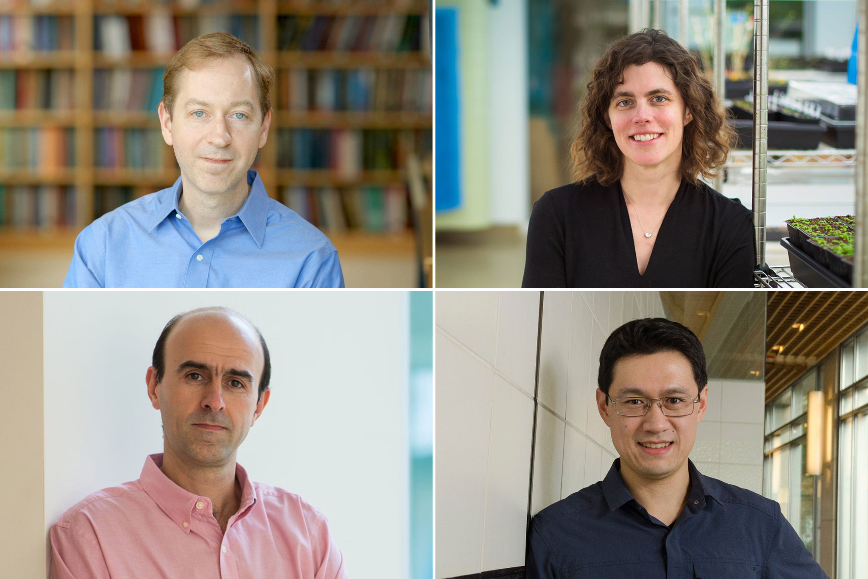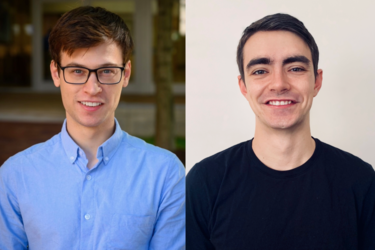
Whitehead Institute graduate student researchers Christopher Giuliano (Lourido Lab) and Julian Roessler (Hrvatin Lab) have been awarded the 2024 Regeneron Prize for Creative Innovation.
Merrill Meadow | Whitehead Institute
July 30, 2024
Whitehead Institute graduate student researchers Christopher Giuliano and Julian Roessler have been awarded the 2024 Regeneron Prize for Creative Innovation. In addition, postdoctoral researcher Chen Weng was selected as a finalist in the postdoctoral fellows competition.
The Regeneron Prize, sponsored by global biotechnology company Regeneron Pharmaceuticals, Inc., is a competitive award designed to recognize and honor exceptional talent and originality in biomedical research. Individual graduate students and postdoctoral fellows in the biomedical sciences are nominated by the nation’s top research universities. Then, nominees outline their “Dream Projects” — potentially groundbreaking research projects that they would pursue given unrestricted access to resources and state-of-the-art technology.
The “Dream Project” proposals, presented by the nominees to a selection committee comprised of Regeneron’s leading scientists, are used to evaluate a trainee’s scientific merit, elegance, precision, and creativity. Novel research ideas and out-of-the-box thinking is encouraged — although the proposal must include a strong rationale, basic methodology and design for the project, and a discussion of how its results could advance the field. Both Giuliano and Roessler have been awarded $50,000 for their proposals, which can be used in any way the winners choose. In addition, Weng was awarded $5,000 as a finalist, and Regeneron has made a $10,000 grant to the Whitehead Institute as the home institute of the winners to support its seminar series.
This year’s awards are distinctive in that the two winners are from the same institution: Both Giuliano and Roessler are pursuing their PhDs at Massachusetts Institute of Technology (MIT) and conducting their doctoral research at Whitehead Institute.
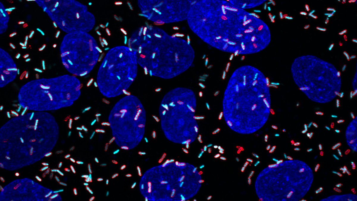
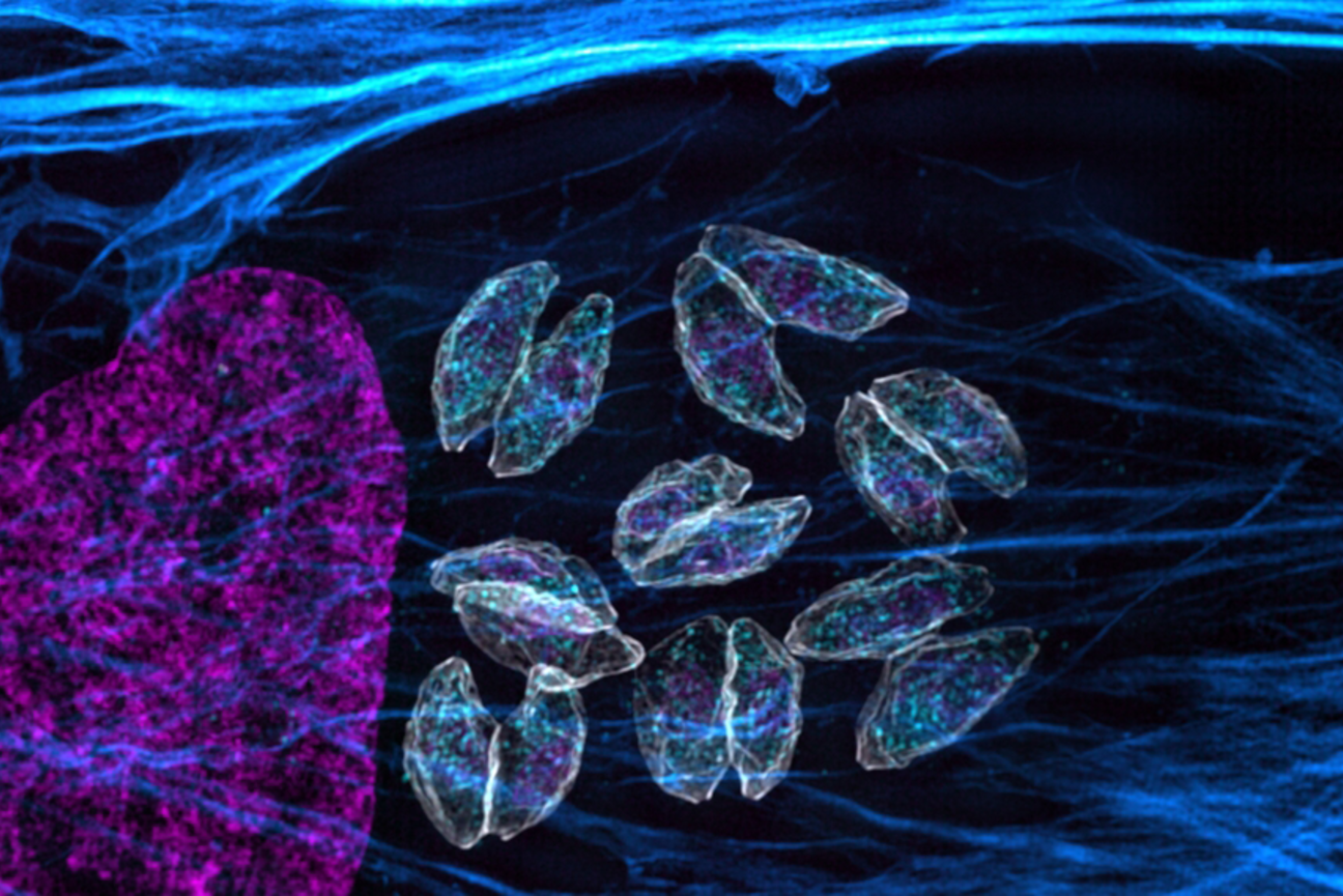
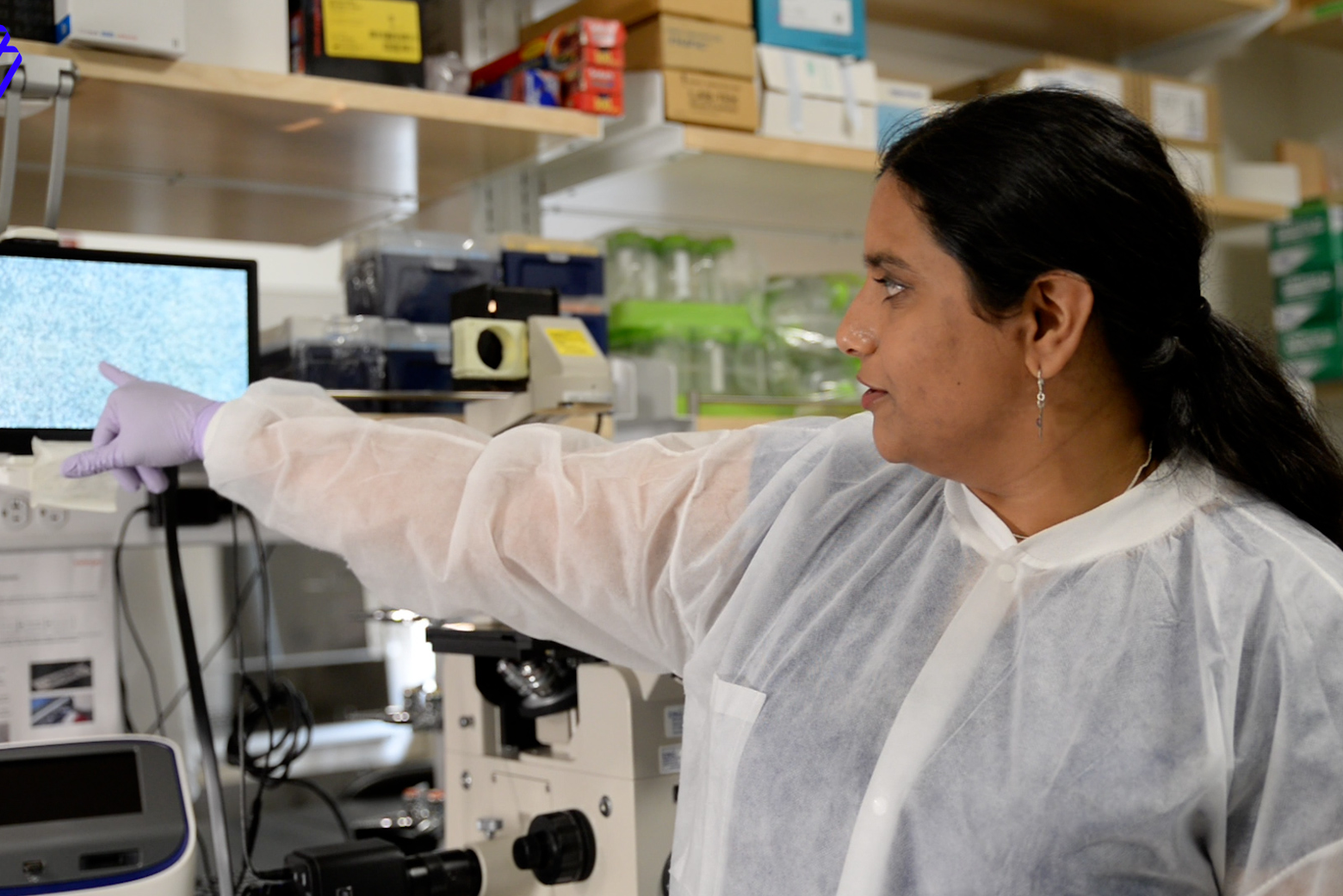
Giuliano is a researcher in the lab of Whitehead Institute Member Sebastian Lourido, who is also an associate professor of biology at MIT and holds the Landon Clay Career Development Chair at Whitehead Institute. Giuliano’s Dream Project seeks to address the unique challenges posed by genetically based muscle disorders. “An obstacle in using current gene therapies to treat these conditions,” he explains, “is that muscle tissue comprises large syncytial cells, which contain hundreds of nuclei in a shared cytoplasm. Even when a gene therapy is able to reach an individual muscle cell, it often isn’t able to spread to every nucleus within that cell.” However, certain parasites, like Toxoplasma gondii, thrive because they have the capacity to successfully gain access to and manipulate muscle cells. T. gondii, the primary focus of the Lourido lab’s work, may infect nearly one third of all humans. “My project,” Giuliano says, “would identify the specific biological mechanisms used by the parasites to spread their virulence factor proteins throughout the cell. Using genetic screens for protein spread, we would work toward applying these protein features to improve the efficiency of muscle-directed gene therapies, and ultimately test our system in a mouse model of Duchenne muscular dystrophy.”
Roessler is a researcher in the lab of Whitehead Institute Member Siniša Hrvatin, who is also an assistant professor of biology at MIT. While Roessler’s doctoral research focuses on the neuronal circuitry underlying torpor and hibernation in small mammals, his Dream Project seeks to identify the sensory circuitry regulating the “diving reflex” displayed in land- and sea-dwelling mammals, including humans. The diving reflex occurs when an animal’s face is immersed in cold water, prompting an array of organs to reduce their function in ways that, scientists believe, privileges the flow of oxygen to the brain and muscles. “That this reflex has been conserved across millions of years of mammalian evolution suggests an extraordinary genetic advantage,” Roessler says. “Yet, researchers have given comparatively little attention to the neuronal circuits underlying this reflex, and we don’t understand even the fundamental mechanisms by which the nervous system coincidently detects both cold temperature and the presence of water.” Beyond elucidating a foundational aspect of mammalian biology, Roessler’s projects could, if pursued, underpin new interventions for conditions ranging from migraine headaches to cardiac arrhythmia that might be ameliorated by artificial stimulation or inhibition of the diving response.
Weng is a postdoctoral researcher in the lab of Whitehead Institute Member Jonathan Weissman, who is also a professor of biology at MIT, the Landon T. Clay Professor of Biology at Whitehead Institute, and an Investigator of the Howard Hughes Medical Institute. His Dream Project — which proposes a new approach to using single-cell genealogy to understand factors driving cell line evolution — is an extension of his current work. Indeed, this past year he co-developed a technology that details the family trees of human blood cells and provides new insights into the differences between lineages of hematopoietic stem cells. The technology gives researchers unprecedented access to any human cells’ histories — and a path to resolving previously unanswerable questions.