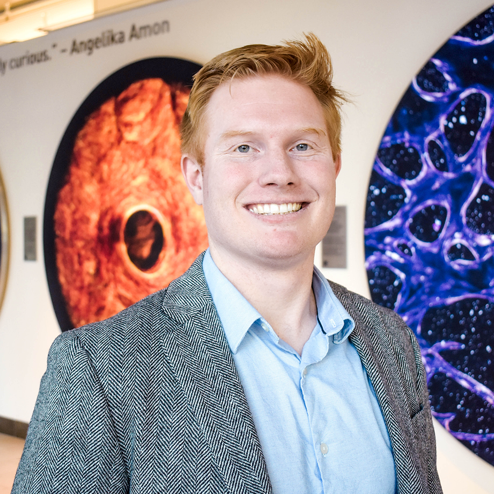
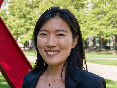
A cell protector collaborates with a killer

New research from the Horvitz Lab reveals what it takes for a protein that is best known for protecting cells against death to take on the opposite role.

Jennifer Michalowski | McGovern Institute
November 1, 2024

From early development to old age, cell death is a part of life. Without enough of a critical type of cell death known as apoptosis, animals wind up with too many cells, which can set the stage for cancer or autoimmune disease. But careful control is essential, because when apoptosis eliminates the wrong cells, the effects can be just as dire, helping to drive many kinds of neurodegenerative disease.

By studying the microscopic roundworm Caenorhabditis elegans—which was honored with its fourth Nobel Prize last month—scientists at MIT’s McGovern Institute have begun to unravel a longstanding mystery about the factors that control apoptosis: how a protein capable of preventing programmed cell death can also promote it. Their study, led by McGovern Investigator Robert Horvitz and reported October 9, 2024, in the journal Science Advances, sheds light on the process of cell death in both health and disease.

“These findings, by graduate student Nolan Tucker and former graduate student, now MIT faculty colleague, Peter Reddien, have revealed that a protein interaction long thought to block apoptosis in C. elegans, likely instead has the opposite effect,” says Horvitz, who shared the 2002 Nobel Prize for discovering and characterizing the genes controlling cell death in C. elegans.

Mechanisms of cell death

Horvitz, Tucker, Reddien and colleagues have provided foundational insights in the field of apoptosis by using C. elegans to analyze the mechanisms that drive apoptosis as well as the mechanisms that determine how cells ensure apoptosis happens when and where it should. Unlike humans and other mammals, which depend on dozens of proteins to control apoptosis, these worms use just a few. And when things go awry, it’s easy to tell: When there’s not enough apoptosis, researchers can see that there are too many cells inside the worms’ translucent bodies. And when there’s too much, the worms lack certain biological functions or, in more extreme cases, can’t reproduce or die during embryonic development.

Work in the Horvitz lab defined the roles of many of the genes and proteins that control apoptosis in worms. These regulators proved to have counterparts in human cells, and for that reason studies of worms have helped reveal how human cells govern cell death and pointed toward potential targets for treating disease.

A protein’s dual role

Three of C. elegans’ primary regulators of apoptosis actively promote cell death, whereas just one, CED-9, reins in the apoptosis-promoting proteins to keep cells alive. As early as the 1990s, however, Horvitz and colleagues recognized that CED-9 was not exclusively a protector of cells. Their experiments indicated that the protector protein also plays a role in promoting cell death. But while researchers thought they knew how CED-9 protected against apoptosis, its pro-apoptotic role was more puzzling.

CED-9’s dual role means that mutations in the gene that encode it can impact apoptosis in multiple ways. Most ced-9 mutations interfere with the protein’s ability to protect against cell death and result in excess cell death. Conversely, mutations that abnormally activate ced-9 cause too little cell death, just like mutations that inactivate any of the three killer genes.

An atypical ced-9 mutation, identified by Reddien when he was a PhD student in Horvitz’s lab, hinted at how CED-9 promotes cell death. That mutation altered the part of the CED-9 protein that interacts with the protein CED-4, which is proapoptotic. Since the mutation specifically leads to a reduction in apoptosis, this suggested that CED-9 might need to interact with CED-4 to promote cell death.

The idea was particularly intriguing because researchers had long thought that CED-9’s interaction with CED-4 had exactly the opposite effect: In the canonical model, CED-9 anchors CED-4 to cells’ mitochondria, sequestering the CED-4 killer protein and preventing it from associating with and activating another key killer, the CED-3 protein —thereby preventing apoptosis.

To test the hypothesis that CED-9’s interactions with the killer CED-4 protein enhance apoptosis, the team needed more evidence. So graduate student Nolan Tucker used CRISPR gene editing tools to create more worms with mutations in CED-9, each one targeting a different spot in the CED-4-binding region. Then he examined the worms. “What I saw with this particular class of mutations was extra cells and viability,” he says—clear signs that the altered CED-9 was still protecting against cell death, but could no longer promote it. “Those observations strongly supported the hypothesis that the ability to bind CED-4 is needed for the pro-apoptotic function of CED-9,” Tucker explains. Their observations also suggested that, contrary to earlier thinking, CED-9 doesn’t need to bind with CED-4 to protect against apoptosis.

When he looked inside the cells of the mutant worms, Tucker found additional evidence that these mutations prevented CED-9’s ability to interact with CED-4. When both CED-9 and CED-4 are intact, CED-4 appears associated with cells’ mitochondria. But in the presence of these mutations, CED-4 was instead at the edge of the cell nucleus. CED-9’s ability to bind CED-4 to mitochondria appeared to be necessary to promote apoptosis, not to protect against it.

Looking ahead

While the team’s findings begin to explain a long-unanswered question about one of the primary regulators of apoptosis, they raise new ones, as well. “I think that this main pathway of apoptosis has been seen by a lot of people as more or less settled science. Our findings should change that view,” Tucker says.

The researchers see important parallels between their findings from this study of worms and what’s known about cell death pathways in mammals. The mammalian counterpart to CED-9 is a protein called BCL-2, mutations in which can lead to cancer.  BCL-2, like CED-9, can both promote and protect against apoptosis. As with CED-9, the pro-apoptotic function of BCL-2 has been mysterious. In mammals, too, mitochondria play a key role in activating apoptosis. The Horvitz lab’s discovery opens opportunities to better understand how apoptosis is regulated not only in worms but also in humans, and how dysregulation of apoptosis in humans can lead to such disorders as cancer, autoimmune disease and neurodegeneration.

An elegant switch regulates production of protein variants during cell division

Cells make variants of thousands of proteins. These variants are not produced indiscriminately, but rather through precise regulatory mechanisms that can meet rapidly changing needs of the cell according to new research from the Cheeseman Lab.

Greta Friar | Whitehead Institute
October 18, 2024

Our cells contain thousands of proteins that have gone largely undetected and unstudied until recent years: these are variants of known proteins, which cells can make when their protein-building machinery interacts differently with the same stretch of genetic code. These protein variants have typically been overlooked as occasional accidents of gene expression, but researchers including Whitehead Institute Member Iain Cheeseman are discovering that they are actually abundant and can play important roles in cell functions. Researchers in Cheeseman’s lab are studying individual protein variants to learn more about them and their roles in health and disease, but they also wanted to understand broader patterns of protein variant production: how do cells control when to make one variant of a protein versus another, and what are the consequences of such switches?

Cheeseman, who is also a professor of biology at the Massachusetts Institute of Technology, and graduate student in his lab Jimmy Ly have now identified how cells switch to a different pattern of protein variant production during mitosis, or cell division. In research published in the journal Nature on October 23, they show that this broad regulatory switch helps cells survive paused cell divisions that can sometimes occur in healthy humans or be triggered by certain chemotherapy treatments. The work confirms that cells make variants of thousands of proteins, and also demonstrates that cells do not do so indiscriminately. Rather, cells use precise regulatory mechanisms to switch between different patterns of protein variant production, in order to rapidly tailor the proteins available to fit the changing needs of the cell.

A plethora of hidden proteins

Hw can our cells contain unknown proteins? In high school biology classes, students learn the rule that each gene codes for exactly one protein, such that if you know an organism’s genetic code, you should know every protein it can make. In fact, there are instead many genes that code for multiple proteins. For a protein to be made, first the genetic code for it is copied from DNA into a messenger RNA (mRNA). Then, a ribosome, the cellular machine that follows the instructions in genetic code to build a protein, locates the coding sequence within the mRNA by scanning for the start codon, a sequence of the three bases A, U, and G – bases are the chemical building blocks of RNA, abbreviated as A, U, C, and G. The ribosome recognizes the AUG start codon as the place to begin following instructions, and builds a protein based on the genetic sequence from there through to another trio of bases called a stop codon. However, one way that different versions of a protein can be produced is that a ribosome may begin reading the instructions from multiple different starting points.

Sometimes, a ribosome may miss the first AUG start codon and skip ahead to another AUG somewhere in the middle of the gene’s code, creating a truncated version of the protein. Sometimes, a ribosome may treat a similar trio of bases, such as CUG or GUG, as a start codon. This can cause it to begin earlier, creating a protein based on an extended genetic sequence. These possibilities mean that cells contain thousands more different proteins, or variants of proteins, than are represented by the dogma of one gene, one protein.

In order to understand protein variant production, the researchers—in collaboration with researchers from Whitehead Institute Member David Bartel’s lab–used a method that let them carefully track ribosomes to compare which start sites ribosomes tended to use. They looked at start site selection during mitosis versus during the rest of the cell cycle and found that a dramatic shift in use occurred for thousands of start sites. Specifically, the researchers found that during mitosis, ribosome scanning becomes more stringent. The ribosome will only begin making proteins at AUG sequences, and even then, only at AUGs that have preferable sequences of bases surrounding them—known as a strong Kozak context. This increased selectivity does not always lead to the familiar version of the protein being made during mitosis; sometimes the first AUG start codon has a weak Kozak context, so a truncated protein gets made from an AUG start codon with a stronger Kozak context that lies within the gene.

“Coming into this project, we knew very little about protein production during mitosis—for a long time, people didn’t think much protein production happened in mitosis at all,” Ly says. “It was satisfying to show not only that it is occurring, but that there’s a shift in which proteins are being made—and that this shift is important for cellular viability.”

How cells switch between protein variant programs

The researchers next identified how the switch to increased stringency is initiated during mitosis. They discovered that the key player is a protein called eIF1, which is one of many partners that can pair with ribosomes to help them select their start site. In particular, increased eIF1 pairing with ribosomes causes the ribosomes to be more stringent in their start codon selection, inhibiting the usage of non-AUG initiation sites or sites with weak Kozak contexts.

During mitosis, ribosome pairing with eIF1 increases sharply, leading to the shift in stringency. This change in pairing rate during mitosis puzzled the researchers: ribosomes and their partners, including eIF1, all typically reside together in the main body of the cell—where ribosomes make proteins—so they should be able to pair freely at any time. The researchers looked for other molecules in the same location that could be altering how ribosomes and eIF1 interact during different parts of the cell cycle, but they couldn’t find anything. Eventually, the researchers realized that the answer to the puzzle lay in a separate location: the nucleus.

They found that cells maintain a large pool of eIF1 inside of the nucleus, locked away from the ribosomes. Then, during cell division, the wall of the nucleus dissolves, mixing its contents with the rest of the cell. This is necessary for the dividing cell to divvy up its DNA, but it also releases the pool of eIF1 to pair with ribosomes, increasing stringency. At the end of mitosis, the nucleus reforms and eIF1 is re-incorporated into the nucleus of each of the two daughter cells, and the cells return to a less stringent program.

“The explanation for increased interaction between eIF1 and ribosomes during mitosis had really stumped us, and so when I saw eIF1 localizing to the nucleus, that was a really exciting ‘aha’ moment,” Ly says. “Discovering this mechanism of nuclear release during mitosis was unexpected, and it’s interesting to think about how else cells might be using it.”

Consequences of increased stringency for the cell

Once the researchers understood the how, they then wanted to understand the why? What they discovered is that when cells have no nuclear pool of eIF1, and so no change in stringency during mitosis, they are more likely to die during mitosis. In particular, these cells fare poorly during mitotic arrest, a state in which cells get stuck in mitosis for hours or even days–much longer than typical mitosis. Arrest occurs when cells detect a possible cell division error and so halt their division until the error is corrected or the cell dies.

One effect of increased stringency during mitosis is related to mitochondria, which are required for energy production in many cell types and are therefore required for maintaining viability. Cells stuck in mitotic arrest need energy to keep them going through this unexpected delay. The researchers found that increased stringency during mitosis led to an increase in the production of important mitochondrial proteins, boosting the cells’ energy supply to get them through arrest.

Increased stringency also gives cells the tools they need to escape arrest, even if they haven’t fixed the error that caused them to pause division. In a Nature paper in 2023, Cheeseman and then-postdoc in his lab Mary-Jane Tsang showed that when cells build up enough of the truncated version of a protein called CDC20, they can escape arrest. Ly’s work adds to this story by showing that the nuclear release of eIF1 increases stringency, leading to more production of truncated CDC20 during mitosis, which explains how cells build up enough of this protein variant during mitosis to trigger their escape. These findings may have important potential implications for some cancer chemotherapy strategies.

Some chemotherapies work by trapping cancer cells in mitotic arrest until they die. Cheeseman, Tsang, and Ly’s work collectively shows that when cancer cells lack sufficient truncated CDC20—as can occur in the absence of nuclear eIF1—the cells cannot escape arrest and so are killed off by these chemotherapies at higher rates. These results could be used to improve the efficacy of antimitotic chemotherapy drugs.

The switch in protein variant production that the researchers found affects thousands of proteins. These newly identified protein variants serve as a foundation for many future projects in the lab.

As the researchers continue to examine the consequences of this switch to stringency during mitosis, they are also searching for other cases in which cells regulate protein variant production outside of mitosis. For example, the researchers are interested in how this switch in stringency affects fertility; immature egg cells spend a long time in a form of arrested cell division without an intact nucleus, and Ly observed eIF1 in the nucleus of the immature female eggs.

“Cells have axes of control that they use to quickly make broad changes in gene expression,” Cheeseman says. “Several of these are central to controlling cell division—for example, the role of phosphorylation as a regulatory switch in mitosis has been well studied. Our work identifies another axis of control, and we’re excited to discover more about when and how cells make use of it.”

Laub Lab News Brief: anti-viral defense system in bacteria modifies mRNA

Killing the messenger

Lillian Eden | Department of Biology
October 23, 2024

Newly characterized anti-viral defense system in bacteria aborts infection through novel mechanism by chemically modifying mRNA.


Like humans and other complex multicellular organisms, single-celled bacteria can fall ill and fight off viral infections. A bacterial virus is known as a bacteriophage, or, more simply, a phage, which is one of the most ubiquitous life forms on Earth. Phages and bacteria are engaged in a constant battle, the virus attempting to circumvent the bacteria’s defenses, and the bacteria racing to find new ways to protect itself.

These anti-phage defense systems are carefully controlled and prudently managed — dormant but always poised to strike. 

New research recently published in Nature from the Laub Lab in the Department of Biology at MIT has characterized an anti-phage defense system in bacteria known as CmdTAC. CmdTAC prevents viral infection by altering mRNA, the single-stranded genetic code used to produce proteins, of both the host and the virus.  

This defense system detects phage infection at a stage when the viral phage has already commandeered the host’s machinery for its own purposes. In the face of annihilation, the ill-fated bacterium activates a defense system that will halt translation, preventing the creation of new proteins and aborting the infection — but dooming itself in the process. 

“When bacteria are in a group, they’re kind of like a multicellular organism that is not connected to one another. It’s an evolutionarily beneficial strategy for one cell to kill itself to save another identical cell,” says Christopher Vassallo, a postdoc and co-author of the study. “You could say it’s like self-sacrifice: one cell dies to protect the other cells.” 

The enzyme responsible for altering the mRNA is called an ADP-ribosyltransferase.  Researchers have characterized hundreds of these enzymes — although only a few are known to target DNA or other types of RNA, all but a handful target proteins. This is the first time these enzymes have been characterized targeting mRNA within cells.

Expanding understanding of anti-phage defense

Co-first author and graduate student Chris Doering noted that it is only within the last decade or so that researchers have begun to appreciate the breadth of diversity and complexity of anti-phage defense systems. For example, CRISPR gene editing, a technique used in everything from medicine to agriculture, is rooted in research on the bacterial CRISPR-Cas9 anti-phage defense system. 

CmdTAC is a subset of a widespread anti-phage defense mechanism called a toxin-antitoxin system. A TA system is just that: a toxin capable of killing or altering the cell’s processes rendered inert by an associated antitoxin. 

Although these TA systems can be identified — if the toxin is expressed by itself, it kills or inhibits the growth of the cell; if the toxin and antitoxin are expressed together, the toxin is neutralized — characterizing the cascade of circumstances that activates these systems requires extensive effort. In recent years, however, many TA systems have been shown to serve as anti-phage defenses. 

Two general questions need to be answered to understand a viral defense system: how do bacteria detect an infection, and how do they respond?

Detecting infection

CmdTAC is a TA system with an additional element, and the three components generally exist in a stable complex: the toxin CmdT, the antitoxin CmdA, and an additional component that mediates the system, the chaperone CmdC. 

If the phage’s protective capsid protein is present, CmdC disassociates from CmdT and CmdA and interacts with the phage capsid protein instead. In the model outlined in the paper, the chaperone CmdC is, therefore, the sensor of the system, responsible for recognizing when an infection is occurring. Structural proteins, such as the capsid that protects the phage genome, are a common trigger because they’re abundant and essential to the phage.

The uncoupling of CmdC leads to the degradation of the neutralizing antitoxin CmdA, which releases the toxin CmdT to do its lethal work.

Toxicity on the loose

Guided by computational tools, the researchers knew that CmdT was likely an ADP-ribosyltransferase due to its similarities to other such enzymes. As the name suggests, the enzyme transfers an ADP ribose onto its target.

To determine how CmdT was altering mRNA, the researchers tested a mix of short sequences of single-stranded RNA to see if the enzyme was drawn to any sequences or positions in particular. RNA has four bases: A, U, G, and C, and the evidence points to the enzyme recognizing GA sequences. 

The CmdT modification of GA sequences in mRNA blocks its translation. The cessation of creating new proteins aborts the infection, preventing the phage from spreading beyond the host to infect other bacteria. 

“Not only is it a new type of bacterial immune system, but the enzyme involved does something that’s never been seen before: the ADP-ribsolyation of mRNA,” Vassallo says. 

Although the paper outlines the broad strokes of the anti-phage defense system, there’s more to learn: it’s unclear how CmdC interacts with the capsid protein, and how the chemical modification of GA sequences prevents translation. 

Beyond Bacteria

While exploring anti-phage defense aligns with the Laub Lab’s overall goal of understanding how bacteria function and evolve, these results may have broader implications beyond bacteria.

Senior author Michael Laub, Salvador E. Luria Professor and HHMI Investigator, says the ADP-ribosyltransferase has homologs in eukaryotes, including human cells. They are not well studied, and not currently among the Laub Lab’s research topics, but they are known to be up-regulated in response to viral infection. 

“There are so many different — and cool — mechanisms by which organisms defend themselves against viral infection,” Laub says. “The notion that there may be some commonality between how bacteria defend themselves and how humans defend themselves is a tantalizing possibility.” 

Establishing boundaries of the genetic kind

The pseudoautosomal region (PAR) is a critical area on the Y chromosome that swaps genetic information with the X chromosome. Recent research from the Page Lab reaffirms the location of PAR and offers a refined understanding of where crossover events occur.

Shafaq Zia | Whitehead Institute
October 14, 2024

At first, the X and the Y sex chromosomes seemed like an unlikely pair. But then, researchers, including Whitehead Institute Member David Page, began finding clues that suggested otherwise: identical DNA sequences on the X and Y chromosomes.

Soon, it became clear that the tips of the X and Y chromosomes join together in a tight embrace, swapping genetic material during the process of sperm production from immature male germ cells. This limited area of genetic exchange between the two sex chromosomes is called the pseudoautosomal region (PAR).

But science is an iterative process—a continuous cycle of questioning, testing, and revising knowledge. Last fall, what had long been considered well established in genetics was called into question when new research suggested that the boundary of the PAR might be half a million base pairs away from the accepted location. Given that a typical human gene is about tens of thousands of base pairs, this length would potentially span multiple genes on the X and Y chromosomes, raising serious concerns about the accuracy and validity of decades of scientific literature.

Fortunately, new work from Page, research scientist Daniel Winston Bellott, and colleagues—published Oct. 14 in the American Journal of Human Genetics—offers clarity. In this study, the group re-examines the size of the PAR using sequencing data presented by outside researchers in their 2023 work, alongside decades of genomic resources, and single-cell sequencing of human sperm. Their findings confirm that the location of the boundary to the PAR, as identified by scientists in 1989, still holds true.

“If one is interested in understanding sex differences in health and disease, the boundary of the pseudoautosomal region is arguably the most fundamental landmark in the genome,” says Page, who is also a professor of biology at the Massachusetts Institute of Technology and an Investigator with Howard Hughes Medical Institute. “Had this boundary been multiple genes off, the field would have been shaken to its foundations.”

Dance of the chromosomes

The X and Y chromosomes evolved from an ancestral pair of chromosomes with identical structures. Over time, the Y chromosome degenerated drastically, losing hundreds of functional genes. Despite their differences, the X and Y chromosomes come together during a special type of cell division called male meiosis, which produces sperm cells.

This process begins with the tips of the sex chromosomes aligning side by side like two strands of rope. As the X and Y chromosomes embrace each other, enzymes create breaks in the DNA. These breaks are repaired using the opposite chromosome as a template, linking the X and Y together. About half of the time, an entire segment of DNA, which often contains multiple genes, will cross over onto the opposite chromosome.

The genetic exchange, called recombination, concludes with the X and Y chromosomes being pulled apart to opposite ends of the dividing cell, ensuring that each chromosome ends up in a different daughter cell. “This intricate dance of the X and Y chromosomes is essential to a sperm getting either an X or a Y—not both, and not neither,” says Page.

This way when the sperm—carrying either an X or a Y—fuses with the egg—carrying an X—during fertilization, the resulting zygote has the right number of chromosomes and a mix of genetic material from both parents.

But that’s not all. The swapping of DNA during recombination also allows for the chromosomes to have the same genes but with slight variations. These unique combinations of genetic material across sex chromosomes are key to genetic diversity within a species, enabling it to survive, adapt, and reproduce successfully.

Beyond the region of recombination, the Y chromosome contains genes that are important for sex determination, for sperm production, and for general cellular functioning. The primary sex-defining gene, SRY, which triggers the development of an embryo into a male, is located only 10,000 bases from the boundary of the PAR.

Advancing together

To determine whether the location of this critical boundary on the human sex chromosomes—where they stop crossing over during meiosis and become X-specific or Y-specific—had been misidentified for over three decades, researchers began by comparing publicly-available DNA sequences from the X and the Y chromosomes of seven primate species: humans, chimpanzees, gorillas, orangutans, siamangs, rhesus macaques, and colobus monkeys.

Based on the patterns of crossover between the X and the Y chromosomes of these species, the researchers constructed an evolutionary tree. Upon analyzing how DNA sequences close to and distant from the PAR boundary group together across species, the researchers found a substitution mutation—where a letter in a long string of letters is swapped for a different one—in the DNA of the human X and Y chromosomes. This change was also present in the chimpanzee Y chromosome, suggesting that the mutation originally occurred in the last common ancestor of humans and chimpanzees and was then transferred to the human X chromosome.

“These alignments between various primates allowed us to observe where the X and the Y chromosomes have preserved identity over millions of years and where they have diverged,” says Bellott. “That [pseudoautosomal] boundary has remained unchanged for 25 million years.”

Next, the group studied crossover events in living humans using a vast dataset of single-cell sequencing of sperm samples. They found 795 sperm with clear swapping of genetic material somewhere between the originally proposed boundary of the PAR and the newly-proposed 2023 boundary.

Once these analyses confirmed that the original location of the PAR boundary remains valid, Page and his team turned their attention to data from the 2023 study that contested this 1989 finding. The researchers focused on 10 male genomes assembled by the outside group, which contained contiguous sequences from the PAR.

Since substitutions on the Y chromosome typically occur at a steady rate, but in the PAR, changes on the X chromosome can transfer to the Y through recombination, the researchers compared the DNA sequences from the ten genomes to determine whether they followed the expected steady rate of change or if they varied.

The team found that close to the originally proposed PAR boundary, the DNA sequences changed at a steady rate. But further away from the boundary, the rate of change varied, suggesting that crossover events likely occurred in this region. Furthermore, the group identified several shared genetic differences between the X and the Y chromosomes of these genomes, which demonstrates that recombination has occurred even closer to the PAR boundary than scientists observed in 1989.

“Ironically, instead of contradicting the original boundary, the 2023 work has helped us refine the location of crossover to an even narrower area near the boundary,” says Page.

Thanks to the efforts of Page’s group at Whitehead Institute, our understanding of the PAR is clearer than ever, and business can go on as usual for researchers investigating sex differences in health and disease.

Bat cells possess a unique antiviral mechanism, preventing the SARS-CoV-2 virus from taking control

Bats have the amazing ability to coexist with viruses that are deadly to humans. New work from the Jaenisch Lab uncovers an antiviral mechanism that allows viruses to enter bat cells but prevents them from replicating.

Shafaq Zia | Whitehead Institute
October 14, 2024

Viruses are masters of stealth. From the moment a virus enters the host’s body, it begins hijacking its cells. First, the virus binds to a specific protein on the cell’s surface through a lock-and-key mechanism. This protein, known as a receptor, facilitates the entry of the virus’s genetic material into the cell. Once inside, this genetic code takes over the cell’s machinery, directing it to produce copies of the virus and assemble new viral particles, which can go on to infect other cells. Upon detecting the invasion, the host’s immune system responds by attacking infected cells in hopes of curbing the virus’s spread.

But in bats, this process unfolds differently. Despite carrying several viruses — Marburg, Ebola, Nipah, among others — bats rarely get sick from these infections. It seems their immune systems are highly specialized, allowing them to live with viruses that would typically be deadly in humans, without any clinical symptoms.

Since the onset of the COVID-19 pandemic, the lab of Whitehead Institute Founding Member Rudolf Jaenisch has been investigating the molecular basis of bats’ extraordinary resilience to viruses like SARS-CoV-2. In their latest study, published in the journal PNAS on Oct. 14 , Jaenisch lab postdoc Punam Bisht and colleagues have uncovered an antiviral mechanism in bat cells that allows viruses to enter the cells but prevents them from replicating their genome and completing the hijacking process.

“These cells have elevated expressions of antiviral genes that act immediately, neutralizing the virus before it can spread,” says Jaenisch, a professor of biology at the Massachusetts Institute of Technology. “What’s particularly interesting is that many of these antiviral genes have counterparts, or orthologs in humans.”

Striking a delicate balance

The innate immune system is the body’s first line of defense against foreign invaders like the SARS-CoV-2 virus. This built-in security system is always on alert, responding swiftly — within minutes to hours — to perceived threats.

Upon detecting danger, immune cells rush to the site of infection, where they target the virus with little precision in attempts to slow it down and buy time for the more specialized adaptive immune system to take over. During this process, these cells release small signaling proteins called cytokines, which coordinate the immune response by recruiting additional immune cells and directing them to the battleground.

If the innate immune response alone isn’t sufficient to defeat the virus, it signals the adaptive immune system for support. The adaptive immune system tailors its attacks to the exact pathogen it is fighting and can even keep records of past infections to launch a faster, more aggressive attack the next time it encounters the same pathogen.

But in some infections, the innate immune response can quickly spiral out of control before the adaptive immune response is activated. This phenomenon, called a cytokine storm, is a life-threatening condition characterized by the overproduction of cytokines. These proteins continue to signal the innate immune system for backup even when it’s not necessary, leading to a flood of immune cells at the site of infection, where they inadvertently begin damaging organs and healthy tissues.

Bats, on the other hand, are uniquely equipped to manage viral infections without triggering an overwhelming immune response or allowing the virus to take control. To understand how their innate immune system achieves this delicate balance, Bisht and her colleagues turned their attention to bat cells.

In this study, researchers compared how the SARS-CoV-2 virus replicates in human and bat stem cells and fibroblasts — a type of cell involved in the formation of connective tissue. While fibroblasts are not immune cells, they can secrete cytokines and guide immune response, particularly to help with tissue repair.

After exposing these cells to the SARS-CoV-2 virus for 48 hours, the researchers used a Green Fluorescent Protein (GFP) tag to track the virus’s activity. GFP is a fluorescent protein whose genetic code can be added as a tag to a gene of interest. This causes the products of that gene to glow, providing researchers with a visual marker of where and when the gene is expressed.

They observed that over 80% of control cells — derived from the kidneys of African green monkeys and known to be highly susceptible to SARS-CoV-2 — showed evidence of the virus replicating. In contrast, they did not detect any viral activity in human and bat stem cells or fibroblasts.

In fact, even after introducing the human ACE2 receptor — which SARS-CoV-2 uses to bind and enter cells — into bat cells, the infected bat fibroblasts were able to replicate viral RNA and produce viral proteins, but at much lower levels compared to infected human fibroblasts.

These bat fibroblasts, however, could not assemble these viral proteins into fully infectious virus particles, suggesting an abortive infection, where the virus is able to initiate replication but fails to complete the process and produce progeny viruses.

Using electron microscopy to look inside bat and human cells, they began to understand why: in human cells, SARS-CoV-2 had created special structures called double-membrane vesicles (DMV). These vesicles acted like a bubble, shielding the viral genome from detection and providing it safe space to replicate more effectively. However, these “viral replication factories” were absent in bat fibroblasts.

When the researchers examined the gene expression profiles of these bat fibroblasts and compared them those of infected human cells, they found that although both human and bat cells have genes regulating the release of a type of cytokine called interferons, these genes are already turned on in bat fibroblasts — unlike in human cells — even before virus infection occurs.

These findings suggest that bat cells are in a constant state of vigilance. This allows their innate immune system to stop the SARS-CoV-2 virus in its tracks early on in the replication process before it can entirely hijack cellular machinery.

Surprisingly, this antiviral mechanism does not protect bat cells against all viruses. When the researchers infected bat fibroblasts with Zika virus, the virus was able to replicate and produce new viral particles.

“This means there are still many questions unanswered about how bat cells resist infection,” says Bisht. “COVID-19 continues to circulate, and the virus is evolving quickly. Filling in these gaps in our knowledge will help us develop better vaccines and antiviral strategies.”
The researchers are now focused on identifying the specific genes involved in this antiviral mechanism, and exploring how they interact with the virus during infection.

Research Reflections: Alison Biester (PhD ’24), Drennan Lab

New snapshots of ancient life

Alison Biester | Department of Chemistry
October 3, 2024

The resolution revolution, beating “blobology”, and shedding light on how ancient microbes thrived in a primordial soup.

The earliest life on earth created biological molecules despite the limited materials available in the primordial soup such as CO2, hydrogen gas, and minerals containing iron, nickel, and sulfur.

As ancient microbes evolved, they developed proteins that sped up chemical reactions, called enzymes. Enzymes were evolutionarily advantageous because they created local environments called active sites optimized for reaction performance.

Although we know that carbon is the building block of life on earth–we wouldn’t exist without carbon-based molecules such as proteins and DNA–much remains unclear about how more complex carbon-based molecules were originally generated from CO2. Proteins and DNA are huge molecules with thousands of carbon atoms, so creating life from CO2 would be no small undertaking.

Catherine Drennan, Professor of Biology and Chemistry and HHMI Investigator and Professor, has long studied the enzymes that perform these crucial reactions wherein CO2 is converted into a form of carbon that cells can use, which requires iron, nickel, and sulfur.

In particular, she uses structural biology to study carbon monoxide dehydrogenase (CODH), which reacts with CO2 to produce CO, and acetyl-CoA synthase (ACS), which uses CO with another single unit of carbon to create a carbon-carbon bond. Crystallographic work by Drennan and others has provided structural snapshots of bacterial CODH and ACS, but its structure in other contexts remains elusive. During my PhD, I worked with Drennan on the structural characterization of CODH and ACS, culminating in a publication in PNAS, published October 3, 2024.

Throughout Drennan’s career, the lab has used a method known as X-ray crystallography to determine enzyme structures at atomic resolution. In recent years, however, cryogenic electron microscopy (cryo-EM) has risen in popularity as a structural biology technique.

Cryo-EM offers some key advantages over X-ray crystallography, such as its ability to capture structures of large and dynamic complexes. However, cryo-EM is limited in its ability to elucidate structures of small proteins, an area where X-ray crystallography continues to excel.

To perform a cryo-EM experiment, proteins are rapidly frozen in a thin layer of ice and imaged on an electron microscope. By capturing images of the protein in various orientations, researchers can generate a 3D model of their protein of interest.

Around 2015, cryo-EM reached a tipping point known as the “resolution revolution.” Due to improvements in both the hardware for collecting cryo-EM data and the software used for data processing, the technique could, for the first time, be used to determine protein structures at near-atomic resolution.

Seeing the potential for this new technique, MIT opened its very own cryo-EM facility with two electron microscopes in 2018. Just a year later, I joined the Drennan lab. When I began my thesis work, Cathy asked “Would you like to do crystallography or cryo-EM?”

Eager to try something that was both novel for researchers and new to me, I chose cryo-EM.

Ancient microbes

An ancient type of microbe, archaea, also uses CODH and ACS. Without information on how these protein chains interact, we cannot understand how these proteins work together within this complex–but it’s a difficult question to answer. In total, the complex contains forty protein chains that interact with one another and adopt various conformations to perform their chemistry.

We don’t know for sure which ACS enzyme came first, the bacterial or archaeal one, but we know they are both very ancient.

Archaeal CODH has been visualized via X-ray crystallography, but that CODH was isolated from the enormous megadalton enzyme complex present in the native archaea.

A CO2 molecule, which reacts with CODH, is 44 daltons; the enzyme complex at 2.2 megadaltons is 50,000 times the size of CO2. The complex consists of several copies of CODH, ACS, and a cobalt-containing enzyme that donates the second one-carbon unit used by ACS. Due to the large and dynamic nature of the complex, it was a great candidate for visualizing with cryo-EM.

Before I joined the lab, a collaboration had been initiated between the Drennan Lab and Dr. David Grahame of the Uniformed Services University of the Health Sciences, an expert in archaeal CODH and ACS.

Just before his retirement, Grahame grew hundreds of liters of archaea and isolated approximately one gram of the enzyme complex that he provided to the Drennan Lab for structural characterization. Each cryo-EM experiment can use as little as a microgram of protein. For a structural biologist, having one gram of protein–in theory, enough for one million experiments–to work with is a dream.

Blobology

With an abundance of protein, I embarked on this project with this exciting new technique on a promising target. I prepared my cryo-EM sample and collected data at the new MIT cryo-EM facility. As I was collecting data, I could see in the images large protein complexes that appeared to be my complex of interest. I could also see some smaller proteins that were consistent with the shape of isolated CODH. When I went on to process my data, I focused on the larger protein complexes, since the structure of isolated CODH was already known.

However, when I finished processing my first dataset, I was a bit disappointed. My resolution was very low–instead of atoms, I was seeing amorphous blobs, and I had no idea which blob matched with which protein, or how the proteins fit together. Rather than post-revolution cryo-EM, I felt like I was performing the “blobology” of the past.

Our cryo-EM data contains detailed structural information that becomes evident after significant data processing. On the left is the initial structure of our proteins of interest, carbon monoxide dehydrogenase (CODH) and acetyl-CoA synthase (ACS), and on the right is our final, detailed one. Photo courtesy of Alison Biester.

But the project was young, and a few failed experiments are par for the course of a PhD.

The next step was sample optimization, and luckily I had plenty of sample to work with. I tried preparing the protein in a different way, changed the protein concentration, used different additives, and scaled up my data collection.

Nothing helped. No matter what I tried, I could not move out of blobology territory. So, as one does when a project is failing, I stepped away. I worked on other projects and stopped thinking about the archaeal CODH and ACS.

A few months later, the cryo-EM facility was seeking users to try a new sample preparation instrument called the chameleon. Chameleon automates the sample preparation process and is intended to improve sample quality. With plenty of sample still to spare, I volunteered to try the instrument.

Just prior to my data collection, the facility had also installed a new software that allows data processing as it is being collected. The software uses automated processes to select proteins within your data; previously, I had only selected large protein complexes consistent with my complex of interest after the fact.

The new software is not very discriminating–but I was surprised when I looked at the results of the live processing. The processing showed that I had a protein complex in my sample that I did not expect – a complex of CODH and ACS!

This complex had just one copy of CODH and one copy of ACS, unlike the full complex that has multiple copies of each. My excitement for the project was reinvigorated. With this new target, could I leave blobology behind and finally join the resolution revolution?

After running more experiments and collecting more data and a few months of data processing, I realized that the sample contained three different states: isolated CODH, CODH with one copy of ACS, and CODH with two copies of ACS. I was able to use the Model-based Analysis of Volume Ensembles (MAVEn) tool developed by the Davis Lab at MIT to sort out these three states. When I finished the data processing, I achieved near-atomic resolution of all three states.

Through this work, for the first time, we can see what the archaeal ACS looks like. The archaeal ACS is fundamentally different from the bacterial one: a huge portion of the enzyme is missing, including part of the enzyme that makes up the active site in bacteria, leaving open the question of what the ACS active site looks like in archaea.

In our structure of archaeal ACS in complex with CODH, we were surprised to see that the active site looks almost identical to the bacterial one. This similarity is enabled by the archaeal CODH, which compensates for the missing part of ACS.

Given how similar the ACS active site environment in bacterial and archaea, we are likely getting a look at an active site that has remained conserved over billions of years of evolution.

Although the project didn’t fulfill its original promise of solving the structure of the large, dynamic protein complex, I did find intriguing insights. The tools available in 2015 would not have enabled me to achieve these results; it is clear to me that the resolution revolution is far from over, and the evolution of structural biology has been fascinating to experience. Cryo-EM has and will continue to evolve, as amazing new tools are still being developed.

Since graduating from MIT, I’ve been working at the Protein Data Bank, the data center that houses all available protein structure information. Working here gives me a front-row view of new discoveries in structural biology. I’m so excited to see where this field will go in the future.

Cancer biologists discover a new mechanism for an old drug

Study reveals the drug, 5-fluorouracil, acts differently in different types of cancer — a finding that could help researchers design better drug combinations.

Anne Trafton | MIT News
October 7, 2024

Since the 1950s, a chemotherapy drug known as 5-fluorouracil has been used to treat many types of cancer, including blood cancers and cancers of the digestive tract.

Doctors have long believed that this drug works by damaging the building blocks of DNA. However, a new study from MIT has found that in cancers of the colon and other gastrointestinal cancers, it actually kills cells by interfering with RNA synthesis.

The findings could have a significant effect on how doctors treat many cancer patients. Usually, 5-fluorouracil is given in combination with chemotherapy drugs that damage DNA, but the new study found that for colon cancer, this combination does not achieve the synergistic effects that were hoped for. Instead, combining 5-FU with drugs that affect RNA synthesis could make it more effective in patients with GI cancers, the researchers say.

“Our work is the most definitive study to date showing that RNA incorporation of the drug, leading to an RNA damage response, is responsible for how the drug works in GI cancers,” says Michael Yaffe, a David H. Koch Professor of Science at MIT, the director of the MIT Center for Precision Cancer Medicine, and a member of MIT’s Koch Institute for Integrative Cancer Research. “Textbooks implicate the DNA effects of the drug as the mechanism in all cancer types, but our data shows that RNA damage is what’s really important for the types of tumors, like GI cancers, where the drug is used clinically.”

Yaffe, the senior author of the new study, hopes to plan clinical trials of 5-fluorouracil with drugs that would enhance its RNA-damaging effects and kill cancer cells more effectively.

Jung-Kuei Chen, a Koch Institute research scientist, and Karl Merrick, a former MIT postdoc, are the lead authors of the paper, which appears today in Cell Reports Medicine.

An unexpected mechanism

Clinicians use 5-fluorouracil (5-FU) as a first-line drug for colon, rectal, and pancreatic cancers. It’s usually given in combination with oxaliplatin or irinotecan, which damage DNA in cancer cells. The combination was thought to be effective because 5-FU can disrupt the synthesis of DNA nucleotides. Without those building blocks, cells with damaged DNA wouldn’t be able to efficiently repair the damage and would undergo cell death.

Yaffe’s lab, which studies cell signaling pathways, wanted to further explore the underlying mechanisms of how these drug combinations preferentially kill cancer cells.

The researchers began by testing 5-FU in combination with oxaliplatin or irinotecan in colon cancer cells grown in the lab. To their surprise, they found that not only were the drugs not synergistic, in many cases they were less effective at killing cancer cells than what one would expect by simply adding together the effects of 5-FU or the DNA-damaging drug given alone.

“One would have expected that these combinations to cause synergistic cancer cell death because you are targeting two different aspects of a shared process: breaking DNA, and making nucleotides,” Yaffe says. “Karl looked at a dozen colon cancer cell lines, and not only were the drugs not synergistic, in most cases they were antagonistic. One drug seemed to be undoing what the other drug was doing.”

Yaffe’s lab then teamed up with Adam Palmer, an assistant professor of pharmacology at the University of North Carolina School of Medicine, who specializes in analyzing data from clinical trials. Palmer’s research group examined data from colon cancer patients who had been on one or more of these drugs and showed that the drugs did not show synergistic effects on survival in most patients.

“This confirmed that when you give these combinations to people, it’s not generally true that the drugs are actually working together in a beneficial way within an individual patient,” Yaffe says. “Instead, it appears that one drug in the combination works well for some patients while another drug in the combination works well in other patients. We just cannot yet predict which drug by itself is best for which patient, so everyone gets the combination.”

These results led the researchers to wonder just how 5-FU was working, if not by disrupting DNA repair. Studies in yeast and mammalian cells had shown that the drug also gets incorporated into RNA nucleotides, but there has been dispute over how much this RNA damage contributes to the drug’s toxic effects on cancer cells.

Inside cells, 5-FU is broken down into two different metabolites. One of these gets incorporated into DNA nucleotides, and other into RNA nucleotides. In studies of colon cancer cells, the researchers found that the metabolite that interferes with RNA was much more effective at killing colon cancer cells than the one that disrupts DNA.

That RNA damage appears to primarily affect ribosomal RNA, a molecule that forms part of the ribosome — a cell organelle responsible for assembling new proteins. If cells can’t form new ribosomes, they can’t produce enough proteins to function. Additionally, the lack of undamaged ribosomal RNA causes cells to destroy a large set of proteins that normally bind up the RNA to make new functional ribosomes.

The researchers are now exploring how this ribosomal RNA damage leads cells to under programmed cell death, or apoptosis. They hypothesize that sensing of the damaged RNAs within cell structures called lysosomes somehow triggers an apoptotic signal.

“My lab is very interested in trying to understand the signaling events during disruption of ribosome biogenesis, particularly in GI cancers and even some ovarian cancers, that cause the cells to die. Somehow, they must be monitoring the quality control of new ribosome synthesis, which somehow is connected to the death pathway machinery,” Yaffe says.

New combinations

The findings suggest that drugs that stimulate ribosome production could work together with 5-FU to make a highly synergistic combination. In their study, the researchers showed that a molecule that inhibits KDM2A, a suppressor of ribosome production, helped to boost the rate of cell death in colon cancer cells treated with 5-FU.

The findings also suggest a possible explanation for why combining 5-FU with a DNA-damaging drug often makes both drugs less effective. Some DNA damaging drugs send a signal to the cell to stop making new ribosomes, which would negate 5-FU’s effect on RNA. A better approach may be to give each drug a few days apart, which would give patients the potential benefits of each drug, without having them cancel each other out.

“Importantly, our data doesn’t say that these combination therapies are wrong. We know they’re effective clinically. It just says that if you adjust how you give these drugs, you could potentially make those therapies even better, with relatively minor changes in the timing of when the drugs are given,” Yaffe says.

He is now hoping to work with collaborators at other institutions to run a phase 2 or 3 clinical trial in which patients receive the drugs on an altered schedule.

“A trial is clearly needed to look for efficacy, but it should be straightforward to initiate because these are already clinically accepted drugs that form the standard of care for GI cancers. All we’re doing is changing the timing with which we give them,” he says.

The researchers also hope that their work could lead to the identification of biomarkers that predict which patients’ tumors will be more susceptible to drug combinations that include 5-FU. One such biomarker could be RNA polymerase I, which is active when cells are producing a lot of ribosomal RNA.

The research was funded by the Damon Runyon Cancer Research Fund, a Ludwig Center at MIT Fellowship, the National Institutes of Health, the Ovarian Cancer Research Fund, the Holloway Foundation, and the STARR Cancer Consortium.

A tour of our cells through time

From chemical reactions occurring in a splinter of a second, to evolution shaping species over billions of years, the processes that make up our biology occur in time frames both short and long. In this video, we will hear from Department of Biology Faculty and Whitehead Institute scientists on how their thinking about different time scales informs scientific discovery.

Produced by Madeleine Turner | Whitehead Institute
September 17, 2024

Hear from the Cheeseman Lab, Weissman Lab, Hravatin Lab, and Yamashita Lab at the Whitehead Institute in the following video.

BSG-MSRP-Bio student profile: Yeongseo Son, Spranger Lab

All It takes to titer: discovering a love of troubleshooting at MIT

Noah Daly | Department of Biology
September 25, 2024

BSG-MSRP-Bio student Yeongseo Son breathed new life into her love of science over the summer in the Spranger Lab studying immune responses in the lung in the Department of Biology at MIT.


When Yeongseo Son was initially invited to join the Spranger Lab as part of the Bernard S. and Sophie G. Gould MIT Research Program in Biology, she thought the email was spam. Having grown up in the South for most of her life, she had never pictured herself at MIT.

Back home at the University of Georgia, Son studies neutrophils, a kind of innate immune cell that serves as the body’s first line of defense against foreign pathogens. After taking a graduate-level course on immunology last semester, Son realized she needed to increase her basic understanding of the broad discipline.

“I knew that coming to work with Professor Spranger would give me a chance to work on cancer immunology and T cell biology, two really cool and important fields I haven’t been exposed to,” Son says.

It took several attempts from the Senior Lecturer and BSG-MSRP-Bio program coordinator Mandana Sassanfar to reach her before Son accepted.  

“Before I arrived, I was worried it would be too intense or that I wouldn’t fit in,” Son says. “I couldn’t have been more wrong: yes, the work is challenging, but everyone is here because they truly love science.”

Vexing Viruses

In the lab of Stefani Spranger, Associate Professor in the Department of Biology and Intramural Faculty of the Koch Institute for Integrative Cancer Research, Son was first tasked with a seemingly simple second project: growing a new strain of influenza to infect mice that had recently recovered from another strain. 

This quest involved multiple steps, such as culturing cells, infecting the cells with the virus, and measuring how lethal it is to host cells, working with a strain that her lab hadn’t used before.   

To test the strength of the virus, the virus is mixed with host cells in order to infect them. Then the host cells are placed on a layer of agar, a gelatinous substance that provides nutrients for the host cells. When a virus-infected cell dies, it creates a hole in the layer of cells called a plaque. The number of plaques is recorded to determine the virus’s titer, or frequency. 

Son excitedly executed her plaque assay after breezing through the first two steps. The next day, to her surprise and disappointment, all her cells — including the negative control — had died. 

“The first time it failed, I was crushed because I had written the protocol over and over,” Son says. 

That initial disappointment, however, turned into excitement to solve the problem. She worked closely with her mentor, Postdoc Taylor Heim, who helped motivate her to keep trying to figure out what had gone wrong.

Son spent weeks designing a process to effectively titer the virus. She laid out a plan of action to assess what could be toxic to the cells and systematically tested each component of the protocol that could affect the growth of her strain of influenza. 

It took Son four attempts before she had a eureka moment: the success of her cell cultures depended on the precise measurement of just one reagent. 

Too much of the reagent meant the cells would all die on arrival, but just a little bit, and they would survive. It took Son three more attempts — seven experiments in total — to fully ensure the success of the assay.

Throughout this process, and despite her many failures, Son realized she finds troubleshooting very enjoyable. Each failure was unique and crucial for her eventual success.  

“I’m making a difference — I’m figuring something out that can really help with future experiments,” Son says. “That moment of success is why I gained such confidence in being a scientist.”

Yeongseo Son and Professor Spranger in the lab at the Koch Institute. Photo credit: Mandana Sassanfar.

Lighting Up the Lungs

In the Spranger Lab, Son’s other summer project focused on the respiratory system. She was examining a type of specialized cell called resident memory CD8+ T cells in the lungs and lymph nodes of mice infected with influenza. These specialized T cells gain a kind of memory of how to fight off a virus and remain in the lungs and lung-draining lymph node tissues long after the tissues have overcome the immune challenge of something like influenza. 

Son’s postdoctoral student mentor Taylor Heim is especially interested in the potential of these cells for cancer immunotherapy.

To better understand how the resident memory T cell populations change over time, Son and Heim conducted a time-point experiment in which mice were studied at different points after being infected with influenza. They do this by injecting antibodies into the mouse’s bloodstream after infection, which mark any immune cells circulating in the blood, allowing the researchers to gauge if the cells are recruited to help fight a virus.

Son’s work this summer goes deeper, examining proteins known as cytokines that enable the immune system to combat germs or other substances that can harm an organism. 

Son used a genetically modified mouse to track the production of interferon-gamma, IFN‐γ. IFN‐γ is a cytokine that plays a key role in regulating immune responses, often helping fight off infection and cancer. Son found evidence that resident memory T cells produce this cytokine in both the lungs and lung-draining lymph nodes. 

The goal of this research is to one day use the information collected on resident memory CD8+ T cell populations and cytokine expression to help systematically target cancerous cells that appear in the body.

“Yeongseo has helped us pioneer a system to track how these cells move within tissues of living mice,” Spranger explains. “By using this approach, we will be able to understand how they are affecting cancer development and how cancer is affecting them, and that’s pretty exciting.”

Learning Outside the Lab

The BSG-MSRP-Bio program also gave Son near-constant access to faculty from across the biology department, both through extracurricular offerings such as dinner seminars and journal clubs as well as departmental retreats. 

She’s also sat down with professors individually and heard more about their stories and research as part of her podcast Let’s Talk Chemistry. Nobel Laureate Phil Sharp, whose office is on the same floor as the Spranger Lab, joined the show after Son dropped by his office to introduce herself. Son learned more about his discoveries in RNA splicing and the behind-the-scenes details of his Nobel Prize ceremonies. 

At MIT, Son has found a welcoming community of enthusiastic scientists working towards common goals, especially in her lab. Every day, members of the Spranger Lab actively seek each other out to have lunch together, and she feels right at home with them.

“I realized that yes, the people in this community are intensely passionate about their work, but they’re also multi-dimensional with a ton of different interests,” Son says. “One of the graduate students in my lab even gave me tennis lessons, and I’m already a better player because of it.”

As she returns to her studies in Georgia and begins the process of applying to graduate schools, Son is excited about her future in science. Armed with new knowledge, confidence, and community, she’s ready for whatever curveball her career in science will throw her next.

Want to know more about our BSG-MSRP-Bio Students? Read more testimonials and stories here.

Improving biology education here, there, and everywhere

At the cutting edge of pedagogy, Mary Ellen Wiltrout has shaped blended and online learning at MIT and beyond.

Samantha Edelen | Department of Biology
September 18, 2024

When she was a child, Mary Ellen Wiltrout PhD ’09 didn’t want to follow in her mother’s footsteps as a K-12 teacher. Growing up in southwestern Pennsylvania, Wiltrout was studious with an early interest in science — and ended up pursuing biology as a career.

But following her doctorate at MIT, she pivoted toward education after all. Now, as the director of blended and online initiatives and a lecturer with the Department of Biology, she’s shaping biology pedagogy at MIT and beyond.

Establishing MOOCs at MIT

To this day, E.C. Whitehead Professor of Biology and Howard Hughes Medical Institute (HHMI) investigator emeritus Tania Baker considers creating a permanent role for Wiltrout one of the most consequential decisions she made as department head.

Since launching the very first MITxBio massive online open course 7.00x (Introduction to Biology – the Secret of Life) with professor of biology Eric Lander in 2013, Wiltrout’s team has worked with MIT Open Learning and biology faculty to build an award-winning repertoire of MITxBio courses.

MITxBio is part of the online learning platform edX, established by MIT and Harvard University in 2012, which today connects 86 million people worldwide to online learning opportunities. Within MITxBio, Wiltrout leads a team of instructional staff and students to develop online learning experiences for MIT students and the public while researching effective methods for learner engagement and course design.

“Mary Ellen’s approach has an element of experimentation that embodies a very MIT ethos: applying rigorous science to creatively address challenges with far-reaching impact,” says Darcy Gordon, instructor of blended and online initiatives.

Mentee to motivator

Wiltrout was inspired to pursue both teaching and research by the late geneticist Elizabeth “Beth” Jones at Carnegie Mellon University, where Wiltrout earned a degree in biological sciences and served as a teaching assistant in lab courses.

“I thought it was a lot of fun to work with students, especially at the higher level of education, and especially with a focus on biology,” Wiltrout recalls, noting she developed her love of teaching in those early experiences.

Though her research advisor at the time discouraged her from teaching, Jones assured Wiltrout that it was possible to pursue both.

Jones, who received her postdoctoral training with late Professor Emeritus Boris Magasanik at MIT, encouraged Wiltrout to apply to the Institute and join American Cancer Society and HHMI Professor Graham Walker’s lab. In 2009, Wiltrout earned a PhD in biology for thesis work in the Walker lab, where she continued to learn from enthusiastic mentors.

“When I joined Graham’s lab, everyone was eager to teach and support a new student,” she reflects. After watching Walker aid a struggling student, Wiltrout was further affirmed in her choice. “I knew I could go to Graham if I ever needed to.”

After graduation, Wiltrout taught molecular biology at Harvard for a few years until Baker facilitated her move back to MIT. Now, she’s a resource for faculty, postdocs, and students.

“She is an incredibly rich source of knowledge for everything from how to implement the increasingly complex tools for running a class to the best practices for ensuring a rigorous and inclusive curriculum,” says Iain Cheeseman, the Herman and Margaret Sokol Professor of Biology and associate head of the biology department.

Stephen Bell, the Uncas and Helen Whitaker Professor of Biology and instructor of the Molecular Biology series of MITxBio courses, notes Wiltrout is known for staying on the “cutting edge of pedagogy.”

“She has a comprehensive knowledge of new online educational tools and is always ready to help any professor to implement them in any way they wish,” he says.

Gordon finds Wiltrout’s experiences as a biologist and learning engineer instrumental to her own professional development and a model for their colleagues in science education.

“Mary Ellen has been an incredibly supportive supervisor. She facilitates a team environment that centers on frequent feedback and iteration,” says Tyler Smith, instructor for pedagogy training and biology.

Prepared for the pandemic, and beyond

Wiltrout believes blended learning, combining in-person and online components, is the best path forward for education at MIT. Building personal relationships in the classroom is critical, but online material and supplemental instruction are also key to providing immediate feedback, formative assessments, and other evidence-based learning practices.

“A lot of people have realized that they can’t ignore online learning anymore,” Wiltrout noted during an interview on The Champions Coffee Podcast in 2023. That couldn’t have been truer than in 2020, when academic institutions were forced to suddenly shift to virtual learning.

“When Covid hit, we already had all the infrastructure in place,” Baker says. “Mary Ellen helped not just our department, but also contributed to MIT education’s survival through the pandemic.”

For Wiltrout’s efforts, she received a COVID-19 Hero Award, a recognition from the School of Science for staff members who went above and beyond during that extraordinarily difficult time.

“Mary Ellen thinks deeply about how to create the best learning opportunities possible,” says Cheeseman, one of almost a dozen faculty members who nominated her for the award.

Recently, Wiltrout expanded beyond higher education and into high schools, taking on several interns in collaboration with Empowr, a nonprofit organization that teaches software development skills to Black students to create a school-to-career pipeline. Wiltrout is proud to report that one of these interns is now a student at MIT in the class of 2028.

Looking forward, Wiltrout aims to stay ahead of the curve with the latest educational technology and is excited to see how modern tools can be incorporated into education.

“Everyone is pretty certain that generative AI is going to change education,” she says. “We need to be experimenting with how to take advantage of technology to improve learning.”

Ultimately, she is grateful to continue developing her career at MIT biology.

“It’s exciting to come back to the department after being a student and to work with people as colleagues to produce something that has an impact on what they’re teaching current MIT students and sharing with the world for further reach,” she says.

As for Wiltrout’s own daughter, she’s declared she would like to follow in her mother’s footsteps — a fitting symbol of Wiltrout’s impact on the future of education.