Computing changes in cell fate

Meena Chakraborty ’19 has spent two years in the lab of Nobel Prize winner Philip Sharp, combining computer science and wet lab techniques to study the impact of microRNAs on gene expression.

Raleigh McElvery
May 2, 2018

When Meena Chakraborty was eleven years old, her parents took her to South Africa to show her what life was like outside her hometown of Lexington, Massachusetts. The trip was first and foremost a family vacation, but what struck Chakraborty, now a junior at MIT, was neither the sights nor safaris, but their visit to a children’s hospital. Looking back, she identifies that experience as the catalyst that spurred her current career path, centered on three years of biology research with implications for human health.

“I remember being astounded that the patients there were my age,” she says. “I had all these things in my life to look forward to, while they were fighting HIV and might not survive. That’s when I started thinking that I could do something to counter disease, and studying biology seemed like the best way to do that.”

Up until that point, she’d intended to be a writer. So when it came time to choose a college, she initially shied away from MIT, fearing it would be too “tech-focused.”

“Even though I was primarily interested in biology, I still wanted diversity in terms of the academic subjects and the people around me,” she says. “But it became clear that MIT really encourages you to step outside your major. Every undergrad has to complete a Humanities, Arts, or Social Sciences concentration, and I chose philosophy. Those classes have become a staple of my undergrad experience, and allowed me to keep in touch with my love for writing while still focusing on my science.”

Given her propensity for math, she declared Course 6-7 (Computer Science and Molecular Biology), as a means to develop analytical tools to decipher large data sets and better understand biological systems. The summer after her freshman year, she had her first chance to marry these two skills in a real-world setting: she began working in the lab of Nobel Prize winner Philip Sharp, located in the Koch Institute for Integrative Cancer Research.

This was her first foray into computational biology, but it wasn’t her first time at the Koch — she’d shadowed two graduates students in the Irvine lab for a summer as a junior in high school. This time, though, as an undergraduate, she was assigned her own project, under the guidance of postdoctoral fellow Salil Garg. Together, they’ve studied a type of RNA known as microRNA (miRNA) for the past two years.

Messenger RNA (mRNA) — perhaps the most well-known of the RNAs — constitutes the intermediate step between DNA and the final product of gene expression: the protein. In contrast, miRNAs are never translated into proteins. Instead, they bind to complementary sequences in target mRNAs, preventing those mRNAs from being turned into proteins, and blocking gene expression.

This miRNA-directed silencing is widespread and complex. In some cases, miRNAs silence single genes. In others, multiple miRNAs coordinate to turn groups of genes on and off in concert, thereby controlling entire sets of genes that interact with one another.  For example, two years ago, Chakraborty’s mentor used computational methods to pinpoint a group of poorly expressed, understudied “nonclassical” miRNAs that appear to coordinate the expression of pluripotency genes. Pluripotency gene levels dictate the behavior and fate of embryonic stem cells — non-specialized cells awaiting instructions to “differentiate” and assume a particular cell type (skin cell, blood cell, neuron, and so on).

Chakraborty then used a technique known as fluorescence-activated cell sorting (FACS) to determine how nonclassical miRNAs affect gene expression in embryonic stem cells. She used a FACS assay to detect miRNA activity, engineering special DNA and inserting it into mouse embryonic stem cells. The DNA contained two genes: one encoding a red fluorescent protein with a place for miRNAs to bind, and another that makes a blue fluorescent protein and lacks this miRNA attachment site. When the miRNA binds to the gene expressing the red fluorescing protein, it is silenced, and the cell makes fewer red proteins compared to blue ones, whose production remains unhindered.

“We know when miRNAs are active, they will reduce the expression of the red florescent protein, but not the blue one,” she says. “And that’s precisely what we’ve seen with these nonclassical miRNAs, suggesting that they are active in the cell.”

Chakraborty is excited about what this finding could mean for cancer research. A growing number of studies have shown that some cancers arise when miRNAs fail to help embryonic stem cells interconvert between cell states.

Although she spends anywhere from four to 20 hours a week in lab, Chakraborty hasn’t lost sight of her extracurriculars. As co-president of the Biology Undergraduate Student Association, she serves as a liaison between biology students and faculty, coordinating events to connect the two. As the discussion chair for the Effective Altruism Club, she promotes dialogue between student club members regarding charities — how these organizations can maximize their donations, and how the public should decide which ones to support. As a volunteer for the non-profit Help at Your Door, she inputs grocery lists from senior citizens and disabled individuals into a computer program, and then coordinates with community members to deliver the specified order.

Last summer, she was accepted into the Johnson & Johnson UROP Scholars Program, joining approximately 20 fellow undergraduate women in STEM research at MIT during the summer term. Her cohort attended faculty presentations, workshops, and networking events geared towards post-graduate careers in the sciences.

“I really appreciated that program, because I think a lot of women are afraid of science due to societal norms,” she says. “I remember originally thinking I wouldn’t be good at computer science or math, and now here I am combining both skills with wet lab techniques in my research.”

Most recently, Chakraborty was a recipient of the 2017-2018 Barry Goldwater Scholarship Award, selected from a nationwide field of candidates nominated by university faculty. She will also remain on campus this coming summer to conduct faculty-mentored research as part of the MIT Amgen Scholars Program.

After she graduates in 2019, Chakraborty intends to pursue a PhD in a biology-related discipline, perhaps computational biology. After that, the options are endless — professor, consultant, research scientist. She’s still weighing the possibilities, and doesn’t seem too concerned about selecting one just yet.

“I know I’m going in the right direction, because it hits me every time I finish a challenging assignment or whenever I figure out a new approach in the lab,” she says. “When I complete a task like that with the help of friends and mentors, there’s this sense of pride and a feeling that I can’t believe how much I’ve learned in just once semester. The way my brain considers problems and finds solutions is just so different from the way it was three years ago when I first started out as a freshman.”

Photo credit: Raleigh McElvery
Study suggests perioperative NSAIDs may prevent early metastatic relapse in post-surgical breast cancer patients
Nicole Giese Rura | Whitehead Institute
April 11, 2018

Cambridge, MA – According to research conducted in mice by Whitehead Institute scientists, surgery in breast cancer patients, which while often curative, may trigger a systemic immunosuppressive response, allowing the outgrowth of dormant cancer cells at distant sites whose ability to generate tumors had previously been kept in check by the immune system. Taking a non-steroidal anti-inflammatory drug (NSAID) around the time of surgery may thwart such early metastatic relapse without impeding post-surgical wound healing.

The team’s work was published in the April 11 issue of the journal Science Translational Medicine.

“This represents the first causative evidence of surgery having this kind of systemic response,” says Jordan Krall, the first author of the paper and a former postdoctoral researcher in the lab of Whitehead Founding Member Robert Weinberg. “Surgery is essential for treating a lot of tumors, especially breast cancer. But there are some side effects of surgery, just as there are side effects to any treatment.  We’re starting to understand what appears to be one of those potential side effects, and this could lead to supportive treatment alongside of surgery that could mitigate some of those effects.”

Although the association between surgery and metastatic relapse has been documented, a causal line between the two has never been established, leading many to consider early metastatic relapse to be the natural disease progression in some patients. Previous studies of breast cancer patients have shown a marked peak in metastatic relapse 12-18 months following surgery. Although the underlying mechanism for such a spike has not been understood, a 2010 retrospective clinical trial conducted in Belgium provides a clue: Breast cancer patients taking a non-steroidal anti-inflammatory (NSAID) for pain following tumor resection had lower rates of this early type of metastatic relapse than patients taking opioids for post-surgical pain. Anti-inflammatory drugs also have previously been shown to directly inhibit tumor growth, but Krall and Weinberg thought that the NSAIDs’ effects in these studies may be independent of the mechanism responsible for the effects noted in the retrospective clinical trial.

To investigate the causes of early metastatic relapse after surgery, the team created a mouse model that seems to mirror the immunological detente keeping in check dormant, disseminated tumor cells in breast cancer patients. In this experimental model, the mice’s T cells stall the growth of tumors that are seeded by injected cancer cells. When mice harboring dormant cancer cells underwent simulated surgeries at sites distant from the tumor cells, tumor incidence and size dramatically increased. Analysis of the blood and tumors from wounded mice showed that wound healing increases levels of cells called inflammatory monocytes, which differentiate into tumor-associated macrophages.  Such macrophages, in turn, can act at distant sites to suppress the actions of T lymphocytes that previously succeeded in keeping the implanted tumors under control. Krall and Weinberg then tested the effects of the NSAID meloxicam (Mobic®), thinking that this anti-inflammatory drug might block the effects of immuno-suppressive effects of wound healing.  In fact, when mice received the NSAID after or at the time of surgery, the drug prevented a systemic inflammatory response created by the wound healing and the meloxicam-treated mice developed significantly smaller tumors than wounded, untreated mice; often these tumors completely disappeared. Notably, meloxicam did not impede the mice’s wound healing

Still, Weinberg cautions that scientists are just beginning to understand the connections between post-surgical wound healing, inflammation, and metastasis.

“This is an important first step in exploring the potential importance of this mechanism in oncology,” says Weinberg, who is also a professor of biology at Massachusetts Institute of Technology (MIT) and director of the MIT/Ludwig Center for Molecular Oncology.

This work was supported by the Advanced Medical Research Foundation, the Transcend Program (a partnership between the Koch Institute and Janssen Pharmaceuticals Inc.), the Breast Cancer Research Foundation, the Ludwig Center for Molecular Oncology at MIT, and the Samuel Waxman Cancer Research Foundation, the American Cancer Society, Hope Funds for Cancer Research, the Charles A. King Trust, the National Health and Medical Research Council of Australia (NHMRC APP1071853), the National Institutes of Health (NIH/NCI 1K99CA201574-01A1), the American Cancer Society Ellison Foundation (PF-15-131-01-CSM), and the U.S. Department of Defense (W81XWH-10-1-0647).

* * *
Robert Weinberg’s primary affiliation is with Whitehead Institute for Biomedical Research, where his laboratory is located and all his research is conducted. He is also a professor of biology at Massachusetts Institute of Technology and director of the MIT/Ludwig Center for Molecular Oncology.
* * *
Full Citation:
“The systemic response to surgery triggers the outgrowth of distant immune-controlled tumors in mouse models of dormancy”
Science Translational Medicine, April 11, 2018.
 Jordan A. Krall (1), Ferenc Reinhardt (1), Oblaise A. Mercury (1), Diwakar R. Pattabiraman (1), Mary W. Brooks (1), Michael Dougan (1,2), Arthur W. Lambert (1), Brian Bierie (1), Hidde L. Ploegh (1,3 *) Stephanie K. Dougan (1,4), Robert A. Weinberg (1,3,5).
1. Whitehead Institute for Biomedical Research, Cambridge, MA 02142, USA.
2. Division of Gastroenterology, Massachusetts General Hospital, Boston, MA 02114, USA.
3. Department of Biology, Massachusetts Institute of Technology, Cambridge, MA 02142, USA.
4. Department of Cancer Immunology and Virology, Dana-Farber Cancer Institute, Boston, MA 02215, USA.
5. Ludwig Center for Molecular Oncology, Massachusetts Institute of Technology, Cambridge, MA 02142, USA.
*Present address: Program in Cellular and Molecular Medicine, Boston Children’s Hospital, Boston, MA 02115, USA.
AACR Announces Special Recognition Awards
American Association for Cancer Research
April 2, 2018

PHILADELPHIA — The American Association for Cancer Research (AACR) will present Special Recognition Awards to four individuals whose work has made extraordinary contributions to the AACR’s mission to accelerate the prevention and cure of all cancers through research, education, communication, and collaboration.

Anna D. Barker, PhD; C. Kent Osborne, MD; Phillip A. Sharp, PhD; and Col. James E. Williams will receive the awards at the AACR Annual Meeting 2018, which is being held from Saturday, April 14, through Wednesday, April 18, at McCormick Place in Chicago.

These AACR Awards recognize groundbreaking, innovative work across the entire cancer community, and they reflect a wide range of contributions to cancer science and medicine. This year’s award recipients represent meritorious work in research, patient care, policymaking, and advocacy.

“It is our great personal honor to present these Special Recognition Awards,” said AACR Chief Executive Officer Margaret Foti, PhD, MD (hc). “This year’s award recipients have made such tremendous contributions to the cancer field. Their extraordinary accomplishments, whether in the lab, the clinic, the halls of Congress, or in their very own community, have truly changed the lives of cancer patients and their loved ones. We are so grateful for their enduring commitments to the cause.”

This year’s winners:

Anna D. Barker, PhD, will receive the 2018 AACR Distinguished Award for Exceptional Leadership in Cancer Science Policy and Advocacy.

Barker is the director of the National Biomarker Development Alliance; the director of Transformative Healthcare Knowledge Networks; co-director, Complex Adaptive Systems; and a professor in the School of Life Sciences at Arizona State University.

Barker has been chairperson of the AACR Scientist↔Survivor Program since she conceptualized the program more than two decades ago. She also provided outstanding leadership in cancer science policy and advocacy for the AACR through her work as Chair of the AACR’s Public Education Committee (now the Science Policy and Government Affairs Committee) from 1993-2002. She continues to serve on this committee, lending her considerable expertise to its initiatives. In addition, she served on the AACR Board of Directors from 1995-1996 and 1998-2001. She was Deputy Director of the National Cancer Institute from 2002-2010.

“Dr. Barker’s innovative leadership in cancer advocacy has driven the success of her brainchild, the AACR Scientist↔Survivor Program, for it brings together cancer scientists and physicians along with cancer advocates at our scientifically vibrant Annual Meeting and at the Science of Cancer Health Disparities Meeting,” Foti said. “This unique program has had an indelible, positive effect on the professional and personal lives of both cancer researchers and advocates, and it has been hailed around the world as the most important initiative of its type.”

C. Kent Osborne, MD, will receive the 2018 AACR Distinguished Award for Extraordinary Scientific Achievement and Leadership in Breast Cancer Research.

Osborne is the director of the Dan L Duncan Comprehensive Cancer Center at Baylor College of Medicine, where he is also a professor and the Dudley and Tina Sharp Chair for Cancer Research. Since 1992, he has been a codirector of the San Antonio Breast Cancer Symposium (SABCS), the world’s largest and most prestigious conference devoted to breast cancer.
Osborne’s own research has focused on improving the effectiveness of endocrine and HER-2 targeted therapies in patients with breast cancer.

“Dr. Osborne has made extraordinary contributions to breast cancer research during his spectacular career as a physician-scientist, producing significant new insights and providing important data that have improved the clinical outcomes of breast cancer patients,” Foti said. “In addition, this award recognizes his exceptional, selfless stewardship of SABCS, which has grown and thrived under his capable leadership. The AACR is proud to be a partner in SABCS, along with UT Health and Baylor College of Medicine, and we look forward to a long and fruitful relationship with Dr. Osborne.”

Phillip A. Sharp, PhD, FAACR, will receive the 2018 AACR Distinguished Award for Extraordinary Scientific Innovation and Exceptional Leadership in Cancer Research and Biomedical Science.

Sharp is an Institute professor and faculty member at Massachusetts Institute of Technology’s David H. Koch Institute for Integrative Cancer Research. A world leader in molecular biology and biochemistry, he won the 1993 Nobel Prize in Physiology or Medicine for his co-discovery of RNA splicing. He was elected as an inaugural Fellow of the AACR Academy in 2013.

Dr. Sharp has been Chair of the Stand Up To Cancer (SU2C) Scientific Advisory Committee over the past decade, leading the selection of 23 “Dream Teams” of top researchers and other SU2C research groups. He served as program chair of the AACR’s Inaugural Special Conference in 1988. That conference, “Gene Regulation and Oncogenes,” has been characterized as a watershed meeting in stimulating novel, transformative thinking about the molecular biology of cancer. In October 2018, he will lead the 30th Anniversary Special Conference on “Convergence: Artificial Intelligence, Big Data, and Prediction in Cancer.”

“During his illustrious career, Dr. Sharp has consistently manifested extraordinary dedication to the AACR and its mission,” Foti said. “He has provided sage advice and counsel to the AACR on numerous important issues, and his loyalty to our organization continues to this day.

“As the Scientific Partner of Stand Up To Cancer, the AACR has had a spectacular vantage point to witness how Dr. Sharp embraces the urgent need for collaboration in cancer research. He has translated his considerable scientific expertise into a dynamic leadership role in cancer science that stimulates innovation and encourages other scientists to bring their best original work to the goal of defeating cancer in all its forms,” Foti said.

Col. James E. Williams will receive the AACR 2018 Distinguished Public Service Award for Exceptional Leadership in Cancer Advocacy.

Williams, a retired Army colonel who served in the Vietnam War, was diagnosed with prostate cancer in 1991. After he beat the disease, he embarked on a passionate effort to educate men about the disease. His advocacy efforts include serving as a member of the Editorial Advisory Board of the AACR’s Cancer Today magazine; serving as Chairman of the Board of The Intercultural Cancer Council (ICC); serving as Chairman of the Pennsylvania Prostate Cancer Coalition; participating on the Patient Advocacy Committee of the Alliance for Clinical Trials in Oncology; and serving as a Board member of the Alliance for Prostate Cancer Prevention.

“Jim Williams is an inspiration and a role model not only to other cancer survivors, but also to the scientific community at large,” Foti said.  “We are indebted to him for his steadfast passion to advocate for increased funding and research dedicated to men’s health issues, with an emphasis on prostate cancer. His selfless efforts are also instrumental in improving outcomes for racial and ethnic minorities and the medically underserved.”

Novel human/mouse model could boost type 1 diabetes research
Nicole Giese Rura | Whitehead Institute
March 27, 2018

Cambridge, MA – About 1.5 million people in the United States have type 1 diabetes, according to the Centers for Disease Control and Prevention (CDC), and yet doctors know very little about what triggers the disease. Now researchers at Whitehead Institute have developed a novel platform with human beta cells that could allow scientists to better understand the mechanisms underlying this disease and what provokes it.

In Type 1 diabetes, an autoimmune disease also called juvenile or insulin-dependent diabetes, the immune system destroys beta cells—the cells in the pancreas that produce insulin. Insulin is required for glucose to enter the body’s cells, so people with type 1 diabetes must closely monitor their glucose levels and take insulin daily. Type 1 diabetes is usually diagnosed during childhood or young adulthood, and possible causes of the disease that are being actively researched include genetics, viral infection, other environmental factors, or some combination of these.

Currently, scientists studying the disease may use animal models, such as non-obese diabetic (NOD) mice that do not include human cells, or mouse and rat models with beta cells derived from human induced pluripotent stem cells (iPSCs)—cells that have been pushed to a pluripotent state—implanted into the animals’ kidney capsules. These models hint at clinical applications that may control glucose levels in type 1 diabetes patients, but because the beta cells do not reside in the pancreas, the models do not reflect the cell-tissue interactions that are likely intrinsic in the development of type 1 diabetes.

To address these shortcomings, a team of researchers led by Haiting Ma, a postdoctoral researcher in Whitehead Founding Member Rudolf Jaenisch’s lab, implanted beta cells derived from iPSCs into the pancreas of neonatal mice. As the mice grow, the human beta cells become integrated into the mice’s pancreases, respond to increased glucose levels, and secrete insulin into the mouse’s bloodstream for several months following implantation. The team’s work is described online in the journal PNAS this week.

Using mice with human beta cells successfully engrafted into their pancreases, scientists will be able to study how beta cells function in normal and disease conditions, and perhaps help identify the causes of type 1 diabetes. Such insights may lead to new approaches to treat this autoimmune disease.

This work was supported by Liliana and Hillel Bachrach, the National Institutes of Health (NIH RO1-CA084198, 5R01-MH104610-16, R37-HD045022, R01-GM114864, RF1-AG048029, U19-AI3115135, and 1R01-1NS088538-01), the Harvard Stem Cell Institute, the JBP Foundation, and Howard Hughes Medical Institute. Jaenisch is co-founder advisor of Fate Therapeutics, Fulcrum Therapeutics, and Omega Therapeutics, and Doug Melton is the founder of Semma Therapeutics.

* * *
Rudolf Jaenisch’s primary affiliation is with Whitehead Institute for Biomedical Research, where his laboratory is located and all his research is conducted. He is also a professor of biology at Massachusetts Institute of Technology.
* * *
Full Citation:
“Establishment of human pluripotent stem cell derived pancreatic β-like cells in the mouse pancreas”
PNAS, online March 26, 2018.
Haiting Ma (1), Katherine Wert (1), Dmitry Shvartsman (2), Douglas Melton (2), and Rudolf Jaenisch (1,3).
1. Whitehead Institute for Biomedical Research, Cambridge, MA 02142, USA
2. Department of Stem Cell and Regenerative Biology, Harvard Stem Cell Institute, Harvard University, 7 Divinity Avenue, Cambridge, MA 02138, USA
3. Department of Biology, Massachusetts Institute of Technology, Cambridge, MA 02142, USA
A Vision for Science

Clare Harding's microscope image of Toxoplasma gondii parasites is one of this year's winners at the Koch Institute Image Awards

Nicole Giese Rura
March 9, 2018

Scientists use a variety of approaches to unravel the functions of organisms, cells, and even molecules, and some of these approaches produce images that are as stunning as they are informative.  Since 2011, the annual Koch Institute Image Awards, conducted by The Koch Institute for Integrative Cancer Research at MIT, has honored outstanding images created by life science and biomedical researchers in the MIT community.

This year, one of the winning pictures was created by Clare Harding, a postdoctoral researcher in the lab of Whitehead Institute Member and MIT assistant professor of biology, Sebastian Lourido. Harding and the other winners were lauded last night at a gala opening of the exhibit on the Koch building’s ground floor where the winning images will be on display for the coming year.

Whitehead has participated in this contest since its inception, with winning images by Gianluca De Rienzo (postdoctoral researcher in Whitehead Member Hazel Sive’s lab) in 2011, Rob Mathis (graduate student in Whitehead Member Piyush Gupta’s lab) in 2013, Daphne Superville (undergraduate student in Gupta’s lab) in 2015, Dexter Jin (graduate student in Gupta’s lab) in 2016, and Samuel A. LoCascio and Kutay Deniz Atabay (graduate students in Whitehead Member Peter Reddien’s lab) in 2017.

In Harding’s striking entry this year, each white and blue “petal” of the rosettes is a single-celled Toxoplasma gondii, the parasite that causes toxoplasmosis infection. This image was taken moments before the individual parasites comprising the daisy-like clusters would have triggered a massive, coordinated “egress”, which would destroy the host cell they had called home. The host’s nucleus is the large blue oblong jutting in from the upper left (host and parasite DNA are marked blue), and the red dapple marks a molecule in the host cell’s internal skeleton, called tubulin.

Toxoplasmosis infects about 25% of the world’s population and can cause serious disease in pregnant women, infants, and immunocompromised people. Not only is the Lourido lab’s work on T. gondii revealing important clues about this disease, but their research can also shed light on T. gondii’s close cousins on the evolutionary tree: Plasmodium spp., which cause malaria and contribute to more than a million deaths each year; and Cryptosporidium spp., which cause cryptosporidiosis, a gastrointestinal illness that can be fatal in those with a compromised immune system.

Harding’s research in the Lourido lab is focused on GAPM1a, a structural protein that forms a layer directly under T. gondii’s outer membrane and plays a similar architectural role in Plasmodium. This protein scaffold (marked as white in the image) is so vital that it is one of the first things established within daughter cells, which appear in the image as two small white spheres within some of the larger parasites. Parasites lacking the GAPM1a scaffold degrade into amorphous blobs that are unable to infect new host cells—a visual testament of how important this protein is to the parasites.

Light microscopy images like Harding’s are created by passing or reflecting light off of a sample and using one or more lenses to magnify the resulting representation. According to Wendy Salmon, the light microscopy specialist at Whitehead’s W.M. Keck Biological Imaging Facility and a two-time Koch Image Awards judge, all microscopy-based images are imperfect representations of the samples that they depict, because light microscopy is limited by the physics of the light shined on the sample and the glass that comprises the lenses. To push beyond the boundaries of physics and reveal the otherwise invisible, Harding employed two techniques: fluorescent markers and structured illumination microscopy.

Using a light microscope alone, the GAPM1a protein is indiscernible within T. gondii parasites.  But by genetically modifying the parasite to produce the GAPM1a protein fused to a green fluorescent protein, Harding could shine a particular wavelength of light onto the sample and cause the fluorescent protein to glow, thereby illuminating GAPM1a’s presence.

In addition to being able to identify the protein she is studying in a sample, Harding has the additional challenge that the parasites are so tiny—5 micrometers in length, or about 1/16th the width of a human hair—that they are beyond the resolution of light microscopy. In order to visualize the GAPM1a scaffold, Harding used a technique called structured illumination microscopy, which takes advantage of the properties of light in order to see things half the size of what is visible with a conventional light microscope. In this technique, the microscope casts a grid of light onto the specimen and takes images as the grid rotates. The resulting data from the images are processed using an algorithm that reconstructs the specimen’s appearance, enhancing its resolution.

Harding has been working with T. gondii for more than three years and microscopy has always played a major role in her work, but her appreciation for the science and art of microscopy has recently flourished.

“I like microscopy partly because it’s beautiful and partly because with a lot of other techniques, you need to interpret the data. With microscopy, you know what you’re looking at is right there,” says Harding, who is thrilled to have her work featured in the Koch Institute Public Galleries. “I definitely fell in love with microscopy right away. The first time I did it, I realized how much there is to a cell. Even just staining the DNA in a cell, suddenly you can see stars.”

Tracking tumorigenesis

Elizabeth Li ’18 has worked in three cancer-related labs over the past six years, and one day intends to start her own.

Raleigh McElvery
March 6, 2018

Senior Elizabeth Li recreates miniature organs — lungs and intestines — outside the body. She does so in order to study cancer progression in both environments, and over the past six years has worked in three separate cancer-focused labs: two at MIT and another beginning her junior year of high school. One day, she aims to run her own.

“I’ve been into math and science ever since I was little,” she explains, “but in third grade I met a friend who was pretty important to me. She was diagnosed with a very malignant form of brain cancer and ended up dying from it. From that point on — even though I was still very young — I knew I wanted to do cancer research.”

In 9th grade, Li began at the School for Science and Math at Vanderbilt, a joint program between the university and Metro Nashville Public Schools. “I got to skip school once a week to learn research techniques, and had the opportunity to join Andries Zijlstra’s lab my junior year,” she recalls. “I’m actually still part of that group, and I’ve been working on the same project related to cancer metastasis for six years now.”

When it came time to select a university, Li was torn between Vanderbilt — where she was already performing research — and MIT, which she describes as “the place of opportunity.” She was ultimately swayed by MIT’s vast array of research areas, and fully sold after an overnight to preview the undergraduate culture.

Li opted for Course 7 in order to continue doing cancer research, and joined Omer Yilmaz’s lab in 2015 to investigate intestinal tumorigenesis. Here, she spent most of her time doing organoid work, studying the progression of colorectal cancer in miniaturized and simplified versions of the intestine. Li removed individual intestinal stem cells — or sometimes the entire “crypts” in which they reside — and grew them inside a 3D gel. This environment allowed the cells to differentiate and interact as they would in the colon, rather than growing on a flat, plastic dish.

Li and other members of the Yilmaz lab watched these cells multiply, observing their shape and the regeneration process. Li’s method of assessment varied depending on the research question: on some days, she stained the cells for proliferation markers, and on others she exposed them to different metabolites or drugs to see how the cells responded.

“On a typical day, I would come in during the morning between classes, and again in the afternoons and evenings,” she says. “The experiments differed, but we tended to do a lot of genetic manipulation. We’d make plasmids, CRISPR-Cas9 knockouts, or test for gene and protein expression using qPCR and Western blots.”

After two years, Li’s primary mentor finished her postdoctoral training, and Li transitioned to Jackie Lees’ lab at the beginning of her senior year. Li is now working with a fellow undergraduate on an independent project, centered on the enzyme protein arginine methyltransferase 5 (PRMT5).

PRMT5 catalyzes the transfer of methyl groups to the amino acid arginine in certain proteins, modifying their function. The enzyme also plays a key role in regulating gene splicing, the process by which segments of pre-mRNA are removed — changing the genetic code so that multiple genes can be encoded by the same initial transcript.

The Lees lab is interested in PRMT5 because it affects glioblastoma formation, the most common form of adult brain malignancy. As Li explains, when PRMT5 expression increases, so does tumor formation. Since there are still relatively few therapeutic options to treat glioblastomas, she’s hoping to use small molecules to inhibit PRMT5 expression and thus impede tumor initiation and progression.

“We’re considering using nanoparticles to deliver them,” she says, “and in doing so, hoping to gain a better understanding of how PRMT5 inhibition might impact cancer progression and tumorigenesis.”

Li is testing one small molecule PRMT5 inhibitor in lung organoids and several 2D cell lines — determining how sensitive the cells are and if the organoids will form, to gauge whether a tumor would still develop in the presence of the drug. “Depending on when you add the drug, you can test different aspects of tumorigenesis,” she explains.

She’s also split the past four years between the Biology Undergraduate Student Association (of which she was faculty liaison, outreach chair, and then co-president), the MIT Pre-Medical Society, MIT Lion DanceAsian Dance TeamWind Ensemble, and Improv-a-Do! She’s also heavily involved in DynaMIT, which organizes an annual, week-long science program for economically disadvantaged middle school students.

“There are a lot of extracurriculars to do,” Li admits. “But it’s pretty easy to get involved in the MIT community and still stay on top of your coursework, if you keep it to four or five classes per semester. It’s worked out for me so far — I’m still alive and happy and have time for eating, sleeping, and friends.”

As Li applies to MD-PhD programs, she hopes one day to practice medicine (perhaps pediatric oncology) while running her own lab.

“My advice for incoming MIT undergrads would be to remember to have fun,” she says. “You only have four years, so take advantage of your time here: hang out with your friends, take the classes you want to take, and do things that you enjoy. Hopefully most of those activities will be one and the same.”

Photo credit: Raleigh McElvery
Rudolf Jaenisch and Richard Young: Adventuring, in science and the world
Written by Merrill Meadow | Video by Ceal Capistrano
February 27, 2018

TabulaSynthase The blog of Whitehead Institute; bringing together ideas and perspectives from the Whitehead community and beyond.

As Rudolf Jaenisch and Rick Young sit side-by-side in conversation, a long reptilian skull is perched behind them. Its intact teeth still menacingly sharp, it was a “salty,” a carnivorous, ferocious, salt water crocodile from the north Australian coast. The skull is both a piece of natural history and the perfect souvenir for two scientists who appear most happy when taking on some new and not-wholly-calculable risk—whether as pioneering researchers or globe-trekking adventurers.

Though born half-a-world apart—Jaenisch in Wölfelsgrund, Germany; Young in Pittsburgh, Pennsylvania—the two have collaborated virtually since each arrived at Whitehead in 1984, leveraging shared passions for discovery research and for exploring the Earth’s least-travelled paths. And they have found a potent-but-unexpected synergy between their scientific ventures in Cambridge and their sometimes-risky expeditions to spots such as Chile’s Patagonia, the Namibian desert, and the Himalayas.

Young has developed ground-breaking technology to map human genome regulatory circuitry and discovered core circuitry of human embryonic stem cells; an educator and bio-tech entrepreneur, as well as bench scientist, he’s been recognized as one of the top 50 leaders in science, technology and business. Jaenisch, a National Medal of Science (NMS) recipient, developed technology to create mice with virtually any genetic mutation an investigator wants to study, and is uncovering “druggable” aspects of the mechanisms underlying infectious, autoimmune, and neurological diseases. Together, the two have pursued the investigations underpinning a dozen of the world’s most cited research papers—including a series of studies that laid the groundwork for understanding the genetic control of stem cell pluripotency.

To paraphrase the NMS award announcement, Jaenisch embodies fearlessness even as he explores the very frontiers of human knowledge. “That’s right on the mark,” Young muses. “The key trait that Rudolf exhibits in both science and travel is that he is fearless.”

“Yes, but I often rely on Rick to get me out of trouble,” Jaenisch jokes, recalling an experience from their trip through the Namibian desert seven years ago. On a whim, they decided to climb a granite mountain that seemed to rise straight out of the desert. Local guides said it was a three-day climb; Jaenisch and Young calculated they could do it in one, if they started well before dawn and finished before dusk (to avoid night-hunting leopards). And so they did, but not without incident. Just after their mid-day break, feeling a concern he couldn’t define, Young insisted that they start back down, even though they were close to the summit. “We argued about it for a while, because I really wanted to reach the top,” Jaenisch says. “But one of our standing rules is that whatever we do, we do together, and Rick was adamant about not going to the top, so we turned around.” Even so, their return proved dicey. They had to minutely ration their water for the last six hours; and Jaenisch was virtually crawling by the time they reached their vehicle. “Rick was probably right,” he says, wryly.

“On our treks, Rudolf has no sense of danger, whereas I’m the guy who gets a little anxious sometimes,” Young says. Those protestations aside, his anxiety sounds like the internal voice of the experienced pilot that he is, used to making instinctual calculations, quickly. And the fact that Young is only somewhat less inclined to risk-taking becomes clear when he discusses his choice on their Australia trip to go for a morning run beside waters harboring fast-running salt crocs.

Helping each other get out of challenging situations—such as when they and fellow climbers were caught in a blinding snow-storm on a Himalayan peak—has created both deep trust and fine-tuned understanding of each other’s capacities. As a result, explains Young, “When we’re back at Whitehead doing science, we have very good instincts about what we can accomplish together and we’re not afraid to do things that are, scientifically, very risky.”

While they make no specific plans to discuss science on their trips—they make as few advance decisions as possible about what they’ll do during a trip—science naturally arises, and some of their most important projects have resulted from these free-wheeling conversations. The most recent include a major research initiative on gene control in diabetes and a proof-of-principle study on a Rett syndrome treatment.

“These trips—whether climbing mountains, kayaking the open ocean, or hiking barely explored forests—are all about four things: exploring places we’ve never seen, learning something new, testing our physical capacities, and wholly unplugging from the rest of the world,” Jaenisch explains.

“We go with our eyes, ears, and minds wide-open. In that context, especially with someone who you really trust, it’s natural that good ideas emerge and that some of them be quite risky. But the science that’s emerged from our excursions has been a wonderful serendipity.”

The Man Who Uncovered a New (Old) Way to Fight Cancer

Matthew Vander Heiden helped revive the forgotten— but critical—study of cancer metabolism.

Sam Apple | MIT Technology Review
February 21, 2018

Person in plaid shirt by windowOne day last October, MIT biology professor Matthew Vander Heiden showed up in one of his trademark plaid shirts to teach his undergraduate course on cancer biology. As usual, he peppered his lecture with questions, filling six sliding chalkboards with arrows mapping cellular pathways; he had to erase the boards halfway through class to make room for more notations. But what might have seemed like an ordinary lecture was far from ordinary in one respect: although Vander Heiden was explaining some of the most fundamental aspects of how tumors grow, most of what he was teaching his students would have been absent from nearly every introductory course on cancer biology a decade ago. The science Vander Heiden discussed that afternoon amounted to a once-lost but recently rediscovered chapter in the history of cancer research.
What he didn’t mention in class is that he’d played as large a role as anyone in bringing it back.

That lost chapter focuses on metabolism, and how cancers use nutrients for energy and as building blocks for new cancer cells. It began with a discovery in the early 1920s that most cancers stuff themselves with glucose and then use it in an unusual way. Whereas normal cells typically break down glucose by burning it with oxygen, cancer cells extract much of its energy through fermentation—essentially the same process microorganisms use to make yogurt, beer, and other foods. Indeed, early-20th-century researchers noticed that cancer cells seemed to behave more like yeast than the cells of an animal. But though it would briefly become a major school of cancer research, metabolism fell by the wayside in the 1960s as researchers turned their attention to how cancer-causing genes signal cells to divide.

Cancer metabolism research appeared to be dead, until Vander Heiden helped launch its revival around two decades ago. Today it’s among the hottest areas of the field, spawning conferences, journals, and promising new therapies. And it has fundamentally changed the way many researchers understand cancer and its origins.

Modest revolutionary

Metabolism’s downfall as a research area in the late 20th century was largely a reflection of the faddish nature of science. It didn’t help that Otto Warburg, the German scientist who discovered the unusual metabolism of cancer cells, was so arrogant that much of the scientific community disliked him. So it’s probably a good thing that Vander Heiden, a down-to-earth type who’s been known to downplay his own role on research papers to give his students and postdocs first-author billing, has been so central to the metabolism revival.

Vander Heiden, 45, grew up in Port Washington, Wisconsin, a small town on Lake Michigan once known for its lawnmower factories, and he lives up to every stereotype of his background. “He carries his Midwestern sensibilities with him everywhere he goes,” says his wife, Brooke Bevis, a biologist and the operations manager for Vander Heiden’s lab at MIT’s Koch Institute for Integrative Cancer Research. “I finally made him give up my old 1995 Honda Civic just a few years ago.”

When Vander Heiden enrolled at the University of Chicago in 1990, medicine was already on his mind. His younger brother had suffered from a rare blood disorder as a child, and Vander Heiden spent much of his own childhood hanging around children’s hospitals. But he had little thought of becoming an academic scientist until he began a work-study job washing out equipment in a University of Chicago biology lab. The work was not glamorous but came with a perk: the graduate students in the lab would let Vander Heiden make solutions for them and show him how they did their experiments.

After graduating, he enrolled in Chicago’s MD-PhD program and landed in the lab of Craig B. Thompson. Today Thompson is the president and CEO of the Memorial Sloan Kettering Cancer Center, but at the time he was studying immunology, looking at how the body eliminated huge numbers of immune cells once they were no longer needed.

When Vander Heiden arrived in Thompson’s lab in 1996, part of the explanation was already understood. Those cells would simply commit suicide, a process known as apoptosis. It was also known that a family of proteins named Bcl-2 could stop a cell from committing suicide—and that they appeared to do so through their impact on mitochondria, tiny organelles known as the powerhouses of the cell for their role in energy production.

Vander Heiden had just joined a cutting–edge immunology lab interested in protein signaling. Yet he had been asked to investigate how Bcl-2 proteins affect mitochondria, a relic of the old, outdated metabolism research. When it became clear that no one in the lab knew much about metabolism, Vander Heiden reread the relevant sections of his undergraduate biochemistry textbook. He also teamed up with Navdeep Chandel, a metabolism researcher at Northwestern University who was then a graduate student in a University of Chicago cellular physiology lab.

When another lab showed that proteins released from the mitochondria could trigger apoptosis, Vander Heiden and -Chandel got an important clue: the decision to commit suicide could now be traced directly to the mitochondria. And yet the deeper question of what was happening inside them remained mysterious until the two researchers arrived at an answer, thanks to a series of elegant experiments designed by Vander Heiden (whom Chandel calls “a world-class experimentalist”) to study how molecules moved through the mitochondrial membrane. They discovered that the release of the mitochondrial proteins was a sign of a failing powerhouse, a notice to the cell that a brownout was under way so it was time to abort. But brownouts weren’t inevitable; the Bcl-2 proteins, like emergency workers called to the scene of an imminent disaster, could resuscitate the metabolic function of the mitochondria and keep things from getting to that point. The suicide signal, in turn, would never be released.

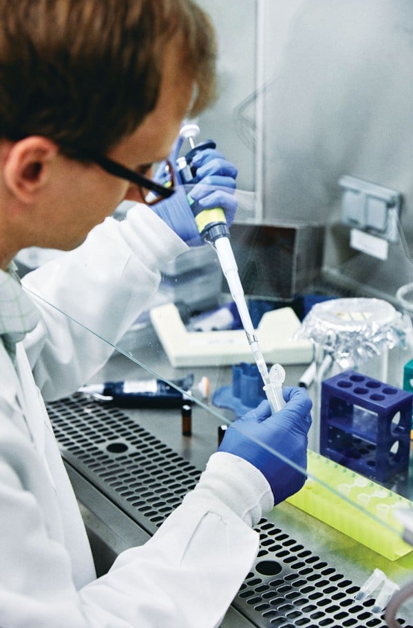
Person pipets in lab hood
Daniel Schmidt, a postdoc in Vander Heiden’s lab, prepares cells to study how metabolism affects cancer cell proliferation. Credit: BUCK SQUIBB
Daniel Schmidt, a postdoc in Vander Heiden’s lab, prepares cells to study how metabolism affects cancer cell proliferation.

BUCK SQUIBB

For Vander Heiden, this was a “watershed moment.” Among other things, it meant that metabolic enzymes weren’t merely supplying energy from food. Metabolism was governing the most fundamental decision a cell has to make—whether to live or die. That meant it had to be interwoven into the signaling cascades that molecular biologists studied. His feeling at the time, he recalls, was “Oh my goodness. We don’t really understand metabolism.”

Vander Heiden might not have envisioned himself delving into research areas that had been discarded decades earlier, but what was more surprising was how little research was then being done in an area that was “as fundamental as you get in terms of how biology works,” he says. “I looked around and no one was studying it.”

Thompson, recognizing the opportunity, shifted the focus of his lab to metabolism. Vander Heiden, meanwhile, continued to pursue Thompson’s broader question of how the body eliminates unwanted immune cells. He already knew that growth factors, messages sent from one cell to the next, kept cells from committing suicide, but how the signals delivered their survival message remained unclear. What he discovered in a series of studies carried out in the late ’90s followed perfectly from his previous research. Growth factors kept cells alive by giving them permission to eat. Without that permission, a cell soon faced an energy crisis, and the mitochondria released their death signals.

The takeaway was clear: our bodies eliminate unwanted cells by starving them to death.

Solving the metabolism mystery

As Vander Heiden’s MD-PhD program was coming to an end, he hadn’t yet begun to focus on cancer, but its possible links with his research on cell suicide were intriguing. Cancer cells were the other side of the coin—cells that were resistant to suicide, that no longer cared about instructions from other cells. So in 2004, after completing a residency in oncology at Brigham and Women’s Hospital in Boston, he was anxious to investigate cancer metabolism for his postdoc research.

Finding the right lab wasn’t easy. At the time, telling leading researchers he wanted to study how cancer cells consumed glucose was like approaching a high-tech manufacturer and announcing you wanted to study the trucks that brought fuel to the factory. It sounded, Vander Heiden says, “like a really ridiculous thing.”

Vander Heiden eventually found a home in the Harvard lab of Lewis Cantley, who now directs the Meyer Cancer Center at Weil Cornell. His research in Cantley’s lab would help solve one of the central riddles of cancer metabolism: why cancer cells are so ravenous for glucose. Researchers had once assumed that cancer cells were turning to fermentation because they’d lost the ability to use oxygen properly and needed some other way to produce energy. But Vander Heiden’s research on a mutated form of the enzyme pyruvate kinase showed something else. Rather than being used for energy, much of the glucose was being shunted into pathways used to build new molecules. What a growing cancer needs most of all from its food, the research suggested, is more spare parts—raw materials for making new DNA, membranes, and proteins.

Rethinking chemotherapy

Vander Heiden’s research with Cantley would also lead to his involvement with Agios Pharmaceuticals, the company behind one of the most promising new drugs to emerge from the metabolism revival. (Cantley says he played a major role in building the company’s science in its early days.) The drug, AG-221, treats acute myelogenous leukemia, a cancer of the blood and bone marrow. It works by blocking the product of a mutated form of the mitochondrial enzyme IDH-2. Approved by the US Food and Drug Administration in August, it has been hailed as the first real advance for the disease in 30 years.

The approval of AG-221 isn’t the only thing generating excitement in the cancer world. Unlike almost all other cancer drugs, AG-221 doesn’t kill the cancer cells but, rather, allows them to develop out of their deranged state into noncancerous, mature, functioning cells. That a single metabolic enzyme could have such profound effects on which genes are expressed in a cell is now one of the many signs that changes in metabolism are not just a response to the needs of a growing cancer. Often, they may actually be causing the cancer itself. It represents a major shift in thought: many cancer-causing genes long known for their ability to signal cells to keep dividing have now been shown to have additional roles in signaling cells to keep eating. Some researchers now believe the overeating typically comes first, driving the transformations that follow.

Since his arrival at MIT and the opening of his lab at the Koch Institute in 2009, Vander Heiden has treated cancer patients and continued to search for better therapies. In recent years he has focused on improving understanding of chemotherapy. Though typically thought of as general poisons, most chemotherapy drugs work because they disrupt metabolic functions. That much has long been known, but less clear is why a particular drug works for some cancers and not for others, even when two cancers carry the same mutations.

It was while explaining to his undergrad cancer biology students how targeted drugs work that Vander Heiden first thought of an answer. As a cancer doctor, he knew that chemotherapies are often chosen on the basis of where in the body a tumor first arose, but what was it about this location that made the difference?

Vander Heiden’s research in mice now suggests that the answer may lie in which foods are available to the cancer as it forms. Melanoma and colon cancer, for example, often have the same mutations, and yet, as he explains, because the two cancers “grow in very different places in the body,” they likely “have access to different nutrients.” He adds, “It has nothing to do with the genetics.” If he turns out to be right, it could lead to a fundamental change in how oncologists think about which drugs to give their patients.

As Vander Heiden turns his attention to old chemotherapy drugs, rethinking why and how they work, he is once again looking to the past for new insights on cancer. It might be more than a coincidence. As Bevis, his wife, says, the outdated Honda Civic isn’t the only item he has struggled to let go of. “The list goes on and on,” she says. “He hates waste and will use items long after someone else would have replaced them with a newer, shinier model.”

Probing a critical player in cancer growth

Alissandra Hillis ’18 has spent all four years at MIT in the same cancer metabolism lab, deciphering the basic science behind pancreatic cancer.

Raleigh McElvery
February 19, 2018

Senior Alissandra Hillis attributes her appetite for the basic sciences to her craving for fundamental knowledge. She’s spent her four years at MIT in the same lab, committed to unraveling the molecular mechanics of pancreatic cancer — the fourth leading cause of cancer death for both men and women, given that symptoms do not often appear until the disease is quite advanced.

“I was always very curious growing up,” she says. “I taught myself how to read at a very young age, just because I wanted to know about things and how they worked. But I didn’t become interested in biology and chemistry specifically until I came to MIT and started taking my General Institute Requirements.”

In doing so, Hillis became enthralled by the prospect of breaking down life into its most fundamental, biological units to decipher cellular function and disease. Originally a Course 7 major with a chemistry minor, she declared Course 5-7 (Chemistry and Biology) as soon as it became available in the fall of 2017 — applying her study of biochemistry and cell metabolism to cancer research.

“When I was quite young, my grandfather was diagnosed with stomach cancer, and ended up having almost three quarters of his stomach removed,” she says. “I was too little to really understand the severity of the situation, but as soon as I came to MIT I started to wonder what was going on at a cellular level. Most people today know someone who is fighting cancer, and yet we’re still lacking effective treatments for its most severe forms.”

Hillis joined Matthew Vander Heiden’s cancer metabolism lab the first semester of her freshman year, and has been there ever since.

Professor Vander Heiden does an excellent job of tailoring the research project to the individual, and there is no hierarchy among lab members,” she says. “I really liked it from the onset, so I stayed.”

For nearly two years, Hillis has been investigating the role of one enzyme, pyruvate kinase muscle isozyme M2 (PKM2), in pancreatic cancer. PKM2 is responsible for catalyzing the final step in glycolysis, which is required to create the energy that fuels cells. Glycolysis is also important in tumor metastasis and growth, since cancer cells demand energy in order to proliferate.

Cancer cells often preferentially express PKM2 over other types of pyruvate kinases such as PKM1. This spurred William Israelsen PhD ’14, a former graduate student in the Vander Heiden lab working in breast cancer models, to delete the PKM2 gene and see what happened. Since PKM2 is critical for glycolysis, and cancer cells require energy to proliferate, he anticipated that removing PKM2 would hinder energy production and thus disrupt tumor development. To his surprise, he found the opposite: deleting PKM2 actually accelerated tumor formation and promoted liver metastasis in mice.

In his 2014 paper, Israelsen concluded that PKM2 might permit cancer cells to maintain their “plasticity,” shifting from one specialized role to another even after they’ve fully matured. In the absence of PKM2, he proposed PKM1 might take over PKM2’s influential role.

Hillis wondered if she could replicate Israelsen’s breast cancer results in a model for pancreatic cancer, especially given the conflicting findings in human data regarding PKM2 expression in the latter. Some studies suggest that high PKM2 expression correlates with accelerated disease, whiles others indicate just the opposite: that high PKM2 expression is associated with better survival rates.

“Going into the project, we were expecting similar effects in both pancreatic and breast cancer models because both cancers preferentially express PKM2, and we were using the same method of PKM2 deletion, just bred into a different cancer model,” Hillis explains. “We anticipated that PKM2 deletion would accelerate pancreatic tumor size and tumor genesis, and decrease the mouse’s lifespan. But we’ve noticed that these effects — if they exist — are very much attenuated in the pancreatic cancer model; there is only a slight decrease in lifespan and increase in tumor size without PKM2.”

Right now, her working hypothesis holds that PKM2’s influence varies depending on the tissue in question. This might explain why her own results don’t exactly parallel what Israelsen found in his breast cancer model. For instance, the method they were using to delete PKM2 is quite effective in the breast and pancreatic cells themselves, but less so in the dense scar-like tissue characteristic of pancreatic tumors in particular. It’s possible, she thinks, that this fibrous tissue may still express some PKM2 even post-knockout, perhaps hindering both a drastic decrease in lifespan and increase in tumor size.

Hillis hopes piecing together PKM2’s mechanism of action will help us better diagnose — and eventually treat — certain cancers. Her most recent results were published in the November 2017 issue Cancer & Metabolism.

Although Hillis enjoys tackling the more fundamental questions concerning cancer, she’s also interested in translating this work from bench to bedside. That’s why she decided to intern with David Ting at the Massachusetts General Hospital Cancer Center this past summer.

“I wanted to try a different type of research before applying to graduate school,” she says. “The Department of Biology frequently sends out emails about job opportunities, and there was one advertising that the Ting lab was looking for a research technician.”

Although she was still a junior at the time, she contacted Ting — an MIT alumnus with a dual degree in 7A and 10 — and together they fashioned a summer position just for her, studying the role of miniscule, fluid-filled transportation structures called exosomes in cancer development and diagnosis.

“That was the first time I’d worked with samples from actual patients,” she says. “Many of the assays were the same, but I felt closer to a clinical application than I ever had before. I really enjoy doing the foundational work to identify the basic problem, but there’s definitely something to be said for experiencing research targeted at creating a diagnostic tool. I can see the pros and cons of both approaches.”

As Hillis begins her final semester at MIT, she’s continuing her work in the Vander Heiden lab, while also finishing up the requirements for her HASS concentration in legal studies. She’s still set on pursuing a PhD in cancer biology, but the propensity to ask tough questions that drew her to science in the first place has led her to realize that the questions she raises in her own research have ramifications far beyond her lab bench. Taking policy-oriented classes in addition to her science-related ones has inspired her to pursue a law degree in conjunction with her PhD — weaving together her love for science with a newfound interest in the rules and regulations that govern how science is funded, performed, shared, applied, and monetized.

“I really enjoy doing research, and that’s something I probably will continue to do,” she says, “but I also want to influence science-related regulations, which is something I couldn’t possibly do without a law degree. I would still be heavily immersed in science, while applying the subjects I love in new and exciting ways.”

Photo credit: Raleigh McElvery