From chemical reactions occurring in a splinter of a second, to evolution shaping species over billions of years, the processes that make up our biology occur in time frames both short and long. In this video, we will hear from Department of Biology Faculty and Whitehead Institute scientists on how their thinking about different time scales informs scientific discovery.
Produced by Madeleine Turner | Whitehead Institute
September 17, 2024
Hear from the Cheeseman Lab, Weissman Lab, Hravatin Lab, and Yamashita Lab at the Whitehead Institute in the following video.
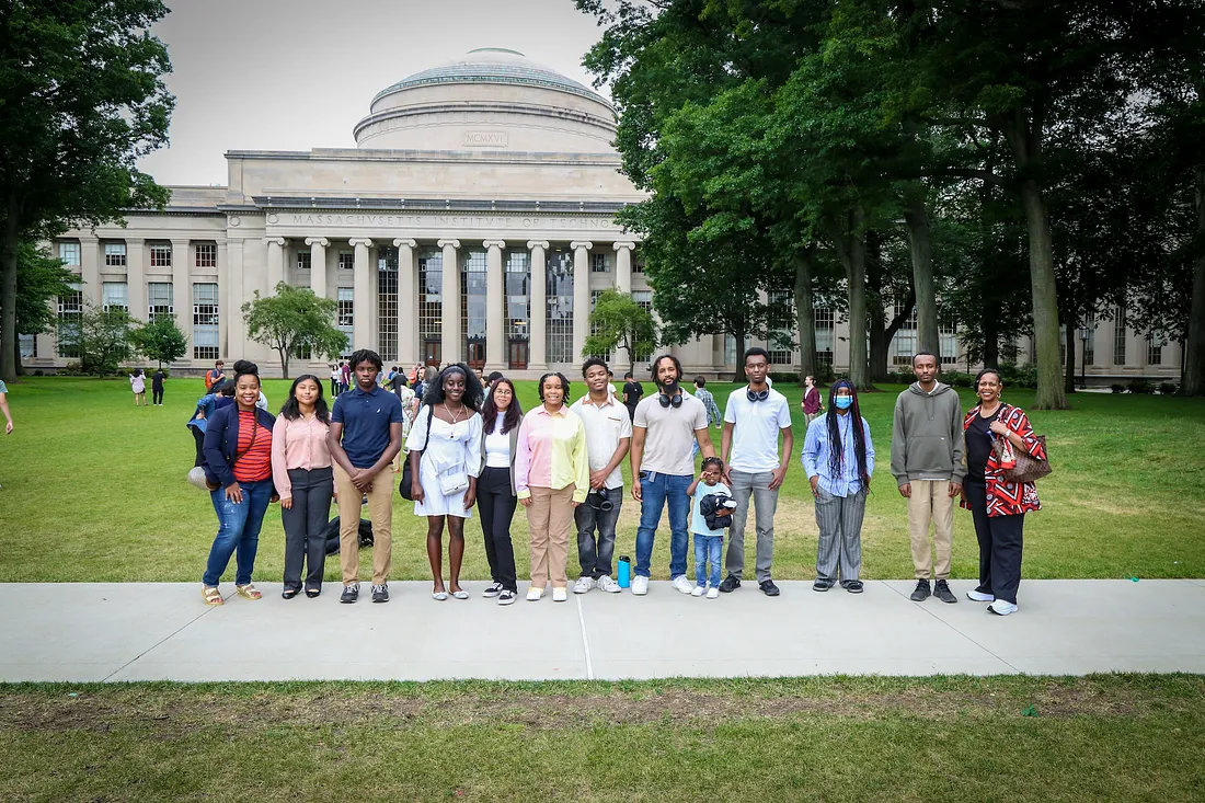
MIT Digital Learning Lab’s high school interns gain professional experience working on the backend of open online MITx courses. The program emerged after Mary Ellen Wiltrout, PhD '09, digital learning scientist at MIT Open Learning, connected with the executive director and founder of Empowr, a nonprofit that serves low-income communities by creating a school-to-career pipeline through software development skills.
Katherine Ouellette | MIT Open Learning
August 26, 2024
Switching programming languages is not as simple as switching word processors. Yet high schooler Thomas Esayas quickly adapted from Swift to Python during his 2023 internship with the MIT Digital Learning Lab, a joint program between MIT Open Learning and the Institute’s academic departments. One year later, Esayas returns to the Institute for a second internship and as a new undergraduate student.
“I felt thoroughly challenged and learned a lot of new skills,” says Esayas.
Through this remote opportunity, interns gain real-world coding experience and practice professional skills by collaborating on MIT’s open online courses. The four interns from Digital Learning Lab’s 2023 and 2024 cohorts also participate in Empowr, a four-year program for low-income high school students that teaches in-demand software development skills and helps them secure paid internships.
The Digital Learning Lab program emerged after Mary Ellen Wiltrout PhD ’09, digital learning scientist at MIT Open Learning, connected with Adrian Devezin, executive director and founder of Empowr, at a conference about making education more accessible and equitable.
“It was affirming to have someone else see what Empowr is trying to do,” says Devezin about the organization’s goal to strengthen the school-to-career pipeline. “Being able to collaborate was beautiful for me, and more importantly, to the students.”
Building technical skills and self-confidence
The Digital Learning Lab internship empowers students to build confidence in their technical abilities, career skills, and the college application process. Interns assist the lab’s digital learning scientists with their work developing and maintaining online MITx courses at Open Learning across multiple academic areas.
“I found myself always busy with something interesting to work on,” says Esayas.
The interactive open education resources that Esayas produced last summer are now being used in live courses. He also helped find and fix bugs on the platform that hosts the MITx courses.
The internship’s flexible design allows projects to be adapted based on the student’s personal progress and interests.
“The students became co-creators of their educational experiences,” says Wiltrout, noting this is beneficial from a pedagogical standpoint.
Devezin adds, “I definitely saw a big improvement in their problem-solving abilities. Having to switch their mindset to a new language, work in new frameworks, and work on teams solving real problems enhanced their ability to adapt to new situations.”
The students’ also strengthened their professional repertoire in areas such as collaboration, communication, and project management. The 2023 cohort, Devezin says, developed the initiative to help other students and take on leadership roles.
Now that Esayas has completed his 2024 internship, he says, “I’m glad that I got to collaborate with more people and work on more projects. Overall, I’m very happy I was able to return.”
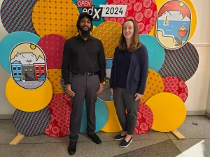
Adrian Devezin, executive director and founder of Empowr (left), and Mary Ellen Wiltrout, digital learning scientist at MIT Open Learning (right), presented their takeaways from the first year of the MIT Digital Learning Lab internship at the 2024 Open edX conference. Photo courtesy of Empowr.
Learning from both sides
Learning occurred for both students and educators alike. Wiltrout says that the Digital Learning Lab values the opportunity to see the interns’ growth day-to-day and week-to-week, since digital learning scientists rarely follow the trajectory of individual learners who are using the course materials they create. Having instant feedback informs how they can adjust their teaching approaches for various problems.
The positive impact of the Digital Learning Lab internship’s hands-on learning experiences has made Devezin rethink the way he teaches class moving forward, and “the problems I want them to be solving,” he says.
Now, Devezin tries to emulate the real-world experience of working on a project for his Empowr students. Instead of assigning coding exercises where he provides the exact methods to solve the problems, he started asking students to determine the correct approach on their own.
The fact that Wiltrout and Devezin are open to adapting their teaching methods based on student feedback is indicative of a key factor to the internship’s success — active participation in students’ growth. It was mutually beneficial for the students and the educators to have determined stakeholders at both Digital Learning Lab and Empowr.
“A lot of dedicated educators understand that there’s a lot of inequities in education, and we need to come together to solve them,” Devezin says.
The Digital Learning Lab internship shows how open source learning materials can make educational and professional opportunities more accessible. The 2024 cohort has been able to increase their annual household income by an average of 75%, a recent Empowr report revealed. Wiltrout says that the two new Empowr students seem more confident with coding and showed enthusiasm and dedication to their tasks as they also consider colleges.
Wiltrout and Devezin presented their takeaways from the internship’s first year at the 2024 Open edX conference.
“I think it’s important to try making sure that more people are aware of tools and resources that are out there,” Wiltrout says. “Then giving people opportunities where they may not have otherwise had that chance.”
Now, Devezin is thinking about how Empowr students can come full circle with their relationship to open educational materials. He’s asking, “How can I help my students contribute to the open source world to give back to others?”
Nine shifts in pedagogical and learning approaches since the global pandemic.
Yvonne Ng | MIT Open Learning
August 7, 2024
The Covid-19 pandemic created radical shifts in approaches to teaching and learning. And while the social, emotional, and mental toll of the pandemic has diminished greatly over the last four years, residual challenges still remain for students and educators. Mary Ellen Wiltrout PhD ’09, director of online and blended learning initiatives, lecturer, and digital learning scientist in Biology at MIT, has identified these shifts in her article, “How to build the future of teaching and learning while growing from the changes and challenges of 2020–21.”
In her article, published in 2022 in Advances in Online Education: A Peer-Reviewed Journal, Wiltrout hypothesized on the lasting impacts of the 2020–2021 events on teaching and learning organized across seven themes: course logistics, tools, activities and assessment for learning, student services and programs, work culture, attitudes, and relationships. Now in 2024 at MIT, Wiltrout can see the positive changes continuing and progressing in these areas:
Flexibility: During the pandemic, instructors were more flexible about coursework requirements, scheduling, grading structure, and expanded the number and types of assignments beyond summative exams. Some enacted policies enabling partial flexibility such as dropping the lowest score on assignments or allowing for late submissions. Now, with student support services approval, instructors remain open to working with students in need of flexibility.
Online learning: Residential colleges relying on completely in-person education now incorporate more blended learning and online courses for students interested in that option. Hybrid instruction and online assignments continue to be part of the curriculum.
Technology: The most valuable functions of the learning management system are the organization of course events and materials and the integration of the multitude of learning tools in one place with one login (for example, web conferencing, discussion forum, grading, video, and calendar). As a result of reducing barriers, more tools like online conferencing, polling, and tablet drawing software to teach, are being used by a larger percentage of teaching staff and students.
Reducing unconscious bias: Grading exams and assignments through an online tool increased the efficiency and consistency of grading with rubrics for every question. And the ability to anonymize submissions in the grading process helps reduce unconscious biases, while students also gain transparency from the rubrics to learn from mistakes and trust the process.
Rethinking in-person sessions: More conversations emerged on how to take advantage of in-person interactions to prioritize activities of value in that mode for learning and work. Instructors and students intentionally kept online approaches that enriched the experience as students returned to campuses. Some digital components enhance student learning, mental well-being, equity, or inclusion and could be as easy as providing a course chat channel for peer-to-peer and peer-to-staff conversations during synchronous sessions. Some instructors maintained more creative, open-ended assignments and online exam policies that seemed experimental during 2020.
Demand for student support services: The effects of the pandemic combined with normalizing taking care of mental health resulted in the sustained high demand for student support services. Institutions continue to invest more in the staffing of these services and programs, such as peer mentoring programs that result in positive academic and attitudinal gains for students. Instructors are generally more aware of how to positively influence students to seek help with simple actions, like speaking in a warm tone and intentionally including a statement about student services.
Belonging and inclusion: Racial and social injustices are being addressed more openly than ever before. Many institutions are recognizing the value and importance of diversity, equity, and inclusion in their students and staff and have invested in funding and training for their community to shift their culture in a positive way. At the course level, instructors have the training and resources available to learn how to become more inclusive teachers (through free resources such as massive open online courses or internal efforts) and have the student and institution pressure to do so.
Mentoring: With the help of online tools and technology, students, educators, and staff are able to foster and create meaningful internship programs. Mentoring in these online programs with students in disparate locations around the world continues to take place and have a positive impact for students and any research that may be part of a program.
Collaborations: Although possible before, more researchers see collaborations across states or countries as less of a hurdle, especially with the everyday use of tools like Zoom. Instructors enhance authentic experiences for students by bringing outside experts into the classroom virtually for discussion — a method that was not used often before the pandemic.
Wiltrout concludes that many opportunities for widespread maintenance of practices that worked well and benefited students during the pandemic can and should continue to persist and grow into the future. Instructors also expanded and improved their curricula and pedagogical approaches to nurture a more inclusive and engaging course for their students and themselves.
“The lasting impacts of the pandemic include profound lessons on what best served both learners and educators,” Wiltrout says. “It’s heartening to see changes and adjustments to pedagogy, student services and programs, attitudes, and relationships that continue to benefit everyone. If these new effective ways endure and grow, then a better future of education for students, staff, and instructors is possible.”
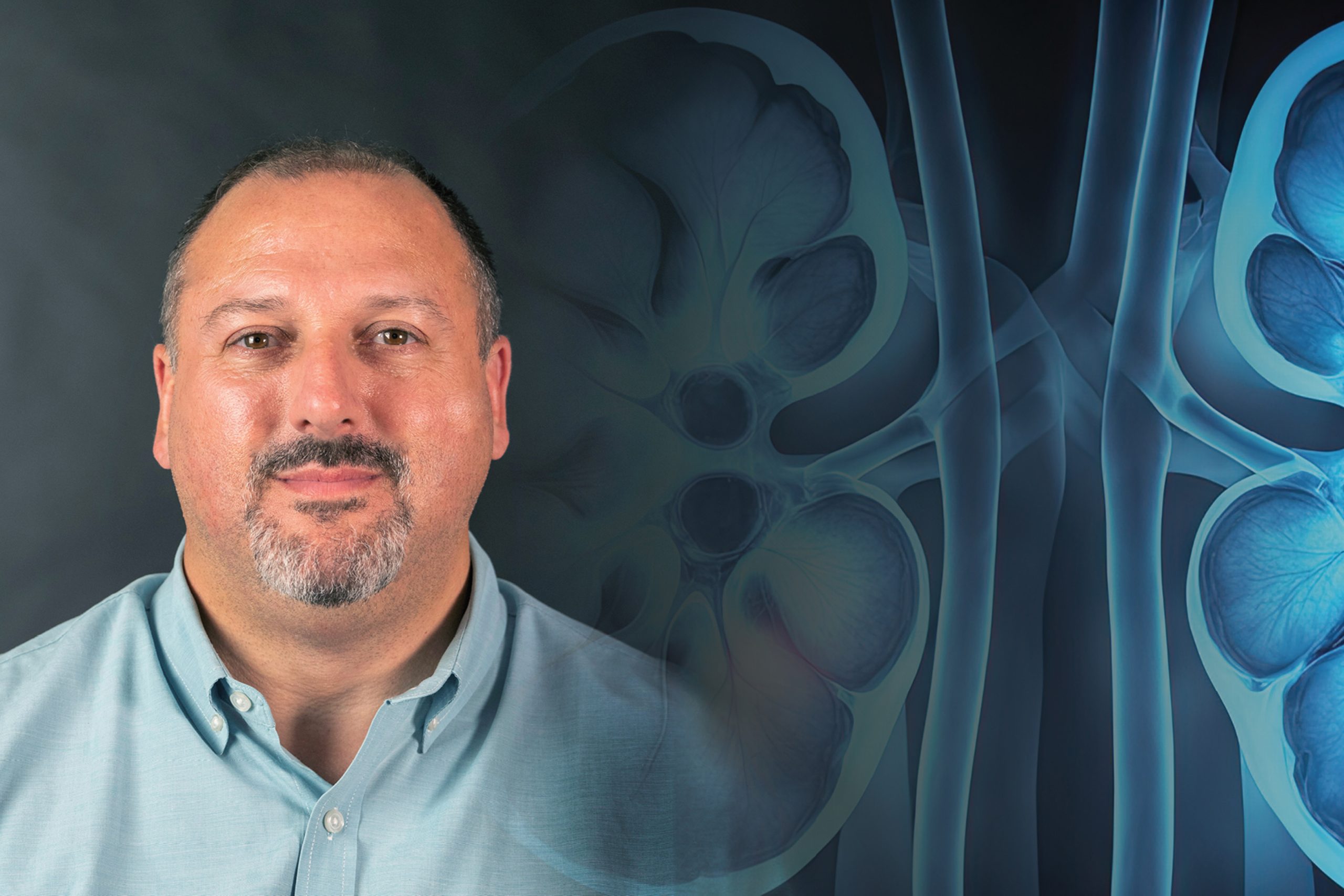
When Reynold I. Lopez-Soler, SB ’94, saw his first kidney transplant, during his medical residency, he found his life’s work.
Kathryn M. O'Neill | MIT Technology Review
September 6, 2024
When Reynold I. Lopez-Soler ’94 saw his first kidney transplant, during his medical residency, he found his life’s work.
“It’s such a magical and incredible thing that you can do this,” says Lopez-Soler, director of the renal transplant program at the Edward Hines Jr. Veterans Affairs Hospital outside Chicago. “You’re watching this organ that was taken out [of the donor], practically lifeless and inert, and through the expertise of surgery it comes to life and becomes pink; it starts to make urine.”
About 100,000 people in the United States are currently waiting for a kidney transplant; on average, they will wait five to seven years. Lopez-Soler is expanding access to this care for veterans.
Kidney transplants are life-changing, he says, not only because kidney disease can make people very sick, but because the main treatment—dialysis, which does some of the kidney’s job outside the body—is so demanding that many patients can’t work or even travel. “Getting a kidney transplant not only fixes the problem, but fixes their lives going forward,” he says. “There is this substantial transformation.”
Growing up in Puerto Rico, Lopez-Soler always expected to become a surgeon (his father is a surgical oncologist). During high school, he discovered the MIT Introduction to Technology, Engineering, and Science (MITES) program, spent a summer on campus, and fell in love with the Institute. “MIT was an incredibly inclusive place,” he says. “Whatever you did, you were welcome. I’ve brought that acceptance with me in my ethos in how I deal with people.”
After majoring in biology at MIT (with a minor in Spanish literature), Lopez-Soler earned his MD and PhD from Northwestern University and completed his surgical residency at Yale New Haven Hospital. Then he practiced in Virginia and New York, where he was director of research at Albany Medical Center.
In 2019, Lopez-Soler was tapped to establish the VA transplant program at Hines, and in its first year, it completed 36 kidney transplants. Last year, the center did 105. He now chairs the Department of Veterans Affairs Transplant Surgery Surgical Advisory Board, which helps develop transplant policies and procedures for the whole VA system.
The grandson of a brigadier general, Lopez-Soler is proud to serve veterans. “I was lucky enough to fall in love with the job because of the people we treat,” he says. “It exposed me to these amazing veterans who have done so much for this country.”
This story also appears in the September/October issue of MIT Alumni News magazine, published by MIT Technology Review.
Photo illustration by Mary Zyskowski; image of Reynold I. Lopez-Soler courtesy of Lopez-Soler.
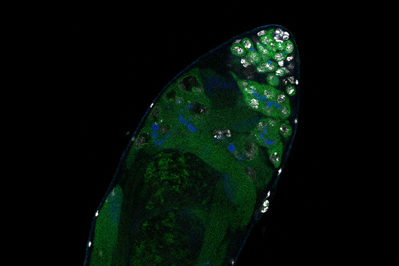
From the intricacies of plant reproduction to genome-wide analyses, Gehring’s lab delves deep into the epigenetic mechanisms shaping plant biology.
Jayashabari Shankar and Alex Tang | The Tech
September 5, 2024
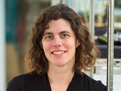
Dr. Mary Gehring is a professor of biology at MIT and a core member of the Whitehead Institute for Biomedical Research. Her research focuses on how epigenetic mechanisms like DNA methylation influence gene regulation during plant reproduction and seed development in the model organism Arabidopsis thaliana. In the classroom, she teaches genetics (7.03), a required course for biology and biological engineering majors.
With her recent appointment as an Howard Hughes Medical Institute (HHMI) investigator, Gehring joins an elite legion of HHMI investigators at the Institute. New cohorts of investigators are only announced once every three years, and they receive $11 million in funding over a seven year term (which can be renewed). Three other MIT faculty received HHMI appointments this year: Gene-Wei Li, associate professor of biology, and brain and cognitive sciences professors Mehrdad Jazayeri and Steven Flavell.
Here, she shares her lab’s research, journey into plant biology, and what she values in undergraduate researchers.
TT: What does your lab conduct research in, and how has being named an HHMI investigator changed your plans, if at all?
My lab focuses on plant biology, particularly on how epigenetic mechanisms like DNA methylation affect gene regulation in plants, especially during reproduction and seed development. We mostly work with Arabidopsis thaliana, a model plant, but we’re also exploring other plant systems.
A typical day in the lab can vary, but it often starts with checking on our plants in the greenhouse. Depending on the day, we might pollinate plants for genetic crosses or genotyping them by isolating DNA and performing PCR. We’re particularly focused on understanding gene expression within seeds: we isolate different seed tissues, sort nuclei based on their properties, and then perform RNA sequencing. We also do a lot of chromatin profiling, histone modifications and DNA methylation analyses across the genome. Since much of our work is genome-wide, bioinformatics plays a big role in our research, with a significant amount of time spent on analyzing data.
It’s still sinking in, but being named an HHMI investigator certainly provides a new level of freedom. It allows us to pursue ideas without the constraints of specific grant funding, which is incredibly liberating. We’re considering expanding our research into new areas beyond epigenetics, like genome structure and chromosome dosage changes, while sticking with plant biology. This recognition has encouraged us to think bigger and explore new directions in our work.
TT: How far back do these interests extend for you?
My interest in plant biology started during my undergraduate years. I majored in biology and was eager to get involved in research. My real fascination with plants began when a new professor, with a background in plant biology, came to my school. I took her course on plant growth and development, which I found incredibly exciting. I was drawn to how plants communicate within their tissues and with each other. This led me to work on a research project for two years, culminating in a senior thesis on root development. After college, I took a year off to work in environmental consulting before heading to graduate school in Plant Biology at UC Berkeley.
TT: What perspectives and characteristics do you appreciate in undergraduate researchers?
Whether it’s undergraduates or postdocs, I value curiosity and dedication. For undergraduates, especially those in UROPs, it’s crucial that they are genuinely interested in the research and willing to ask questions when they don’t understand something. Balancing research with coursework and extracurriculars at MIT is challenging, so I also look for students who can manage their time well. It’s about being curious, dedicated, and communicative.
I hope there are students at MIT who are excited about plant research. It’s a vital area of biology, especially with the growing focus on climate change. While there isn’t a large presence of plant biology at MIT yet, I’m hopeful that it will expand in the coming years, and I’d love to see more students getting involved in this important field.
The Koch Institute at MIT is pleased to announce the winners of the 2024 Angelika Amon Young Scientist Award, Anna Uzonyi and Lukas Teoman Henneberg.
Koch Institute
September 3, 2024
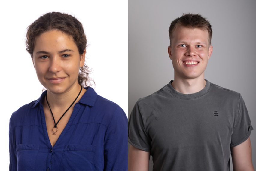
The Koch Institute at MIT is pleased to announce the winners of the 2024 Angelika Amon Young Scientist Award, Anna Uzonyi and Lukas Teoman Henneberg.
The prize was established in 2021 to recognize graduate students in the life sciences or biomedical research from institutions outside the United States who embody Dr. Amon’s infectious enthusiasm for discovery science.
Both of this year’s winners work to unravel the fundamental biology of chromatin, the densely structured complex of DNA, RNA, and proteins that makes up a cell’s genetic material.
Uzonyi is pursuing her PhD at the Weizmann Institute of Science in Israel under the supervision of Schraga Schwartz and Yonatan Stelzer. In her thesis, Uzonyi focuses on deciphering the principles of RNA editing code via large-scale systematic probing.
Henneberg is a doctoral candidatein the Department of Molecular Machines and Signaling, at the Max Planck Institute of Biochemistry in Germany, works under the supervision of Professor Brenda Schulman and Professor Matthias Mann. For his research project, he probes active ubiquitin E3 ligase networks within cells. He works on the development of probes targeting active ubiquitin E3 ligases within cells and utilizing them in mass spectrometry-based workflows to explore the response of these ligase networks to cellular signaling pathways and therapeutics.
This fall, Anna Uzonyi and Lukas Teoman Henneberg, will visit the Koch Institute. The MIT community and Amon Lab alumni are invited to attend their scientific presentations on Thursday, November 14 at 2:00 p.m. in the Luria Auditorium, followed by a 3:30 p.m. reception in the KI Galleries.
Uzonyi will present on “Inosine and m6A: Deciphering the deposition and function of adenosine modifications” and Henneberg will present on “Capturing active cellular destroyers: Probing dynamic ubiquitin E3 ligase networks.“
New research from Jaenisch Lab postdoc Danielle Tomasello focuses on an understudied question: how Rett Syndrome affects cell types in the human brain other than neurons.
Greta Friar | Whitehead Institute
September 6, 2024
Copied to clipboard
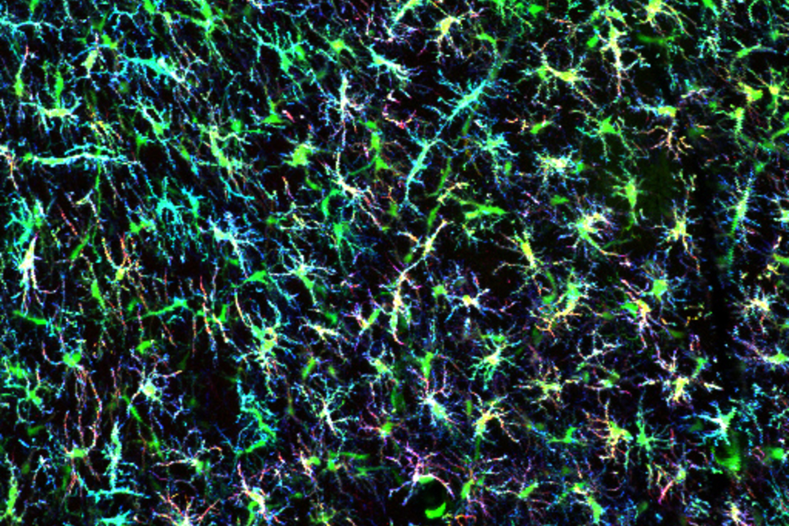
Rett Syndrome is a X-chromosome-linked neurodevelopmental disorder; it can lead to loss of coordination, mobility, ability to speak, and use of the hands, among other symptoms. The syndrome is typically caused by mutations within the gene MECP2. Researchers in Whitehead Institute Founding Member Rudolf Jaenisch’s lab have studied Rett Syndrome for many years in order to understand the biological mechanisms that cause disease symptoms, and to identify possible avenues for treatments or a cure. Jaenisch and colleagues have gained many insights intothe biology of Rett syndrome and developed tools that canrescue neurons from Rett syndrome symptoms in lab models.
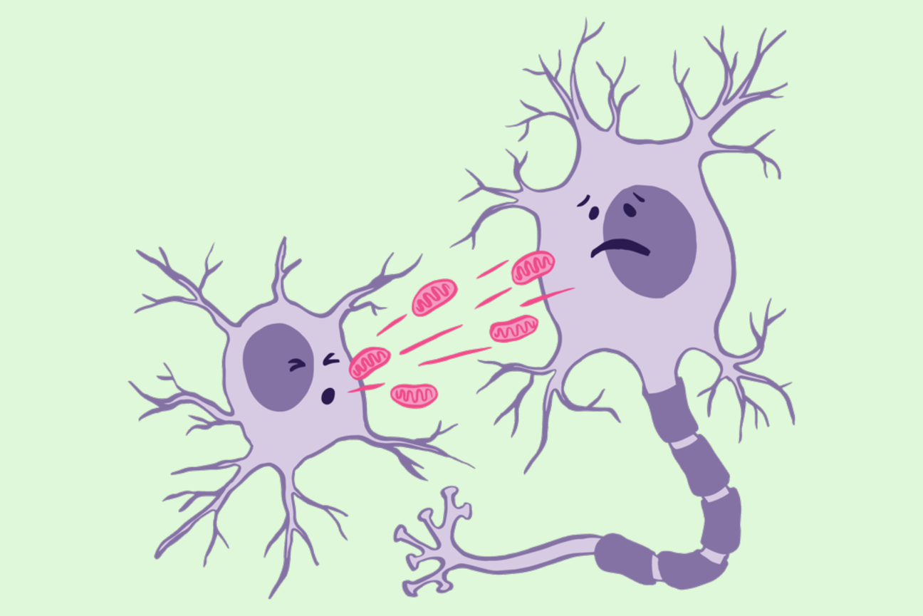
However, much about the biology of Rett Syndrome remains unknown. New research from Jaenisch and postdoc in his lab Danielle Tomasello focuses on an understudied question: how Rett Syndrome affects cell types in the human brain other than neurons. Specifically, Tomasello investigated the effects of Rett Syndrome on astrocytes, a type of brain cell that supports and provides energy for neurons. The work, shared in the journal Scientific Reportson September 6, details changes that occur in Rett syndrome astrocytes, in particular in relation to their mitochondria, and shows how these changes directly impact neurons. The findings provide a new framework for thinking about Rett Syndrome and possible new avenues for therapies.
“By considering Rett Syndrome from a different perspective, this project expands our understanding of a multifaceted and thus far incurable disease,” says Jaenisch, who is also a professor of biology at the Massachusetts Institute of Technology.
Energy metabolism in Rett Syndrome
Mitochondria are organelles that generate energy, which cells use to carry out their functions, and mitochondrial dysfunction was known to occur in Rett Syndrome. Jaenisch and Tomasello found that mitochondria in astrocytes are particularly affected, even more so than mitochondria in neurons. Tomasello grew human stem-cell-derived astrocytes in 2D cultures and also grew 3D organoids: mini brain-like tissues that contain multiple cell types growing in a structure that resembles actual brain anatomy. This approach allowed Tomasello to use human cells, rather than an animal model, and to study how cells behave within a brain-like environment.
When the researchers observed Rett astrocytes grown in these conditions, they found that the mitochondria were misshapen: short, small circles instead of large, long ovals. Additional studies showed evidence of the mitochondria experiencing stress and not being able to generate enough energy through their usual processes. The mitochondria did not have enough of the typical proteins they use to make energy, and so began to break down the cell’s supply of the building blocks of proteins, amino acids, for parts to make up for the missing material. Additionally, the researchers observed an increase in reactive oxygen species, byproducts of mitochondrial metabolism that are toxic to the cell.
Further experiments suggested that the cells try to compensate for this mitochondrial stress by increasing transcription of mitochondrial genes. For example, Tomasello found that regions of DNA called promoters that can increase expression of key mitochondrial genes were more open for the cell to use in Rett astrocytes. Altogether, these findings paint a picture of severe mitochondrial dysfunction in Rett astrocytes.
Although mitochondria in Rett neurons did not have such severe defects, astrocytes and neurons have a close relationship. Not only do neurons rely on astrocytes to supply them with energy, they even accept mitochondria from astrocytes to use for themselves. Jaenisch and Tomasello found that neurons take up dysfunctional mitochondria from Rett astrocytes at a higher rate than they take up mitochondria from unaffected astrocytes. This means that the effects of Rett syndrome on astrocytes have a direct effect on neurons: the dysfunctional mitochondria from the astrocytes end up in the neurons, where they cause damage. Tomasello took mitochondria from Rett astrocytes and placed them on both healthy and Rett neurons. In either case, the neurons took up the dysfunctional mitochondria in large numbers and then experienced significant problems. The neurons entered a hyperexcitable state that is ultimately toxic to the brain. The neurons also contained higher levels of reactive oxygen species, the toxic byproducts of mitochondrial metabolism, which can cause widespread damage. These effects occurred even in otherwise healthy neurons that did not themselves contain a Rett-causing MECP2 mutation.
“This shows that in order to understand Rett Syndrome, we need to look beyond what’s happening in neurons to other cell types,” Tomasello says.
Learning about the role that astrocytes play in Rett Syndrome could provide new avenues for therapies. The researchers found that supplying affected astrocytes with healthy mitochondria helped them to recover normal mitochondrial function. This suggests to Tomasello that one possibility for future Rett Syndrome therapies could be something that either targets mitochondria, or supplies additional mitochondria through the bloodstream.
Together, these insights and their possible medical implications demonstrate the importance of taking a broader look at the foundational biology underlying a disease.
Renee Barbosa, a Schimmel scholar and a graduate student in the Soto-Feliciano Lab, uses a multidisciplinary approach to understand the epigenetic factors in gene expression.
Bendta Schroeder | Koch Institute
July 29, 2024
Professor Emeritus of Biology Paul Schimmel PhD ’67 and his wife Cleo Schimmel are among the biggest champions and supporters of graduate students conducting life science research in the Department of Biology at MIT, as well as in departments such as the Department of Brain and Cognitive Sciences, the Department of Biological Engineering, and the Department of Chemistry, and in cross-disciplinary degree programs including the Computational and Systems Biology Program, the Molecular and Cellular Neuroscience Program, and the Microbiology Graduate Program. In addition to the Cleo and Paul Schimmel (1967) Scholars Fund to support graduate women students in the Department of Biology, in 2021, the Schimmels established the MIT Schimmel Family Program for Life Sciences.
Their generous pledge of $50 million in matching funds called for other donors to join them in supporting the training of graduate students who will tackle some of the world’s most urgent challenges. Driven by their unwavering belief that graduate studentsare the driving force behind much life science research and witnessing a decline in federal funding for graduate education, the Schimmel family established their one-to-one match program. They reached the ambitious goal of $100 million in endowed support in just two years.
The discovery that mutations in genes can drive cancer revolutionized cancer research. In the decades following the identification of the first “oncogene” in a chicken retrovirus in 1970 and the first human oncogene in 1982 by Robert Weinberg at MIT’s Center for Cancer Research, scientists uncovered hundreds more oncogenes, transformed our understanding of how cancer begins and progresses, and developed sophisticated gene-targeted cancer therapies.
A majority of oncogenes were identified in factors controlling cell signaling, proliferation, and differentiation. However, a growing understanding of epigenetics has shown that many cancers, such as some leukemias and sarcomas, are not driven by mutations to these factors themselves, but by disruptions to the molecular pathways that regulate their expression. About 10 percent of all leukemias are driven by abnormal versions of the protein MLL1, one cog in the epigenetic machinery controlling these factors.
Renee Barbosa, a graduate student in the laboratory of Howard S. (1953) and Linda B. Stern Career Development Professor Yadira Soto-Feliciano in the Department of Biology, is joining this next wave of research, using leukemia as a model. A member of MIT’s Koch Institute for Integrative Cancer Research, Soto-Feliciano and her lab study chromatin, the densely coiled structures of DNA and scaffolding proteins that make up our genomes and help ensure genes are expressed at the right times and in the right amounts.
Barbosa focuses on the role of RNA processing and the precisely choreographed alterations to chromatin that govern gene expression. RNA molecules serve as messengers between DNA and its final product, protein, and are subject to extensive processing and regulation. However, not much is known about the interplay between RNA processing and epigenetic machinery, particularly in cancer.
“I hope that my work will uncover additional layers of complexity in the dynamic landscape of gene regulation,” says Barbosa. “It might also identify new mechanisms that can be targeted to help treat leukemia and other cancers.”
Before Barbosa arrived at the Soto-Feliciano Lab, she was already steeped in the molecular intricacies of cancer.
While at the University of Pennsylvania, she earned a BA in biochemistry and biophysics concurrently with a master’s degree in chemistry. Early on, she joined the lab of Ronen Marmorstein, which used molecular approaches to characterize MEK and ERK, two cancer-relevant members of a class of signaling proteins. Upon starting graduate school, she was excited to branch out into other disciplines.
Barbosa has always taken every opportunity she can to learn. Beginning in grade school, science and math were her favorite subjects, but she also explored music, dance, and foreign languages. At the University of Pennsylvania, she even squeezed in a minor in neuroscience.
With its interdisciplinary approach, the Soto-Feliciano Lab provides Barbosa ample opportunities to learn. Because epigenetic factors can elude traditional approaches, the Soto-Feliciano Lab uses a multidisciplinary strategy, ranging from molecular, to large-scale omics analyses, to disease modeling.
“When I was a grad student, we saw the arrival of powerful new massive sequencing and gene editing technologies — and were enabled to ask big new questions,” says Soto- Feliciano. “I am excited that Renee will have even more resources and opportunities, as we enter the next stage of cancer genetics and epigenetics.”
With the support of a Schimmel Fellowship, Barbosa will be ready to take advantage of new developments in her field.
“Support for research early on in graduate school is an incredible opportunity,” says Barbosa. “It means time to delve deep into the literature of the field and identify challenging open questions that I can pursue in my project. Though exploring these unknown areas requires taking bigger risks, I hope that we will get invaluable insight from an understanding of these nuanced and complex mechanisms.”
Graduate student and Schimmel Scholar Annette Jun Diao uses a minimal system to parse the mechanisms underlying gene expression
Lillian Eden | Department of Biology
July 29, 2024
Professor Emeritus of Biology Paul Schimmel PhD ’67 and his wife Cleo Schimmel are among the biggest champions and supporters of graduate students conducting life science research in the Department of Biology at MIT, as well as in departments such as the Department of Brain and Cognitive Sciences, the Department of Biological Engineering, and the Department of Chemistry, and in cross-disciplinary degree programs including the Computational and Systems Biology Program, the Molecular and Cellular Neuroscience Program, and the Microbiology Graduate Program. In addition to the Cleo and Paul Schimmel (1967) Scholars Fund to support graduate women students in the Department of Biology, in 2021, the Schimmels established the MIT Schimmel Family Program for Life Sciences.
Their generous pledge of $50 million in matching funds called for other donors to join them in supporting the training of graduate students who will tackle some of the world’s most urgent challenges. Driven by their unwavering belief that graduate studentsare the driving force behind much life science research and witnessing a decline in federal funding for graduate education, the Schimmel family established their one-to-one match program. They reached the ambitious goal of $100 million in endowed support in just two years.
Annette Jun Diao’s mother loves to tell the story of Diao’s childhood aversion to the study of life — the gross and the squishy. Unlike some future biologists, Diao wasn’t the type to stomp through creeks or investigate the life of frogs. Instead, she was interested in astronomy and only ended up in a high school biology class because of a bureaucratic snafu. The physics course she’d been hoping to take was canceled due to low enrollment, and she was informed molecular biology was being offered instead.
She attended the University of Toronto and joined the molecular genetics department because of the numerous opportunities for hands-on research. She’s now a third-year graduate student in the Department of Biology at MIT.
“I’m fascinated by the mechanisms that underlie the regulation of gene expression,” Diao says. “All of our genetic information is in DNA, and that DNA is an actual molecule with chemical properties that allow it to be passed from one generation to the next.”
Every cell in our bodies contains a genome of approximately 20,000 genes, but the cells in our retinas are vastly different than the cells in our hearts — not all genes are in action simultaneously, and cell fates vary depending on how which genes are active.
“What is really awesome about the department — and what was attractive to me when I was applying to graduate school — is that I wasn’t sure exactly what methods I wanted to use to answer the questions I was interested in,” Diao says. “A huge advantage of the program was that I had a lot to choose from.”
Diao chose to pursue her thesis work with Seychelle Vos, the Robert A. Swanson (1969) Career Development Professor of Life Sciences and HHMI Freeman Hrabowski Scholar. Diao has been recognized with a Natural Sciences and Engineering Research Council of Canada Fellowship, which is similar to a National Science Foundation graduate fellowship in the United States.
Vos’s lab is generally interested in understanding how transcription is regulated, the interplay of genome organization and gene expression, and the molecular machinery involved. Diao has been working with an enzyme called RNA polymerase II (RNAP II), the molecular machine that reads DNA and creates an RNA copy called mRNA. That mRNA goes on to be read by ribosomes to create proteins.
Many questions remain about RNAP II, including what signals instruct it to begin transcription and, once engaged, whether it will transcribe and how quickly it moves.
RNAP II doesn’t work alone. Diao is working to understand how a transcription factor called negative elongation factor associates with RNAP II and whether the DNA sequence affects that interaction.
Within the broader context of the genome, DNA is packaged extremely tightly; if it were allowed to unfold, its total length could stretch from Cambridge to Connecticut. What RNAP II has access to at any given time is therefore quite restricted, which Diao is also exploring.
She has been exploring this topic in what she refers to as a “reductionist approach.” By creating a minimal system — a strand of DNA and the precise addition of certain other isolated components — she can potentially parse out what ingredients and what sequence of events are essential “in order to really get to the nitty-gritty of how genes are regulated.”
Outside of her work in the lab, Diao is part of BioREFS, a peer support group for graduate students, and gwiBio. Both organizations bring members of the department together for scientific talks and socializing activities outside of the lab, and gwiBio also participates in community outreach.
Diao is also a Schimmel Scholar, supported by Professor Emeritus of Biology Paul Schimmel PhD ’67 and his wife Cleo Schimmel.
“It was really great to learn that I was being supported by a scientist who has done a lot of awesome work that’s relevant to my world,” Diao says.
“It is awesome that they are so committed to supporting the graduate program at MIT, especially when federal resources have become more limited,” Vos says. “With their support, our lab can train basic scientists who can then use their knowledge to transform our study of disease. I hope others follow Paul and Cleo’s example.”
In Parkinson's disease, a mutation that causes protein misfolding can also turn the brain’s immune cells from friends to foes, possibly accelerating the progression of the disease. New Research from the Jaenisch Lab aims to uncover mechanisms that go awry in the brain, which may inform the development of new therapies that can halt or even reverse the progression of neurological conditions such as Parkinson's.
Shafaq Zia | Whitehead Institute
August 29, 2024
Copied to clipboard
Dopamine is more than the “rush molecule”. This chemical messenger, produced by neurons in the midbrain, acts as a traffic controller that regulates the flow of electrical signals between neurons, assisting with brain functions like cognition, attention, movement, and behavior. But, in instances of Parkinson’s disease (PD), a progressive brain disorder, dopamine-producing neurons begin to die at an unprecedented rate, leading to dwindling levels of this vital chemical and impaired neural communication.
The lab of Whitehead Institute’s Founding Member Rudolf Jaenisch studies genetic and epigenetic factors — changes in gene expression that control which genes are turned on and off, and to what extent, without altering the DNA sequence itself — underlying neurological disorders like PD, Alzheimer’s disease, and Rett Syndrome. Their work aims to uncover the mechanisms that go awry in the brain, which may inform the development of new therapies that can halt or even reverse the progression of these conditions.
In their latest work, Jaenisch and former postdoctoral associate Marine Krzisch examine how a mutation in the gene that encodes for alpha-synuclein, a protein regulating the release of dopamine, affects the resident immune cells of the brain called microglia. The researchers’ detailed findings, published in the journal Biological Psychiatry on August 29, reveal that the mutation renders microglia extremely sensitive, worsening the problem of inflammation in the brain and potentially exacerbating damage to neurons in Parkinson’s disease.
“In fact, even when these mutant microglia are transplanted into a healthy, young brain, they have heightened activation upon stimulation, and low levels of the protective antioxidant catalase,” Krzisch says. “This tells us that in Familial Parkinson’s disease, which is due to genetic mutations, these microglia may be playing an important role in neuron degeneration.”
When nature’s origami falters
The human body is home to tens of thousands of unique proteins, each essential for processes sustaining life. These proteins are composed of linear chains of smaller building blocks called amino acids that are linked together in a specific sequence. For the proteins to perform their functions, the amino acid chains must crumple, rotate, and twist into stable three-dimensional structures. The stakes are high — just as precise folds and creases are crucial to the art of origami, even minor errors in the protein folding process can result in dysfunctional proteins that contribute to disease.
To date, scientists have identified over 20 causative genes in which mutations can result in Familial Parkinson’s disease, a rare, genetically inherited form of PD affecting individuals under or around the age of 50. Among them is SNCA, which encodes for alpha-synuclein, a small protein abundant in dopamine-producing neurons.
The A53T mutation in SNCA promotes the formation of dysfunctional alpha-synuclein proteins that clump together — almost like a ball of yarn — within dopamine-producing neurons. The accumulation of these protein clumps, also known as Lewy bodies, triggers inflammatory signaling in the brain, eventually killing the affected neurons. However, prior research has also shown that the A53T mutation accelerates the progression of PD, or the rate at which neurons die, although the full molecular mechanisms underlying this process are not yet fully understood.
To uncover pathways involved in this progression, researchers in the Jaenisch Lab turned their attention to star-shaped patrollers called microglia that protect the brain from foreign invaders and respond to injuries, including protein aggregates within neurons. This immune response includes activated microglia trying to clear out Lewy bodies by digesting them, recruiting additional immune cells to the site of neurons with protein aggregates, and even killing off diseased neurons to limit damage to the brain.
But these friends can quickly turn to foes. Over-activated microglia can also degrade healthy neurons in the brain, prompting Jaenisch, Krzisch, and colleagues to investigate if excessive microglia activation is one pathway that contributes to progression in PD.
Microglia go rogue
To explore how the A53T mutation in the SNCA gene affects microglia function in PD, scientists at the Jaenisch Lab began by growing human myeloid precursors — the cells that eventually develop into microglia — in lab culture and transplanting them into the brains of immune-deprived mice.
Given the complexity of the brain, it’s common for researchers to study brain cells in the Petri dish. “But in cell cultures, microglia do not have the same morphology [form] as in the brain, show signs of chronic activation, and they don’t survive for a very long time,” says Krzisch. “When we transplant them in mice, the precursors differentiate into microglia that look and function like those in the human brain, and survive for the mouse’s lifespan.”
Using this method, the researchers compared the gene expression profiles of A53T-mutant microglia with those that did not carry the mutation, revealing differences in pathways linked to inflammation, microglia activation, and DNA repair. Additionally, when A53T-mutant microglia were exposed to an immune activator called lipopolysaccharide, they exhibited a heightened inflammatory response compared to non-mutant microglia.
In fact, even in non-inflammatory conditions, A53T-mutant microglia had decreased expression of catalase, an enzyme that helps break down harmful reactive oxygen species produced in response to protein aggregates in PD.
Understanding the molecular basis of progression in PD is challenging, which explains why there are currently no drugs to alter the disease’s course. With these findings in hand, researchers at the Jaenisch Lab are now eager to explore how factors like aging also influence microglia function and contribute to an increased rate of progression in PD.
“Overactivation of microglia isn’t the only cause of neuron death in Parkinson’s,” says Jaenisch. “But if we can decrease their activation, it will help us get to the point where we can slow down or actually stop the disease.”