Growing to greatness: Professor Mary Gehring on plant epigenetics and becoming an HHMI Investigator

From the intricacies of plant reproduction to genome-wide analyses, Gehring’s lab delves deep into the epigenetic mechanisms shaping plant biology.

Jayashabari Shankar and Alex Tang | The Tech
September 5, 2024

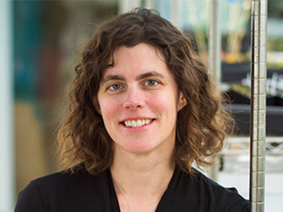
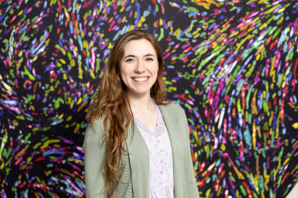
Dr. Mary Gehring is a professor of biology at MIT and a core member of the Whitehead Institute for Biomedical Research. Her research focuses on how epigenetic mechanisms like DNA methylation influence gene regulation during plant reproduction and seed development in the model organism Arabidopsis thaliana. In the classroom, she teaches genetics (7.03), a required course for biology and biological engineering majors.

With her recent appointment as an Howard Hughes Medical Institute (HHMI) investigator, Gehring joins an elite legion of HHMI investigators at the Institute. New cohorts of investigators are only announced once every three years, and they receive $11 million in funding over a seven year term (which can be renewed). Three other MIT faculty received HHMI appointments this year: Gene-Wei Li, associate professor of biology, and brain and cognitive sciences professors Mehrdad Jazayeri and Steven Flavell.

Here, she shares her lab’s research, journey into plant biology, and what she values in undergraduate researchers.

TT: What does your lab conduct research in, and how has being named an HHMI investigator changed your plans, if at all?

My lab focuses on plant biology, particularly on how epigenetic mechanisms like DNA methylation affect gene regulation in plants, especially during reproduction and seed development. We mostly work with Arabidopsis thaliana, a model plant, but we’re also exploring other plant systems.

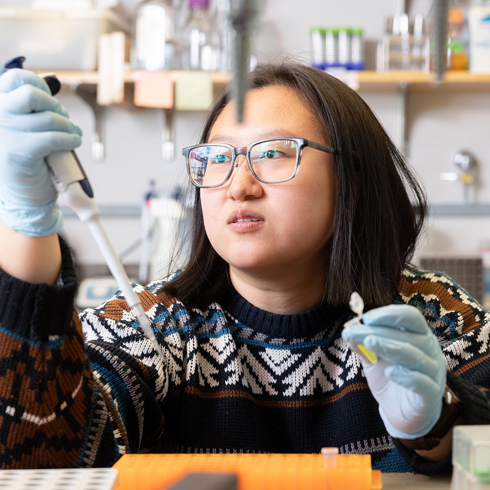
A typical day in the lab can vary, but it often starts with checking on our plants in the greenhouse. Depending on the day, we might pollinate plants for genetic crosses or genotyping them by isolating DNA and performing PCR. We’re particularly focused on understanding gene expression within seeds: we isolate different seed tissues, sort nuclei based on their properties, and then perform RNA sequencing. We also do a lot of chromatin profiling, histone modifications and DNA methylation analyses across the genome. Since much of our work is genome-wide, bioinformatics plays a big role in our research, with a significant amount of time spent on analyzing data.

It’s still sinking in, but being named an HHMI investigator certainly provides a new level of freedom. It allows us to pursue ideas without the constraints of specific grant funding, which is incredibly liberating. We’re considering expanding our research into new areas beyond epigenetics, like genome structure and chromosome dosage changes, while sticking with plant biology. This recognition has encouraged us to think bigger and explore new directions in our work.

TT: How far back do these interests extend for you?

My interest in plant biology started during my undergraduate years. I majored in biology and was eager to get involved in research. My real fascination with plants began when a new professor, with a background in plant biology, came to my school. I took her course on plant growth and development, which I found incredibly exciting. I was drawn to how plants communicate within their tissues and with each other. This led me to work on a research project for two years, culminating in a senior thesis on root development. After college, I took a year off to work in environmental consulting before heading to graduate school in Plant Biology at UC Berkeley.

TT: What perspectives and characteristics do you appreciate in undergraduate researchers?

Whether it’s undergraduates or postdocs, I value curiosity and dedication. For undergraduates, especially those in UROPs, it’s crucial that they are genuinely interested in the research and willing to ask questions when they don’t understand something. Balancing research with coursework and extracurriculars at MIT is challenging, so I also look for students who can manage their time well. It’s about being curious, dedicated, and communicative.

I hope there are students at MIT who are excited about plant research. It’s a vital area of biology, especially with the growing focus on climate change. While there isn’t a large presence of plant biology at MIT yet, I’m hopeful that it will expand in the coming years, and I’d love to see more students getting involved in this important field.

2024 Angelika Amon Young Scientist award winners announced

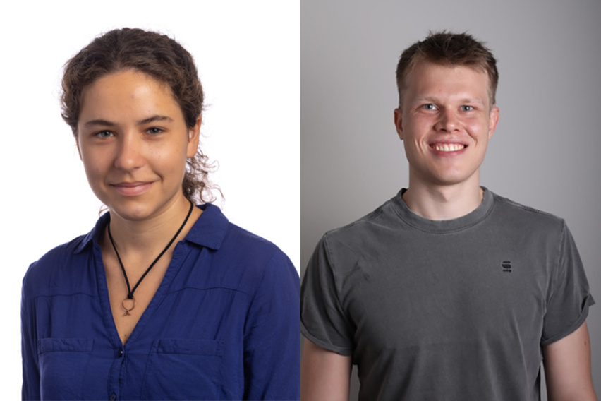
The Koch Institute at MIT is pleased to announce the winners of the 2024 Angelika Amon Young Scientist Award, Anna Uzonyi and Lukas Teoman Henneberg.

Koch Institute
September 3, 2024

The Koch Institute at MIT is pleased to announce the winners of the 2024 Angelika Amon Young Scientist Award, Anna Uzonyi and Lukas Teoman Henneberg.

The prize was established in 2021 to recognize graduate students in the life sciences or biomedical research from institutions outside the United States who embody Dr. Amon’s infectious enthusiasm for discovery science.

Both of this year’s winners work to unravel the fundamental biology of chromatin, the densely structured complex of DNA, RNA, and proteins that makes up a cell’s genetic material.

Uzonyi is pursuing her PhD at the Weizmann Institute of Science in Israel under the supervision of Schraga Schwartz and Yonatan Stelzer. In her thesis, Uzonyi focuses on deciphering the principles of RNA editing code via large-scale systematic probing.

Henneberg is a doctoral candidate in the Department of Molecular Machines and Signaling, at the Max Planck Institute of Biochemistry in Germany, works under the supervision of Professor Brenda Schulman and Professor Matthias Mann. For his research project, he probes active ubiquitin E3 ligase networks within cells. He works on the development of probes targeting active ubiquitin E3 ligases within cells and utilizing them in mass spectrometry-based workflows to explore the response of these ligase networks to cellular signaling pathways and therapeutics.

This fall, Anna Uzonyi and Lukas Teoman Henneberg, will visit the Koch Institute. The MIT community and Amon Lab alumni are invited to attend their scientific presentations on Thursday, November 14 at 2:00 p.m. in the Luria Auditorium, followed by a 3:30 p.m. reception in the KI Galleries.

Uzonyi will present on “Inosine and m6A: Deciphering the deposition and function of adenosine modifications” and Henneberg will present on “Capturing active cellular destroyers: Probing dynamic ubiquitin E3 ligase networks.

Brain cell types are affected differently by Rett Syndrome mutation

New research from Jaenisch Lab postdoc Danielle Tomasello focuses on an understudied question: how Rett Syndrome affects cell types in the human brain other than neurons.

Greta Friar | Whitehead Institute
September 6, 2024

Rett Syndrome is a X-chromosome-linked neurodevelopmental disorder; it can lead to loss of coordination, mobility, ability to speak, and use of the hands, among other symptoms. The syndrome is typically caused by mutations within the gene MECP2. Researchers in Whitehead Institute Founding Member Rudolf Jaenisch’s lab have studied Rett Syndrome for many years in order to understand the biological mechanisms that cause disease symptoms, and to identify possible avenues for treatments or a cure. Jaenisch and colleagues have gained many insights into the biology of Rett syndrome and developed tools that can rescue neurons from Rett syndrome symptoms in lab models.

However, much about the biology of Rett Syndrome remains unknown. New research from Jaenisch and postdoc in his lab Danielle Tomasello focuses on an understudied question: how Rett Syndrome affects cell types in the human brain other than neurons. Specifically, Tomasello investigated the effects of Rett Syndrome on astrocytes, a type of brain cell that supports and provides energy for neurons. The work, shared in the journal Scientific Reports on September 6, details changes that occur in Rett syndrome astrocytes, in particular in relation to their mitochondria, and shows how these changes directly impact neurons. The findings provide a new framework for thinking about Rett Syndrome and possible new avenues for therapies.

“By considering Rett Syndrome from a different perspective, this project expands our understanding of a multifaceted and thus far incurable disease,” says Jaenisch, who is also a professor of biology at the Massachusetts Institute of Technology.

Energy metabolism in Rett Syndrome

Mitochondria are organelles that generate energy, which cells use to carry out their functions, and mitochondrial dysfunction was known to occur in Rett Syndrome. Jaenisch and Tomasello found that mitochondria in astrocytes are particularly affected, even more so than mitochondria in neurons. Tomasello grew human stem-cell-derived astrocytes in 2D cultures and also grew 3D organoids: mini brain-like tissues that contain multiple cell types growing in a structure that resembles actual brain anatomy. This approach allowed Tomasello to use human cells, rather than an animal model, and to study how cells behave within a brain-like environment.

When the researchers observed Rett astrocytes grown in these conditions, they found that the mitochondria were misshapen: short, small circles instead of large, long ovals. Additional studies showed evidence of the mitochondria experiencing stress and not being able to generate enough energy through their usual processes. The mitochondria did not have enough of the typical proteins they use to make energy, and so began to break down the cell’s supply of the building blocks of proteins, amino acids, for parts to make up for the missing material. Additionally, the researchers observed an increase in reactive oxygen species, byproducts of mitochondrial metabolism that are toxic to the cell.

Further experiments suggested that the cells try to compensate for this mitochondrial stress by increasing transcription of mitochondrial genes. For example, Tomasello found that regions of DNA called promoters that can increase expression of key mitochondrial genes were more open for the cell to use in Rett astrocytes. Altogether, these findings paint a picture of severe mitochondrial dysfunction in Rett astrocytes.

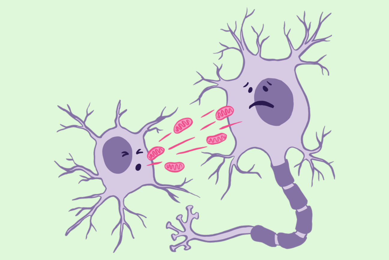
Although mitochondria in Rett neurons did not have such severe defects, astrocytes and neurons have a close relationship. Not only do neurons rely on astrocytes to supply them with energy, they even accept mitochondria from astrocytes to use for themselves. Jaenisch and Tomasello found that neurons take up dysfunctional mitochondria from Rett astrocytes at a higher rate than they take up mitochondria from unaffected astrocytes. This means that the effects of Rett syndrome on astrocytes have a direct effect on neurons: the dysfunctional mitochondria from the astrocytes end up in the neurons, where they cause damage. Tomasello took mitochondria from Rett astrocytes and placed them on both healthy and Rett neurons. In either case, the neurons took up the dysfunctional mitochondria in large numbers and then experienced significant problems. The neurons entered a hyperexcitable state that is ultimately toxic to the brain. The neurons also contained higher levels of reactive oxygen species, the toxic byproducts of mitochondrial metabolism, which can cause widespread damage. These effects occurred even in otherwise healthy neurons that did not themselves contain a Rett-causing MECP2 mutation.

“This shows that in order to understand Rett Syndrome, we need to look beyond what’s happening in neurons to other cell types,” Tomasello says.

Learning about the role that astrocytes play in Rett Syndrome could provide new avenues for therapies. The researchers found that supplying affected astrocytes with healthy mitochondria helped them to recover normal mitochondrial function. This suggests to Tomasello that one possibility for future Rett Syndrome therapies could be something that either targets mitochondria, or supplies additional mitochondria through the bloodstream.

Together, these insights and their possible medical implications demonstrate the importance of taking a broader look at the foundational biology underlying a disease.

RNA processing and gene expression governing

Renee Barbosa, a Schimmel scholar and a graduate student in the Soto-Feliciano Lab, uses a multidisciplinary approach to understand the epigenetic factors in gene expression.

Bendta Schroeder | Koch Institute
July 29, 2024

Professor Emeritus of Biology Paul Schimmel PhD ’67 and his wife Cleo Schimmel are among the biggest champions and supporters of graduate students conducting life science research in the Department of Biology at MIT, as well as in departments such as the Department of Brain and Cognitive Sciences, the Department of Biological Engineering, and the Department of Chemistry, and in cross-disciplinary degree programs including the Computational and Systems Biology Program, the Molecular and Cellular Neuroscience Program, and the Microbiology Graduate Program. In addition to the Cleo and Paul Schimmel (1967) Scholars Fund to support graduate women students in the Department of Biology, in 2021, the Schimmels established the MIT Schimmel Family Program for Life Sciences.

Their generous pledge of $50 million in matching funds called for other donors to join them in supporting the training of graduate students who will tackle some of the world’s most urgent challenges. Driven by their unwavering belief that graduate students are the driving force behind much life science research and witnessing a decline in federal funding for graduate education, the Schimmel family established their one-to-one match program. They reached the ambitious goal of $100 million in endowed support in just two years.

The discovery that mutations in genes can drive cancer revolutionized cancer research. In the decades following the identification of the first “oncogene” in a chicken retrovirus in 1970 and the first human oncogene in 1982 by Robert Weinberg at MIT’s Center for Cancer Research, scientists uncovered hundreds more oncogenes, transformed our understanding of how cancer begins and progresses, and developed sophisticated gene-targeted cancer therapies.

A majority of oncogenes were identified in factors controlling cell signaling, proliferation, and differentiation. However, a growing understanding of epigenetics has shown that many cancers, such as some leukemias and sarcomas, are not driven by mutations to these factors themselves, but by disruptions to the molecular pathways that regulate their expression. About 10 percent of all leukemias are driven by abnormal versions of the protein MLL1, one cog in the epigenetic machinery controlling these factors.

Renee Barbosa, a graduate student in the laboratory of Howard S. (1953) and Linda B. Stern Career Development Professor Yadira Soto-Feliciano in the Department of Biology, is joining this next wave of research, using leukemia as a model. A member of MIT’s Koch Institute for Integrative Cancer Research, Soto-Feliciano and her lab study chromatin, the densely coiled structures of DNA and scaffolding proteins that make up our genomes and help ensure genes are expressed at the right times and in the right amounts.

Barbosa focuses on the role of RNA processing and the precisely choreographed alterations to chromatin that govern gene expression. RNA molecules serve as messengers between DNA and its final product, protein, and are subject to extensive processing and regulation. However, not much is known about the interplay between RNA processing and epigenetic machinery, particularly in cancer.

“I hope that my work will uncover additional layers of complexity in the dynamic landscape of gene regulation,” says Barbosa. “It might also identify new mechanisms that can be targeted to help treat leukemia and other cancers.”

Before Barbosa arrived at the Soto-Feliciano Lab, she was already steeped in the molecular intricacies of cancer.

While at the University of Pennsylvania, she earned a BA in biochemistry and biophysics concurrently with a master’s degree in chemistry. Early on, she joined the lab of Ronen Marmorstein, which used molecular approaches to characterize MEK and ERK, two cancer-relevant members of a class of signaling proteins. Upon starting graduate school, she was excited to branch out into other disciplines.

Barbosa has always taken every opportunity she can to learn. Beginning in grade school, science and math were her favorite subjects, but she also explored music, dance, and foreign languages. At the University of Pennsylvania, she even squeezed in a minor in neuroscience.

With its interdisciplinary approach, the Soto-Feliciano Lab provides Barbosa ample opportunities to learn. Because epigenetic factors can elude traditional approaches, the Soto-Feliciano Lab uses a multidisciplinary strategy, ranging from molecular, to large-scale omics analyses, to disease modeling.

“When I was a grad student, we saw the arrival of powerful new massive sequencing and gene editing technologies — and were enabled to ask big new questions,” says Soto- Feliciano. “I am excited that Renee will have even more resources and opportunities, as we enter the next stage of cancer genetics and epigenetics.”

With the support of a Schimmel Fellowship, Barbosa will be ready to take advantage of new developments in her field.

“Support for research early on in graduate school is an incredible opportunity,” says Barbosa. “It means time to delve deep into the literature of the field and identify challenging open questions that I can pursue in my project. Though exploring these unknown areas requires taking bigger risks, I hope that we will get invaluable insight from an understanding of these nuanced and complex mechanisms.”

Back to the basics of gene regulation

Graduate student and Schimmel Scholar Annette Jun Diao uses a minimal system to parse the mechanisms underlying gene expression

Lillian Eden | Department of Biology
July 29, 2024

Professor Emeritus of Biology Paul Schimmel PhD ’67 and his wife Cleo Schimmel are among the biggest champions and supporters of graduate students conducting life science research in the Department of Biology at MIT, as well as in departments such as the Department of Brain and Cognitive Sciences, the Department of Biological Engineering, and the Department of Chemistry, and in cross-disciplinary degree programs including the Computational and Systems Biology Program, the Molecular and Cellular Neuroscience Program, and the Microbiology Graduate Program. In addition to the Cleo and Paul Schimmel (1967) Scholars Fund to support graduate women students in the Department of Biology, in 2021, the Schimmels established the MIT Schimmel Family Program for Life Sciences.

Their generous pledge of $50 million in matching funds called for other donors to join them in supporting the training of graduate students who will tackle some of the world’s most urgent challenges. Driven by their unwavering belief that graduate students are the driving force behind much life science research and witnessing a decline in federal funding for graduate education, the Schimmel family established their one-to-one match program. They reached the ambitious goal of $100 million in endowed support in just two years.

Annette Jun Diao’s mother loves to tell the story of Diao’s childhood aversion to the study of life — the gross and the squishy. Unlike some future biologists, Diao wasn’t the type to stomp through creeks or investigate the life of frogs. Instead, she was interested in astronomy and only ended up in a high school biology class because of a bureaucratic snafu. The physics course she’d been hoping to take was canceled due to low enrollment, and she was informed molecular biology was being offered instead.

She attended the University of Toronto and joined the molecular genetics department because of the numerous opportunities for hands-on research. She’s now a third-year graduate student in the Department of Biology at MIT.

“I’m fascinated by the mechanisms that underlie the regulation of gene expression,” Diao says. “All of our genetic information is in DNA, and that DNA is an actual molecule with chemical properties that allow it to be passed from one generation to the next.”

Every cell in our bodies contains a genome of approximately 20,000 genes, but the cells in our retinas are vastly different than the cells in our hearts — not all genes are in action simultaneously, and cell fates vary depending on how which genes are active.

“What is really awesome about the department — and what was attractive to me when I was applying to graduate school — is that I wasn’t sure exactly what methods I wanted to use to answer the questions I was interested in,” Diao says. “A huge advantage of the program was that I had a lot to choose from.”

Diao chose to pursue her thesis work with Seychelle Vos, the Robert A. Swanson (1969) Career Development Professor of Life Sciences and HHMI Freeman Hrabowski Scholar. Diao has been recognized with a Natural Sciences and Engineering Research Council of Canada Fellowship, which is similar to a National Science Foundation graduate fellowship in the United States.

Vos’s lab is generally interested in understanding how transcription is regulated, the interplay of genome organization and gene expression, and the molecular machinery involved. Diao has been working with an enzyme called RNA polymerase II (RNAP II), the molecular machine that reads DNA and creates an RNA copy called mRNA. That mRNA goes on to be read by ribosomes to create proteins.

Many questions remain about RNAP II, including what signals instruct it to begin transcription and, once engaged, whether it will transcribe and how quickly it moves.

RNAP II doesn’t work alone. Diao is working to understand how a transcription factor called negative elongation factor associates with RNAP II and whether the DNA sequence affects that interaction.

Within the broader context of the genome, DNA is packaged extremely tightly; if it were allowed to unfold, its total length could stretch from Cambridge to Connecticut. What RNAP II has access to at any given time is therefore quite restricted, which Diao is also exploring.

She has been exploring this topic in what she refers to as a “reductionist approach.” By creating a minimal system — a strand of DNA and the precise addition of certain other isolated components — she can potentially parse out what ingredients and what sequence of events are essential “in order to really get to the nitty-gritty of how genes are regulated.”

Outside of her work in the lab, Diao is part of BioREFS, a peer support group for graduate students, and gwiBio. Both organizations bring members of the department together for scientific talks and socializing activities outside of the lab, and gwiBio also participates in community outreach.

Diao is also a Schimmel Scholar, supported by Professor Emeritus of Biology Paul Schimmel PhD ’67 and his wife Cleo Schimmel.

“It was really great to learn that I was being supported by a scientist who has done a lot of awesome work that’s relevant to my world,” Diao says.

“It is awesome that they are so committed to supporting the graduate program at MIT, especially when federal resources have become more limited,” Vos says. “With their support, our lab can train basic scientists who can then use their knowledge to transform our study of disease. I hope others follow Paul and Cleo’s example.”

Whitehead Institute researchers uncover a new clue toward understanding the molecular basis of Parkinson’s disease

In Parkinson's disease, a mutation that causes protein misfolding can also turn the brain’s immune cells from friends to foes, possibly accelerating the progression of the disease. New Research from the Jaenisch Lab aims to uncover mechanisms that go awry in the brain, which may inform the development of new therapies that can halt or even reverse the progression of neurological conditions such as Parkinson's.

Shafaq Zia | Whitehead Institute
August 29, 2024

Dopamine is more than the “rush molecule”. This chemical messenger, produced by neurons in the midbrain, acts as a traffic controller that regulates the flow of electrical signals between neurons, assisting with brain functions like cognition, attention, movement, and behavior. But, in instances of Parkinson’s disease (PD), a progressive brain disorder, dopamine-producing neurons begin to die at an unprecedented rate, leading to dwindling levels of this vital chemical and impaired neural communication.

The lab of Whitehead Institute’s Founding Member Rudolf Jaenisch studies genetic and epigenetic factors — changes in gene expression that control which genes are turned on and off, and to what extent, without altering the DNA sequence itself — underlying neurological disorders like PD, Alzheimer’s disease, and Rett Syndrome. Their work aims to uncover the mechanisms that go awry in the brain, which may inform the development of new therapies that can halt or even reverse the progression of these conditions.

In their latest work, Jaenisch and former postdoctoral associate Marine Krzisch examine how a mutation in the gene that encodes for alpha-synuclein, a protein regulating the release of dopamine, affects the resident immune cells of the brain called microglia. The researchers’ detailed findings, published in the journal Biological Psychiatry on August 29, reveal that the mutation renders microglia extremely sensitive, worsening the problem of inflammation in the brain and potentially exacerbating damage to neurons in Parkinson’s disease.

“In fact, even when these mutant microglia are transplanted into a healthy, young brain, they have heightened activation upon stimulation, and low levels of the protective antioxidant catalase,” Krzisch says. “This tells us that in Familial Parkinson’s disease, which is due to genetic mutations, these microglia may be playing an important role in neuron degeneration.”

When nature’s origami falters

The human body is home to tens of thousands of unique proteins, each essential for processes sustaining life. These proteins are composed of linear chains of smaller building blocks called amino acids that are linked together in a specific sequence. For the proteins to perform their functions, the amino acid chains must crumple, rotate, and twist into stable three-dimensional structures. The stakes are high — just as precise folds and creases are crucial to the art of origami, even minor errors in the protein folding process can result in dysfunctional proteins that contribute to disease.

To date, scientists have identified over 20 causative genes in which mutations can result in Familial Parkinson’s disease, a rare, genetically inherited form of PD affecting individuals under or around the age of 50. Among them is SNCA, which encodes for alpha-synuclein, a small protein abundant in dopamine-producing neurons.

The A53T mutation in SNCA promotes the formation of dysfunctional alpha-synuclein proteins that clump together — almost like a ball of yarn — within dopamine-producing neurons. The accumulation of these protein clumps, also known as Lewy bodies, triggers inflammatory signaling in the brain, eventually killing the affected neurons. However, prior research has also shown that the A53T mutation accelerates the progression of PD, or the rate at which neurons die, although the full molecular mechanisms underlying this process are not yet fully understood.

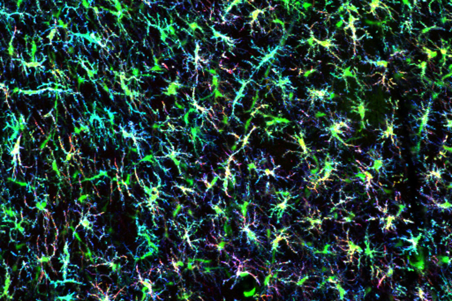
To uncover pathways involved in this progression, researchers in the Jaenisch Lab turned their attention to star-shaped patrollers called microglia that protect the brain from foreign invaders and respond to injuries, including protein aggregates within neurons. This immune response includes activated microglia trying to clear out Lewy bodies by digesting them, recruiting additional immune cells to the site of neurons with protein aggregates, and even killing off diseased neurons to limit damage to the brain.

But these friends can quickly turn to foes. Over-activated microglia can also degrade healthy neurons in the brain, prompting Jaenisch, Krzisch, and colleagues to investigate if excessive microglia activation is one pathway that contributes to progression in PD.

Microglia go rogue

To explore how the A53T mutation in the SNCA gene affects microglia function in PD, scientists at the Jaenisch Lab began by growing human myeloid precursors — the cells that eventually develop into microglia — in lab culture and transplanting them into the brains of immune-deprived mice.

Given the complexity of the brain, it’s common for researchers to study brain cells in the Petri dish. “But in cell cultures, microglia do not have the same morphology [form] as in the brain, show signs of chronic activation, and they don’t survive for a very long time,” says Krzisch. “When we transplant them in mice, the precursors differentiate into microglia that look and function like those in the human brain, and survive for the mouse’s lifespan.”

Using this method, the researchers compared the gene expression profiles of A53T-mutant microglia with those that did not carry the mutation, revealing differences in pathways linked to inflammation, microglia activation, and DNA repair. Additionally, when A53T-mutant microglia were exposed to an immune activator called lipopolysaccharide, they exhibited a heightened inflammatory response compared to non-mutant microglia.

In fact, even in non-inflammatory conditions, A53T-mutant microglia had decreased expression of catalase, an enzyme that helps break down harmful reactive oxygen species produced in response to protein aggregates in PD.

Understanding the molecular basis of progression in PD is challenging, which explains why there are currently no drugs to alter the disease’s course. With these findings in hand, researchers at the Jaenisch Lab are now eager to explore how factors like aging also influence microglia function and contribute to an increased rate of progression in PD.

“Overactivation of microglia isn’t the only cause of neuron death in Parkinson’s,” says Jaenisch. “But if we can decrease their activation, it will help us get to the point where we can slow down or actually stop the disease.”


Alumni News: Mission: Protecting the Planet

MIT Alum Catharine Conley, SB ’88, who earned two bachelor's degrees in biology and the humanities, spent more than a decade as NASA's planetary protection officer, working on protocols to prevent biological contamination on Earth and beyond.

Kathryn M. O'Neill | MIT Technology Review
August 20, 2024

When the space shuttle Columbia disintegrated during reentry in 2003, the disaster killed the human crew of seven—but not every creature onboard.

A collection of roundworms (a.k.a. nematodes) survived and was found in the debris, surprising everyone and prompting Catharine Conley ’88—principal investigator on the experiment—to publish a paper on the implications for astrobiology. It also led Conley to a new NASA role: planetary protection officer.

“Planetary protection is about trying to prevent Earth organisms from getting to other planets and, more importantly, making sure there’s nothing nasty when you bring material back to Earth,” says Conley, who held the job from 2006 to 2017 and helped ensure US compliance with the Outer Space Treaty, the international agreement that governs space exploration.

Conley got an early start on science thanks to a geneticist mother and mathematician father, and then completed two MIT majors—in biology and the humanities, focusing on Russian and French translation—and two bachelor’s degrees. That language study would prove useful: “Translation is essential when communicating with people from very different backgrounds—politicians, managers, bureaucrats, engineers, scientists—so for being planetary protection officer that was probably my most valuable training.”

After earning a PhD in plant sciences from Cornell, Conley studied a protein involved in muscle contraction as a postdoc at the Scripps Research Institute. That work led to NASA, where the Columbia experiment was designed to test the effects of low gravity on nematodes’ muscle tissue (muscle atrophy is a known problem for astronauts).

As the nematodes showed, Earth organisms are hard to kill. So a planetary protection officer must develop protocols not only to prevent biological contamination here but also to ensure that any “alien” life forms discovered elsewhere aren’t actually from Earth. “We have found signs of intelligent life on Mars,” Conley notes wryly. “But it’s us.”

Some scientists theorize that life on Earth actually came from Mars, Conley points out, which would increase the risk of importing something infectious: novel yet related organisms can quickly wreak havoc, as the recent pandemic illustrated.

Conley is currently visiting at the Carnegie Institution for Science, working to develop an analytical framework for assessing whether a space sample is indigenous life, Earth contamination, or just chemistry.

New approach enables a closer look at brain cell organelle

Microglia are involved in brain development, as well as neurodegeneration and brain cancer. A new approach from the Jaenisch Lab allows researchers to isolate and analyze microglia phagosomes.

Greta Friar | Whitehead Institute
August 14, 2024

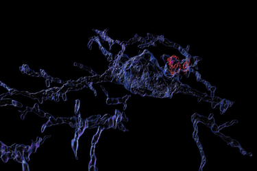
Microglia are the immune system’s front-line enforcers in the brain. They are cells that patrol the brain and destroy anything harmful that they encounter, from invading bacteria to cellular debris. They also remove plaques and prune dysfunctional synapses between neurons. Microglia eliminate their targets by eating them: they envelope material and seal it in bubble-like organelles called phagosomes. A phagosome can then fuse with other organelles that break down its contents.

Microglial phagosomes play important roles in brain development, brain function and a plethora of brain diseases, including neurodegeneration and brain cancer. Therefore, understanding microglial phagosome biology could help to develop new therapies for currently untreatable brain diseases. However, microglia and their organelles have been difficult to study because existing stem cell and animal models insufficiently resemble microglia in the human brain, and because microglia, as vigilant immune patrollers, react to even subtle stimuli and so experimental conditions can trigger changes in the cells that confound analyses.

To overcome those issues, Whitehead Institute Founding Member Rudolf Jaenisch, also a professor of biology at the Massachusetts Institute of Technology; University of Freiburg Professor of Neuropathology Marco Prinz; and University of Freiburg neuropathologist Emile Wogram, who began this project as a postdoctoral researcher in Jaenisch’s lab, have developed a method to isolate and analyze microglia phagosomes in a rapid, gentle, and unbiased fashion.

In research shared in the journal Immunity on August 15, the researchers describe how they can isolate and profile phagosomes from stem cell-derived microglia and fresh human brain tissue. They also share new insights into phagosome biology in the human brain, regarding synaptic pruning and generation of NAD+, a broadly used molecule in the brain, by microglia.

The method that the researchers developed to isolate phagosomes from cells uses immunoprecipitation, in which antibodies latch on to a specific target protein on an organelle’s surface. When the antibodies are collected, they pull the organelles with them. This technique avoids many chemical perturbations that might alter the microglial profile. Sometimes researchers genetically engineer a target for the antibodies, but in order to isolate phagosomes from human brain tissue, Wogram had to find a naturally expressed target. Eventually, he and colleagues found one: the protein CD68.

The researchers first isolated phagosomes from stem cell-derived microglia. They co-cultured the microglia with other brain cell types to create a more brain-like environment, which led to a better match between brain and stem cell-derived microglia gene expression. They triggered some of the microglia to enter an inflammatory or disease-like state to see how that affected the phagosomes. Additionally, Wogram collaborated with the neurosurgery department at the University of Freiburg to get access to brain tissues immediately after their removal during surgery. He isolated phagosomes from brain tissue within a half hour of its removal, allowing him to profile the organelles before their contents could change much.

The profiles that the researchers built included what proteins and metabolites the phagosomes contained, and the whole-cell gene expression profile. The profiles differed significantly between sets of phagosomes, but the researchers identified a core of consistent proteins, including many known and also some unknown phagosome proteins. The results showed that phagosomes contain sensitive signaling molecules that allow them to react quickly to even subtle environmental stimuli.

Additionally, the protein contents of the co-cultured microglia provided strong evidence that when microglia prune synapses, they predominantly prune the side that sends a signal and not the side that receives one. This insight could be useful for understanding how microglia interact with synapses in health and disease.

The researchers also gained insights into a key metabolic pathway that occurs inside of microglia. In excess, the molecule quinolinic acid can be toxic to neurons; it is implicated as involved in many neurodegenerative diseases. However, cells can use quinolinic acid to make NAD+, a molecule broadly used to carry out essential cellular functions. Microglia are the only brain cells that generate NAD+. Wogram and colleagues found that key steps in this process occur in phagosomes. Phagosomes are therefore necessary both for removing excess quinolinic acid to prevent toxicity and for helping to generate NAD+ in the brain.

Finally, Wogram used brain tissues to compare phagosomes from within a tumor to those in the surrounding healthy tissue. The phagosomes in the tumor contained excess quinolinic acid. Although follow-up studies would be needed to confirm the results, these findings are consistent with research that suggests cancer cells use quinolinic acid to fuel their growth.

Collectively, these findings illuminate aspects of phagosome biology and the roles that phagosomes may play in normal brain development and maintenance, as well as in cancer and neurodegeneration. The researchers also anticipate that their method could prove useful for profiling other organelles, especially when the organelles need to be rapidly isolated from human tissue.

Talented high schoolers excel while they explore the brain

Over six years of operation, pre-college outreach programs administered by Mandana Sassanfar, Senior Lecturer and Director of Diversity and Outreach, have placed seven exceptional pre-college students, often from underserved or underrepresented backgrounds, with research groups in The Picower Institute.

David Orenstein | The Picower Institute for Learning and Memory
August 14, 2024

During the pandemic, when many classes delivered online could barely hold students’ attention, Presley Simelus became captivated by the subject of biology thanks to their boundless curiosity and their uncommonly engaging teacher at Prospect Hill Academy Charter School in Cambridge. Meanwhile for Eli Hanechak, the science bug must have bit her very early. She’s wanted to be a doctor for as long as she can remember and in fifth grade built a model of a space station the size of a car out of duct tape, cardboard and broomsticks.

Not every teenager is expected to want to spend their summer breaks exploring science at a bench in an MIT lab, but each year students like Simelus and Hanechak, who have a distinct passion for research, can bring that to The Picower Institute and other research entities around MIT. Over six years of operation, pre-college outreach programs administered by Mandana Sassanfar, Director of Diversity and Outreach, have placed seven exceptional pre-college students, often from underserved or underrepresented backgrounds, with research groups in The Picower Institute. Despite their relative lack of experience compared to the technicians, graduate students, postdocs and professors around them, the students typically thrive.

“Eli has been a wonderful addition to our lab for the summer,” said Kendyll Burnell, the graduate student in the lab of Professor Elly Nedivi who has been working closely with Hanechak. “She is a hard worker, has caught on to techniques quickly, and is constantly asking excellent questions about science and doing research.”

Simelus, too, has been not only learning but also contributing, said their summer host, Yire Jeong, a postdoc in the lab of Associate Professor Gloria Choi.

“Presley has been amazing in our lab, and I was impressed by Presley’s eagerness to learn so much about neuroscience,” Jeong said. “Even when facing technical difficulties, Presley diligently worked to overcome them and achieved meaningful results.”

‘Dive into it’

Simelus, who hails from Everett, Mass., and will be enrolling in Swarthmore College this fall to study biochemistry, first came to MIT through the Leah Knox Scholars Program. Friends who’d been in the program before encouraged them to apply and they got in. During five weeks last summer Simelus and their cohort of fellow Leah Knox high-schoolers had the geeky pleasure of extracting bacteria out of the Charles River and performing a battery of tests to genetically characterize the novel organisms they found. Sassanfar noted that Simelus did the lab work exceptionally well, which is something she looks for when determining whom she might invite back the next summer to do research in an MIT Brain and Cognitive Sciences or Biology lab.

This spring when it came time for Simelus to decide where they might like to take that opportunity, they chose the Choi lab, which studies how the central nervous systems and immune systems interact, sometimes with consequences relevant to disorders including autism. Those keywords intrigued Simelus but really they made the choice because of the potential to learn something entirely new.

It was all this stuff I just simply wasn’t familiar with and I wanted to learn more about it,” Simelus said. “With Gloria’s lab I was truly mystified and I wanted to dive into it. That’s the reason I chose it.”

This summer Simelus has been working with Jeong on a study of how brain cell activity differs when mice are sick vs. when they are well. The project has involved imaging neurons in the brain to detect telltale signs of recent activation, expression of a protein called c-fos. Learning about neuroscience and gaining skills like preparing, staining and imaging tissue have been a very fulfilling outcome of the internship, Simelus said.

“I truly have learned so much about neuroscience,” they said. “I feel like the field, anything related to the brain or neuroscience, is always under this sort of veil and nobody really knows what’s going on. But I feel like my time at the Choi lab has really allowed me to see what neuroscience is about. It’s taught be more about the brain itself and also more about different biology techniques and skills I might need.”

Now the only problem, Simelus said, is that there are even more things to be deeply curious about. Simelus feels committed to harnessing the life sciences in some way in the future to sustain human life and experience. And as someone who not only plays the viola but also composes, they’ve begun thinking more about how the brain responds to music.

There will no doubt be many chances to continue exploring these interests at Swarthmore, but during the summer at MIT, Simelus said they’ve expanded their horizons while still hanging out with friends, some of whom have been working in other nearby labs.

“I don’t think I would have changed my summer,” Simelus said.

‘The perfect opportunity’

Hanechak lives in the tiny Western Massachusetts town of Russell (population: 1,643) and commutes 45 minutes to Pope Francis Preparatory School in Springfield, where she is a rising senior.

In her freshman year at a different school, she yearned for an extra challenge so she got involved in science fair. Interested in medicine, but eager for a project in which she could make a difference without having clinical credentials, she chose to work on reducing pollution by developing a microbe-derived enzyme that could biodegrade plastics. She had read about such enzymes in the research literature and learned that they don’t work as well as engineers have hoped. In successive years she has scrounged lab space and general supervision in labs at Westfield State University and UMass Amherst to create and screen beneficial mutations in the enzyme and to synthesize structures that might help the enzyme work better. The enzyme she presented at the International Science and Engineering Fair last year can degrade plastics in 24 hours.

Sasssanfar, who also directs the Massachusetts Junior Academy of Science (MassJAS), learned of Hanechak’s award-winning science fair presentation and invited her to present at the MassJAS symposium, held at MIT last October. Hanechak did so well, Sassanfar said, she earned a spot present at the American Junior Academy of Science meeting (adjacent to the American Association for the Advancement of Science Annual Meeting) in Denver in February. She also earned Sassanfar’s invitation to join a lab this summer at MIT.

Hanechak has long had an MIT pennant on her wall at home and has admired MIT as a place where regardless of one’s background, if one has a passion for science and technology, that’s what matters.

“No one in my family has gone to college and no one has been involved in a science-related career of any kind,” she said. “One of the reasons MIT has always stood out to me is that there are especially great minds here, but they didn’t all come from established families or super prestigious backgrounds or anything like that. They kind of just were able to make their own way.”

Moreover, the chance to come to MIT to learn about the brain in the Nedivi lab seemed like a great step to take toward that longer-term goal of medicine.

“It seemed like the perfect opportunity to start transitioning into what I want my career to look like and to get some experience doing neuroscience research,” Hanechak said. “I’m very glad I’m able to have this summer experience, like learning the techniques. When I go into my college major of neuroscience, I will have a good background of what I’m doing, besides just my environmental research.”

With Burnell, Hanechak is working on finding a DNA promoter specific for a rare but interesting kind of neuron in the visual cortex, where the brain processes what the eyes see. Finding this genetic signature would allow the lab to label these cells and image them under the microscope, so that they could see how the cells contribute to visual processing.

Hanechak acknowledged she was anxious at first about joining a bigger lab with scientists who have much more experience.

“But my entire summer has been incredibly gratifying and exciting—just being able to work in Cambridge, and live in this area, and experience city life, and then also be in a lab environment where it’s so collaborative and everyone’s very friendly,” she said.

For many teens, summer provides a chance to do what they want to do. Simelus and Hanechak chose the opportunity to explore the brain at The Picower Institute and have made the most of it.

Two Whitehead Institute graduate researchers awarded the 2024 Regeneron Prize for Creative Innovation

Whitehead Institute graduate student researchers Christopher Giuliano (Lourido Lab) and Julian Roessler (Hrvatin Lab) have been awarded the 2024 Regeneron Prize for Creative Innovation.

Merrill Meadow | Whitehead Institute
July 30, 2024

Whitehead Institute graduate student researchers Christopher Giuliano and Julian Roessler have been awarded the 2024 Regeneron Prize for Creative Innovation. In addition, postdoctoral researcher Chen Weng was selected as a finalist in the postdoctoral fellows competition.

The Regeneron Prize, sponsored by global biotechnology company Regeneron Pharmaceuticals, Inc., is a competitive award designed to recognize and honor exceptional talent and originality in biomedical research. Individual graduate students and postdoctoral fellows in the biomedical sciences are nominated by the nation’s top research universities. Then, nominees outline their “Dream Projects” — potentially groundbreaking research projects that they would pursue given unrestricted access to resources and state-of-the-art technology.

The “Dream Project” proposals, presented by the nominees to a selection committee comprised of Regeneron’s leading scientists, are used to evaluate a trainee’s scientific merit, elegance, precision, and creativity. Novel research ideas and out-of-the-box thinking is encouraged — although the proposal must include a strong rationale, basic methodology and design for the project, and a discussion of how its results could advance the field. Both Giuliano and Roessler have been awarded $50,000 for their proposals, which can be used in any way the winners choose. In addition, Weng was awarded $5,000 as a finalist, and Regeneron has made a $10,000 grant to the Whitehead Institute as the home institute of the winners to support its seminar series.

This year’s awards are distinctive in that the two winners are from the same institution: Both Giuliano and Roessler are pursuing their PhDs at Massachusetts Institute of Technology (MIT) and conducting their doctoral research at Whitehead Institute.

Giuliano is a researcher in the lab of Whitehead Institute Member Sebastian Lourido, who is also an associate professor of biology at MIT and holds the Landon Clay Career Development Chair at Whitehead Institute. Giuliano’s Dream Project seeks to address the unique challenges posed by genetically based muscle disorders. “An obstacle in using current gene therapies to treat these conditions,” he explains, “is that muscle tissue comprises large syncytial cells, which contain hundreds of nuclei in a shared cytoplasm. Even when a gene therapy is able to reach an individual muscle cell, it often isn’t able to spread to every nucleus within that cell.” However, certain parasites, like Toxoplasma gondii, thrive because they have the capacity to successfully gain access to and manipulate muscle cells. T. gondii, the primary focus of the Lourido lab’s work, may infect nearly one third of all humans. “My project,” Giuliano says, “would identify the specific biological mechanisms used by the parasites to spread their virulence factor proteins throughout the cell. Using genetic screens for protein spread, we would work toward applying these protein features to improve the efficiency of muscle-directed gene therapies, and ultimately test our system in a mouse model of Duchenne muscular dystrophy.”

Roessler is a researcher in the lab of Whitehead Institute Member Siniša Hrvatin, who is also an assistant professor of biology at MIT. While Roessler’s doctoral research focuses on the neuronal circuitry underlying torpor and hibernation in small mammals, his Dream Project seeks to identify the sensory circuitry regulating the “diving reflex” displayed in land- and sea-dwelling mammals, including humans. The diving reflex occurs when an animal’s face is immersed in cold water, prompting an array of organs to reduce their function in ways that, scientists believe, privileges the flow of oxygen to the brain and muscles. “That this reflex has been conserved across millions of years of mammalian evolution suggests an extraordinary genetic advantage,” Roessler says. “Yet, researchers have given comparatively little attention to the neuronal circuits underlying this reflex, and we don’t understand even the fundamental mechanisms by which the nervous system coincidently detects both cold temperature and the presence of water.” Beyond elucidating a foundational aspect of mammalian biology, Roessler’s projects could, if pursued, underpin new interventions for conditions ranging from migraine headaches to cardiac arrhythmia that might be ameliorated by artificial stimulation or inhibition of the diving response.

Weng is a postdoctoral researcher in the lab of Whitehead Institute Member Jonathan Weissman, who is also a professor of biology at MIT, the Landon T. Clay Professor of Biology at Whitehead Institute, and an Investigator of the Howard Hughes Medical Institute. His Dream Project — which proposes a new approach to using single-cell genealogy to understand factors driving cell line evolution — is an extension of his current work. Indeed, this past year he co-developed a technology that details the family trees of human blood cells and provides new insights into the differences between lineages of hematopoietic stem cells. The technology gives researchers unprecedented access to any human cells’ histories — and a path to resolving previously unanswerable questions.