Undergraduate student and Gould Fellow discusses choosing a summer research lab, living in the Greater Boston Area, and managing imposter syndrome.
Lillian Eden | Department of Biology
August 28, 2025
Mariely Morales Burgos first fell in love with MIT while participating in the Quantitative Methods Workshop, a weeklong intensive offered in January to prepare students to analyze data in biology and neuroscience. Those skills have come in handy this summer while participating in the Bernard S. and Sophie G. Gould MIT Summer Research Program in Biology (BSG-MSRP-Bio), a ten-week training program for non-MIT undergraduate students interested in pursuing an academic career.
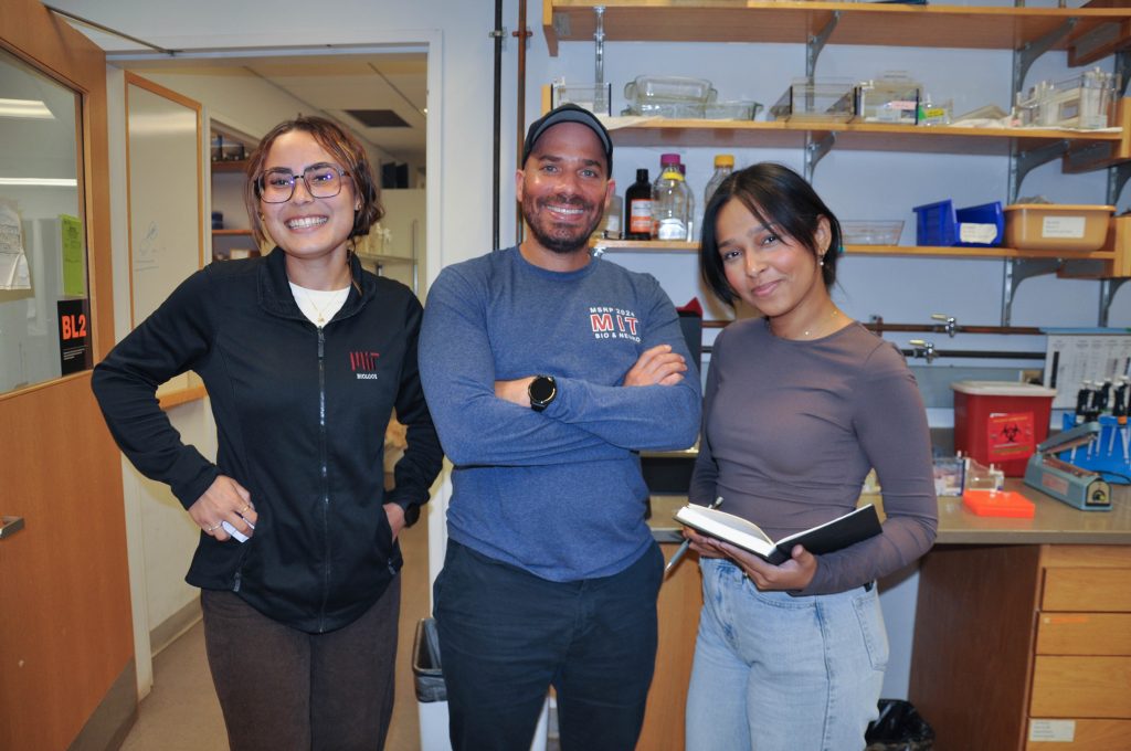
A Gould Fellow and McNair Scholar, Morales Burgos spent the summer mentored by Associate Professor of Biology Eliezer Calo, for whom the program served as a critical stepping stone in his own career. Calo is the first BSG-MSRP-Bio program alum to receive tenure at MIT.
A rising senior at the University of Puerto Rico at Aguadilla, Morales Burgos spent the summer using zebrafish to study the molecular machinery responsible for making proteins.
Q: How did you select your lab, and what have you been working on?
A: I knew I wanted to work in Eliezer’s lab after meeting him during a QMW faculty lunch. I felt like we really connected because of his genuine passion for science, commitment to his trainees, and the way he spoke about his lab and the care he puts into mentoring.
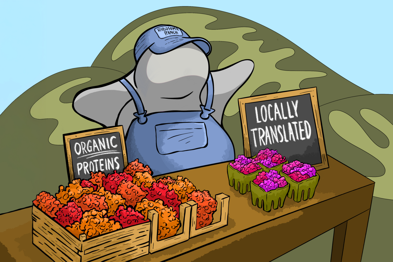
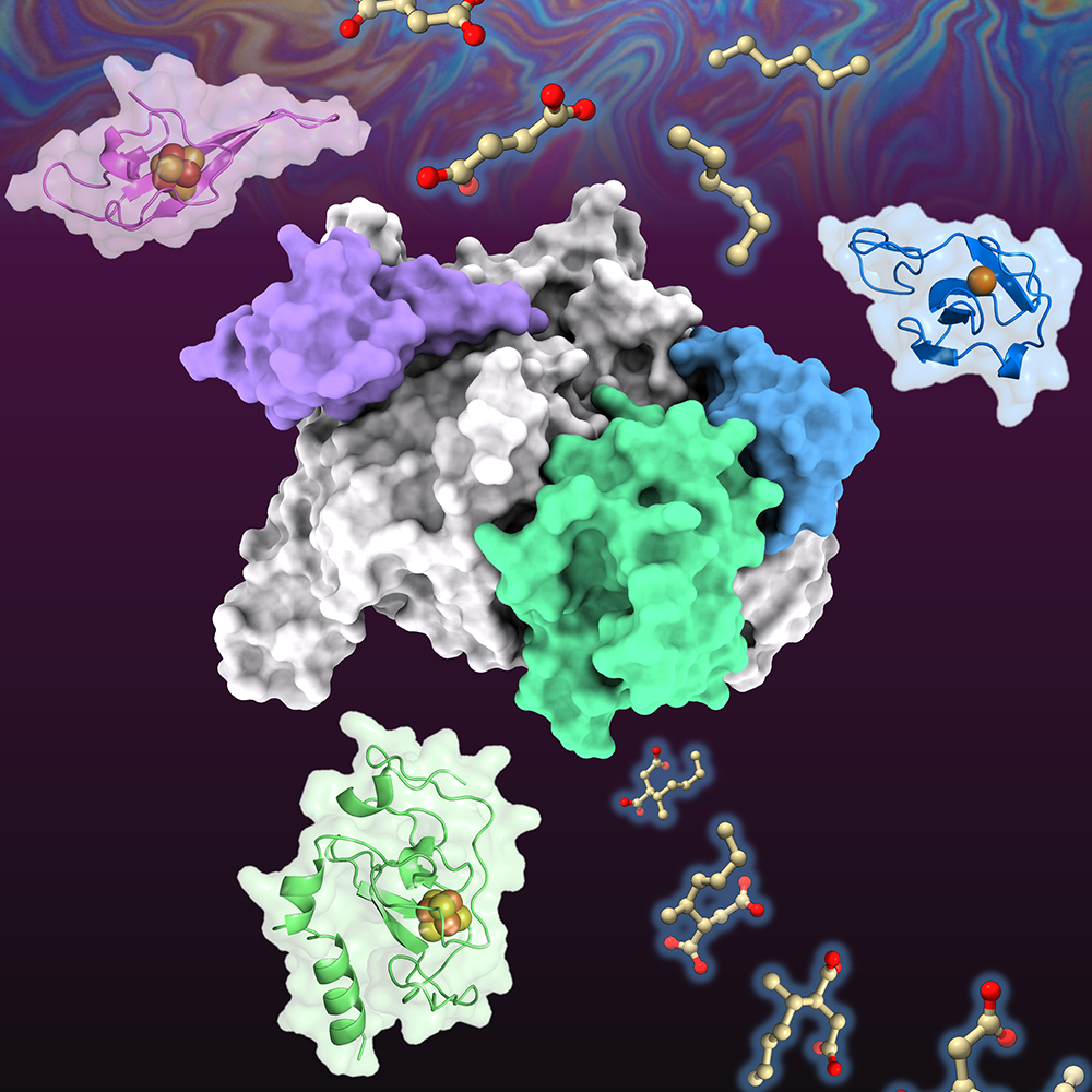
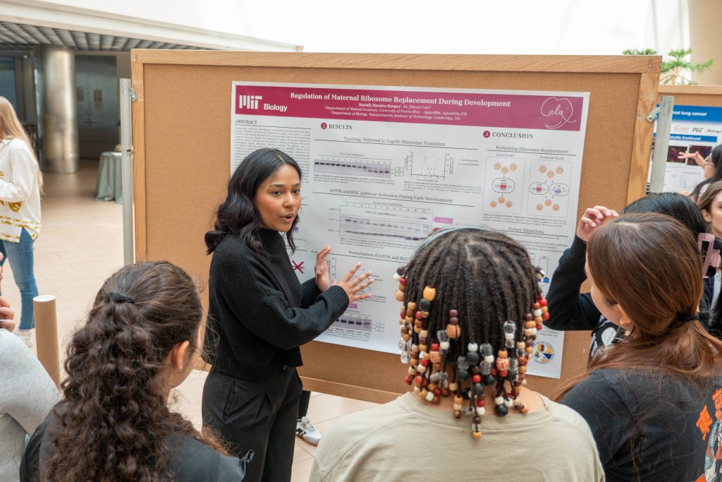
My research focuses on ribosomes, which are the protein factories of the cell, and they’re essential to make what the cell needs to go through different developmental stages and through its most crucial processes. In early development, zebrafish and numerous other organisms depend on maternally deposited ribosomes and associated molecular components inherited directly from the oocyte. As time goes on, their own genomes activate, and they start being able to make their own ribosomes. What I’m studying is this transition from maternal to zygotic ribosomes during early development. We know this transition happens, but we don’t know how this transition is regulated, whether it happens passively, through dilution, or actively, through targeted cellular mechanisms.
One skill that I’ve been able to learn here, other than just learning and applying techniques, is how to develop a whole project independently, how to think critically about the next step of my project, and what other questions I can ask.
Being able to work with a live animal organism and see the developmental stages in real-time, I thought that was really cool. And it really makes me appreciate the beauty of developmental biology, and just life in general.
Q: How did you prepare for the program, and what has it been like living and working in Boston and Cambridge? As a Gould Fellow, you also met with program supporters Mike Gould and Sara Moss, who established the Bernard S. and Sophie G. Gould fund to honor the memory of Mike’s parents. What was it like to meet and talk to Mike and Sara?
A: Once we get accepted, we’re encouraged to start communication with our faculty. I had a few meetings with Eliezer to discuss some papers, and based on our discussion and the expectations for the project, I was able to read more and start preparing before I arrived.
Every few weeks beforehand, we had a meeting with Mandana and the rest of the cohort on Zoom, and we were talking on an app called GroupMe, and we exchanged socials, so when we came here, we weren’t complete, total strangers.
When I’m not in the lab, I spend a lot of time with my roommates, and we like walking around Boston. It’s a very walkable city and has a lot of unique architecture, but Boston weather is very unpredictable. I’m from a tropical island, so I wish someone had told me to prepare for the rain and cold, but the July weather has been so nice.
In Puerto Rico, you don’t have public transportation, so I’ve really enjoyed commuting. Our dorms are at Northeastern, so I take the bus, and it goes over the Charles, and it’s so beautiful.
I’m a person who feels a lot of emotions, so I was the only one who cried when we met the Goulds. It was a bit embarrassing, but that’s okay. They told me to never lose the empathy that I have, no matter how hard my journey is, to keep on holding on to my sentimental side and keep working hard, and they’re so excited to see where we end up and what we end up doing.
Q: This program’s aim is to make research available for students who don’t have access to hands-on experience at their home institutions, so many students, including you, are embarking on independent research projects for the first time, which could trigger “imposter syndrome.” What was that experience like for you, and what advice would you give to future BSG-MSRP-Bio program participants?
A: I was a little bit intimidated by the program, and didn’t apply the first time I had the opportunity. Then I did the Quantitative Methods Workshop, and those eight days were beautiful. I got to see how everybody loves collaborating and that the community here is very supportive. I met many wonderful faculty who were passionate about their research, and that exposure made me realize I would love to be part of a place like this.
Imposter syndrome is something that I feel like most everybody deals with, but MSRP is a place that, if you’re willing to put in the work, everyone is willing to help you reach the places that you dream of being. It might feel intimidating to ask questions, and you could be scared of feeling like you don’t deserve to be in these spaces. But somebody who wants you to grow will answer your questions. I wanted to be able to work independently as soon as possible, because that really showcases your abilities, but no matter what, Eliezer, who’s mentoring me, his door is always open.
What I advise is to really dive into your project and take advantage of everything this program offers. Working hard on your project, you get to fall in love with the process and the questions you’re trying to answer and science as a whole, and there’s nothing better than to spend the summer on a project that you love.