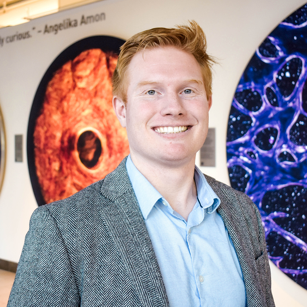
Computational biologist Sergei Kotelnikov is working to develop new methods in protein modeling as part of the School of Science Dean’s Postdoctoral Fellowship.
Lyn Nanticha Ocharoenchai | School of Science
March 31, 2026
Billions of years ago, simple organic molecules drifted across Earth’s primordial landscape — nothing more than basic chemical compounds. But as natural forces shaped the planet over hundreds of millions of years, these molecules began to interact and bond in increasingly complex ways. Along the way, something spectacular emerged: life.
“Life is, to some degree, magical,” says computational biologist Sergei Kotelnikov. Simple organic compounds congregate into polymers, which assemble into living cells and ultimately organisms — the whole being greater than the sum of its parts.
“You can write formulas on how a molecule behaves,” he says, referring to the world of quantum mechanics. “But yet somehow, a few orders of magnitude above, on a bigger scale, it gives rise to such a mystery.”
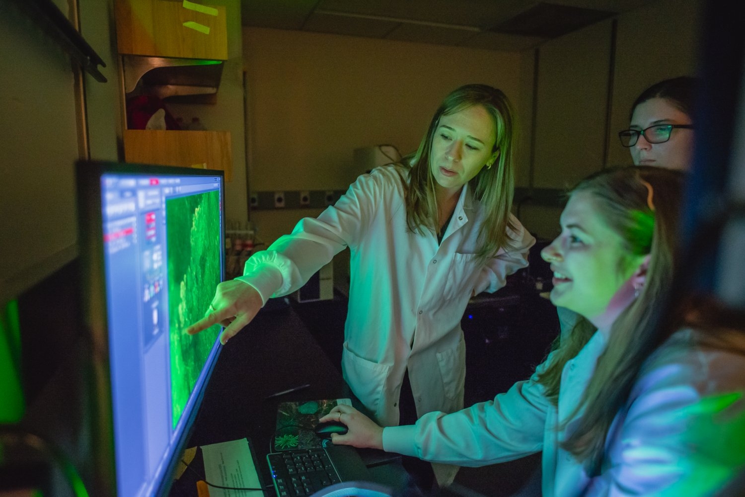
Kotelnikov builds models to analyze and predict the structure of these biomolecules, particularly proteins, the fundamental building blocks of every organism. This year, he joined MIT as part of the School of Science Dean’s Postdoctoral Fellowship to work with the Keating Lab, where researchers focus on protein structure, function, and interaction. Using machine learning, his goal is to develop new methods in protein modeling with potential applications that span from medicine to agriculture.
A hunger for problems to solve
Kotelnikov grew up in Abakan, Russia, a small city sitting right in the center of Eurasia. As a child, one of his favorite pastimes was playing with Lego bricks.
“It encouraged me to build new things, rather than just following instructions,” he says. “You can do anything.”
Kotelnikov’s father, whose background lies in engineering and economics, would often challenge him with math problems.
“Your brain — you can feel some kind of expansion of understanding how things work, and that’s a very satisfactory feeling,” Kotelnikov says.
This itch to solve problems led him to join science Olympiad competitions, and later, a science-focused public boarding school located near the Russian Academy of Sciences, from which he often encountered scientists.
“It was like a candy shop,” he recalls, describing the period as a life-changing experience.
In 2012, Kotelnikov began his bachelor of science in physics and applied mathematics at the Moscow Institute of Physics and Technology — considered one of the leading STEM universities in Russia, and globally — and continued there for his master’s degree. It was there that biology came into the picture.
During a course on statistical physics, Kotelnikov was first introduced to the idea of the “emergence of complexity.” He became fascinated by this “mysterious and attractive manifestation of biology … this evolution that sharpens the physical phenomenon” to create, drive, and shape life as we know it today. By the time he completed his master’s degree, he realized he had only scratched surface of the field of computational biology.
In 2018, he began his PhD at Stony Brook University in New York and began working with Dima Kozakov, who is recognized as one of the world’s leaders in predicting protein interactions and complex structures.
Studying the architecture of life
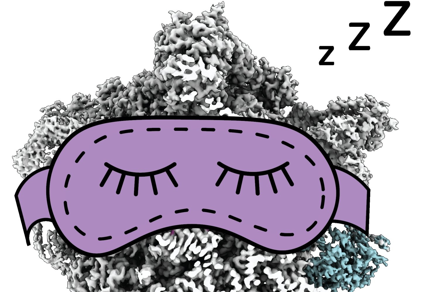
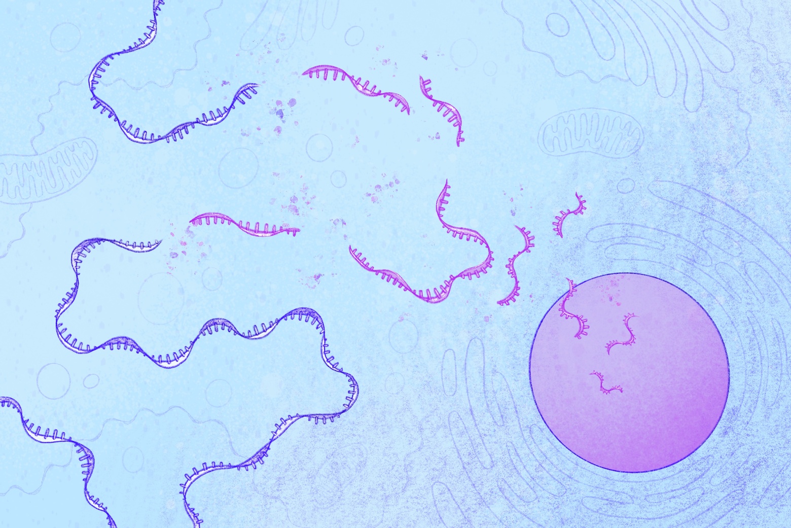
Proteins act like the bricks that construct an organism, underpinning almost every cellular process from tissue repair to hormone production. Like pieces of a Lego tower, their structures and interactions determine the functions that they carry out in a body.
However, diseases arise when they’re folded, curled, twisted, or connected in unusual ways. To develop medical interventions, scientists break down the tower and examine each individual piece to find the culprit and correct their shape and pairing. With limited experimental data on protein structures and interactions currently available, simulations developed by computational biologists like Kotelnikov provide crucial insight that inform fundamental understanding and applications like drug discovery.
With the guidance of Kozakov at Stony Brook’s Laufer Center for Physical and Quantitative Biology, Kotelnikov carried over his understanding of physics to create modeling methods that are more effective, efficient, reliable, and generalizable. Among them, he developed a new way of predicting the protein complex structures mediated by proteolysis-targeting chimeras, or PROTACs, a new class of molecules that can trigger the breakdown of specific proteins previously considered undruggable, such as those found in cancer.
PROTACs have been challenging to model, in part because they are composed of proteins that don’t naturally interact with each other, and because the linker that connects them is flexible. Imagine trying to guess the overall shape of a bendy Lego piece attached to two other pieces of different irregular, unmatched shapes. To efficiently find all possible configurations, Kotelnikov’s method conceptually cuts the linker into two halves and models each separately, then reformulates the problem and calculates it using a powerful algorithm called Fast Fourier Transform.
“It’s kind of like applied math judo that you sometimes need to do in order to make certain intractable computations tractable,” he says.
Kotelnikov’s state-of-the-art methods have been instrumental to his team’s top performance in numerous international challenges including the Critical Assessment of protein Structure Prediction (CASP) competition — the same contest in which the Nobel Prize-winning AlphaFold system for protein 3D structure prediction was presented.
Physics and machine learning
At MIT, Kotelnikov is working with Amy Keating, the Jay A. Stein (1968) Professor of Biology, biology department head, and professor of biological engineering, to study protein structure, function, and interactions.
A recognized leader in the field, Keating employs both computational and experimental methods to study proteins, their interactions, as well as how this can impact disease. By infusing physics with machine learning, Kotelnikov’s goal is to advance modeling methods that can vastly inform applications such as cancer immunology and crop protection.
“Kotelnikov stands to gain a lot from working closely with wet lab researchers who are doing the experiments that will complement and test his predictions, and my lab will benefit from his experience developing and applying advanced computational analyses,” says Keating.
Kotelnikov is also planning to work with professors Tommi Jaakkola and Tess Smidt in MIT’s Department of Electrical Engineering and Computer Science to explore a field called geometric deep learning. In particular, he aims to integrate physical and geometric knowledge about biomolecules into neural network architectures and learning procedures. This approach can significantly reduce the amount of data needed for learning, and improve the generalizability of resulting models.
Beyond the two departments, Kotelnikov is also excited to see how the diversity and interdisciplinary mix of MIT’s community will help him come up with ideas.
“When you’re building a model, you’re entering this imaginary world of assumptions and simplifications and it might feel challenging because of this disconnect with reality,” Kotelnikov says. “Being able to efficiently communicate with experimentalists is of high value.”