Junk DNA makes a comeback

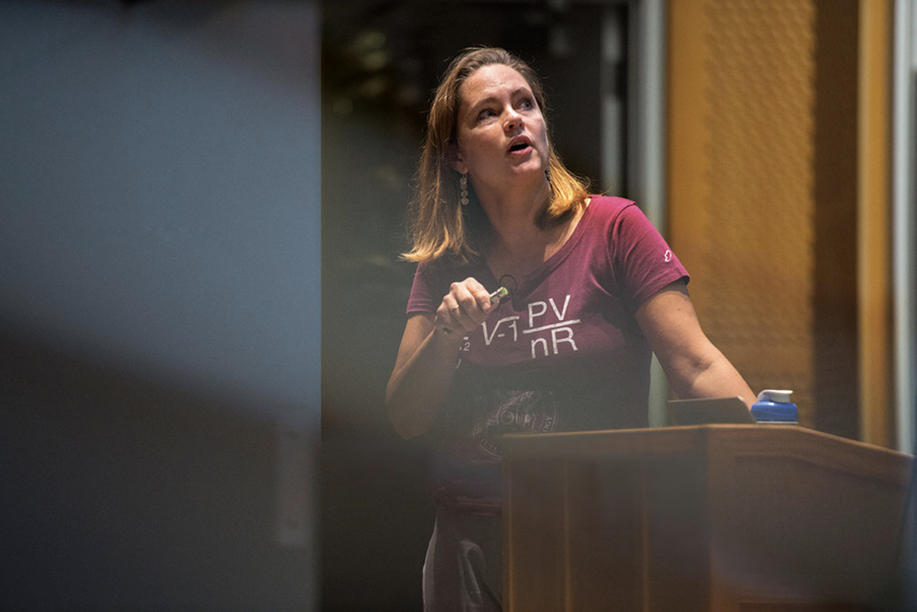
Third-year graduate student Emma Kowal is searching DNA for sequences that regulate gene expression.

Saima Sidik
July 8, 2019

“I went into science because of a certain obsession with the romance of it,” says Emma Kowal, a third-year graduate student in Chris Burge’s lab in the MIT Department of Biology. “I loved the idea of the scientist as an adventurer exploring the frontiers of knowledge and the universe. And I haven’t let go of that yet.”

Kowal has always been an avid science fiction reader, and now she’s living out a real-life scientific odyssey. The quest she’s taken on for her PhD research involves an understudied type of DNA sequence called an intron, and the roles that introns might play in regulating gene expression.

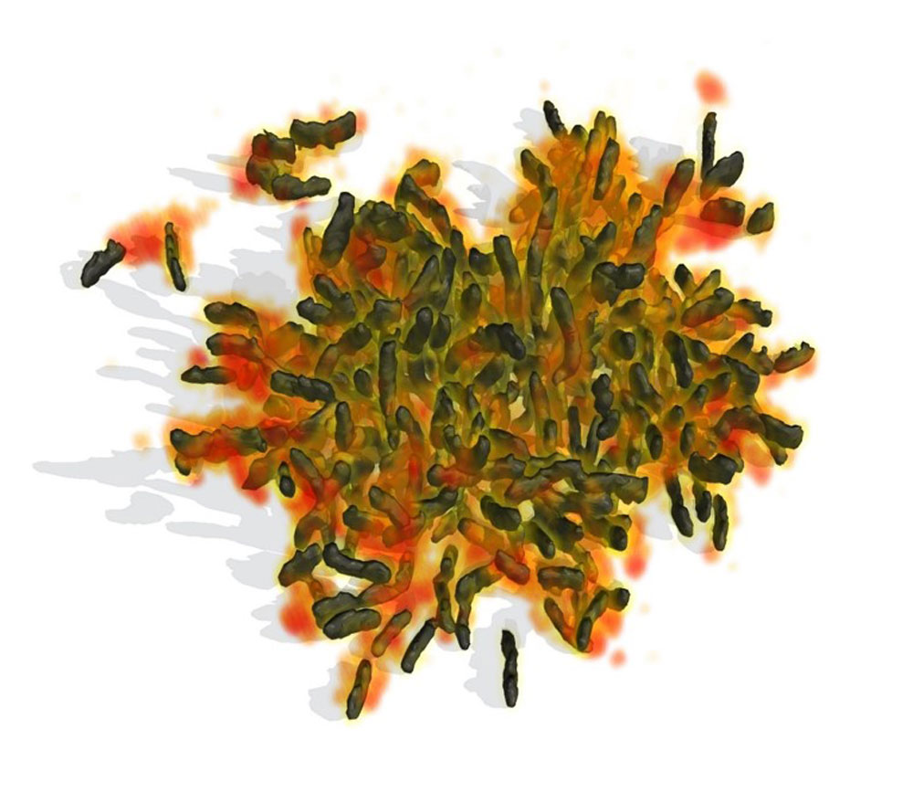
Introns lie between the DNA sequences that cells use for protein production, and are initially incorporated into the messenger RNA, or mRNA, that cells produce as an intermediate step in synthesizing proteins from DNA. But before they complete protein synthesis, cells remove introns from mRNA through a process called splicing, which has led many people to view introns as junk DNA with splicing acting like a garbage disposal.

“Introns appeal to me as the underdog genomic region,” Kowal says. Although they’re often seen as unimportant, introns are ubiquitous and plentiful, collectively making up 24% of the human genome. All eukaryotes have them, and, on average, each human gene encodes eight. Many researchers, including Kowal, think that introns have been underestimated, and that they may play an important role in regulating gene expression.

Introns are only the latest chapter in Kowal’s RNA story. She began her research career as a Harvard University undergraduate student working in the Szostak Lab at Massachusetts General Hospital, where she studied how RNA catalyzed the evolution of cells on the early earth. Although studying primordial life was intellectually stimulating, Kowal wanted to work on something more applied, and so she joined the Church Lab in the Harvard Department of Genetics. There she developed methods for purifying and imaging enigmatic RNA-containing lipid compartments called extracellular vesicles, which cells release into their surrounding environments possibly to communicate with one another.

For the sequel to her bachelor’s degree, Kowal chose to attend MIT Biology because she’d heard that, “at MIT, everyone is one standard deviation nerdier, on average, than they are at other schools.” In this sense, she has not been disappointed. Kowal calls the energy at MIT “unparalleled,” and she says, “people are jazzed about what they’re doing, and the whole campus reflects that.”

In some ways, these reflections are physical. Much of the artwork around MIT pays homage to major scientific discoveries, and Kowal says this reverence for science is one factor that attracted her to MIT. From the mural of DNA in the Biology Department to the golden neurons that descend alongside the staircase in the McGovern Institute for Brain Research, it’s as if the community is saying, “look at how awesome the universe is!” as Kowal puts it.

In other ways, this energy is reflected in the people she converses with daily. “I really like the students here,” Kowal says. “Everyone is enthusiastic, but also down to earth.” When she’s not exploring the realms of science, Kowal sometimes has more fanciful adventures with the Dungeons and Dragons group that she’s formed with some of her classmates.

Kowal didn’t necessarily intend to continue working on RNA at MIT Biology, but when she heard about Chris Burge’s lab, which focuses on RNA and the proteins that mediate its production and stability, she felt a call to action.

The Burge Lab combines high throughput experimental techniques with bioinformatics, and Kowal wants to develop expertise in both these fields. “If you’re skilled in generating and analyzing big data sets, you can ask questions that other people can’t,” she says. The Burge lab seemed like the perfect setting for her PhD.

Over and over, scientists have noticed that cells produce more protein from genes that contain introns than when those same introns are removed. Intron mediated enhancement (IME), as this effect is called, is a “stunningly broad phenomenon,” Kowal says, and scientists have observed it in a wide range of organisms, from yeast to plants to humans.Burge asks his students to begin their degrees with a month-long reading period during which they sift through the literature to find a topic that they want to study. “You’re not allowed to pick up a pipette or do any analysis during your reading period,” Kowal says. “You just read and discuss your ideas and let things percolate.” As she read, Kowal came across a number of studies that discussed the influence that introns have on gene expression levels.

Splicing machinery, which removes introns from mRNA, likely plays a role in IME. This machinery binds mRNA as it’s being produced from DNA, then interacts with, and influences, the RNA production machinery. However, researchers have created mutant introns that can’t be recognized by splicing machinery, and sometimes these introns still enhance gene expression, so splicing isn’t the only factor that drives IME. Moreover, replacing one intron with another of the same size containing a different DNA sequence can change its effect, implying that the exact DNA sequences within introns may dictate their effects on gene expression. Kowal is intrigued by this last point, and wants to find these intronic sequences and figure out which have the largest effects on gene expression and why.

“This is an old mystery that’s ripe for new tools,” Kowal says. Over the last decade, researchers have begun using a technique called RNAseq to count the copies of mRNA that are made from each gene in a population of cells. Instead of replacing an intron with a single alternative DNA sequence, Kowal plans to replace an intron with a myriad of random DNA sequences, then use RNAseq to count how many copies of mRNA cells make when they encode each of these random introns.

Preparing to test these random sequences has been an odyssey in and of itself, and Kowal has spent the last year building the system that she’ll use. First, she needed to decide which intron to replace. She chose one from a gene called UbC. Removing this intron reduces expression of UbC by ten-fold.

Besides contributing strongly to IME, the UbC intron is a great candidate for Kowal’s experiment because it lies in a regulatory region of the UbC mRNA that precedes the portion that’s translated into protein. This let her replace the UbC protein coding region with a fluorescent protein that she’ll use to visualize how much protein cells make when they encode each random intron sequence.

Kowal has spent the last year meticulously incorporating a library of random introns into this synthetic version of the UbC gene. She anticipates being able to introduce them into cells soon, to see which random introns result in the highest levels of mRNA and protein production. Thanks to RNAseq, she’ll be able to monitor how much each random intron contributes to mRNA expression. Because she can measure how brightly the fluorescent protein glows, she can correlate these mRNA levels with protein levels. From this, she’ll learn which intron sequences enhance gene expression most strongly, and she’ll also know whether these introns lead to higher levels of mRNA production, or if the same amount of mRNA is made into more protein. This distinction will offer her clues about the mechanism that introns use to enhance gene expression.

Once Kowal knows which intron sequences promote gene expression most effectively, she’ll take advantage of the Burge lab’s bioinformatics expertise to analyze the distribution of these sequences throughout genomes and predict how they affect global gene expression. Kowal suspects certain intron sequences are bound by proteins that mediate mRNA production and stability, and she thinks her work will identify these protein-intron pairs.

Kowal balances her scientific adventures with outdoor adventures. Specifically, she’s recently fallen in love with rock climbing. “Climbing is a great counterpart to science because it’s something you can chip away at, and then there’s this huge satisfaction when you finally achieve a climb,” she says. “And also, between climbing and pipetting, I have really strong fingers.”

As for her love of science fiction, Kowal hopes to one day pen a science-based adventure of her own, but not before she’s made her mark as a scientist, either as a professor or in industry. ”It makes sense for me to focus most of my energy on science right now,” she says. “But after I’ve led a spectacular, adventurous life in science, maybe I’ll use my reflections to write a novel.”

Posted 7.8.19
JoAnne Stubbe named 2020 Priestley Medalist

MIT biochemist is being honored for her work in understanding enzyme mechanisms

Celia Arnaud | Chemical & Engineering News
June 24, 2019

JoAnne Stubbe, the Novartis Professor of Chemistry and Biology, emerita, at the Massachusetts Institute of Technology, will receive the 2020 Priestley Medal, the American Chemical Society’s highest honor.

“JoAnne is the top mechanistic biochemist of her generation,” says Stephen J. Lippard, one of Stubbe’s colleagues in the MIT chemistry department. “Among her major achievements is understanding the controlled generation of radicals in biology.”

“Throughout her career, JoAnne has taken on some of the experimentally most challenging problems, and time and time again, she provided insights that, while sometimes controversial when she first introduced them, have stood the test of time,” says Wilfred van der Donk, a chemistry professor at the University of Illinois at Urbana-Champaign who was a postdoc in Stubbe’s lab in the 1990s.

Stubbe is best known for figuring out the mechanism of ribonucleotide reductase, an enzyme that catalyzes the conversion of ribonucleotides used in RNA to deoxyribonucleotides used in DNA. That reaction is the only route in nature for making deoxyribonucleotides.

Stubbe showed that the reduction at the 2′ position on the ribose sugar ring involves hydrogen removal at the 3′ position, which was unexpected because a 3′ hydrogen still exists in the final structure. The reaction is particularly unusual because later crystal structures showed a metal cofactor initiates the electron transfer required to power the reduction from more than 35 Å from the reactive site. Such a long distance between the two sites was unexpected because it was too far for conventional electron transfer. Stubbe proposed and demonstrated that the transfer happens in multiple steps.

“The remarkable part of this now widely accepted mechanism is that no crystallographic information was available when JoAnne proposed it,” van der Donk says. “When the structure of the enzyme was reported years later, her predictions proved to be correct, and she herself later provided experimental evidence of many of the radical intermediates.” Donald Hilvert, a chemistry professor at the Swiss Federal Institute of Technology Zurich, says, “Her groundbreaking studies of ribonucleotide reductases revolutionized the field of enzymology.”

Stubbe also uncovered details of the mechanism of action of bleomycin, a cancer drug that works by cleaving double-stranded DNA. “Her group determined the mechanism of this unusual process and solved the NMR structure of cobalt-substituted bleomycin bound to double-stranded DNA, a true tour de force,” van der Donk says.

In search of nature’s winning recipe

Graduate student Darren Parker aims to understand the ratio of ingredients that constitutes the optimal cell.

Raleigh McElvery
May 31, 2019

Fifth-year graduate student Darren Parker is as much a baker as he is a biologist — at least metaphorically speaking. He’s on a mission to understand the ratio of ingredients required to concoct nature’s winning recipe for the optimal cell. Researchers have a solid understanding of which components are essential for cellular function, but they have yet to determine whether it’s critical for cells to generate exactly the right amount of protein.

“In that way, my graduate work is actually pretty simple,” Parker says. “I just want to know if changing the amounts of a specific ingredient has an effect on the overall product.”

The oldest of four brothers, Parker grew up in a suburb just outside of Chicago. When he enrolled in the University of Illinois Urbana-Champaign for his undergraduate studies in 2009, he was considering a major in environmental science. “My high school biology classes were mostly rote memorization,” he explains, “so the molecular aspects just didn’t resonate with me. I was more interested in studying life on a larger scale.”

After his first year, he entered the Integrated Biology program, which essentially “encompassed all biology that wasn’t molecular biology.” He was still required to take an introductory molecular biology course, though, as part of the major. But this time around, something clicked.

He remembers performing his first genetic knockdown experiment, decreasing the level of dopamine receptors in roundworms and witnessing the behavioral ramifications in real time. “I finally had a handle on the molecular concepts enough to really get what was going on,” he says.

He attributed his newfound appreciation for the basic mechanisms underlying life to his fortified chemistry skills. At the beginning of his third year, he officially declared a biochemistry major, and joined a lab in the Department of Chemistry studying nucleic acid enzymes.

Parker’s job was to sift through trillions of short DNA strands, selecting only those that could act like enzymes and cut RNA. He would then home in on the nucleotide sequences within those strands that were best suited to carry out the reaction. After a year-and-a-half, he’d successfully identified a few DNA sequences that could cut RNA molecules with a distinct chemistry. After this point he was excited to try “studying life” as opposed to synthetic reactions.

Mid-way through his fourth year, he joined a biology lab in the College of Medicine probing alternative splicing in liver and heart development. It was a new group with only a few members, and Parker had more experience as an undergraduate than some of the first-year graduate students, so he hit the ground running. His last-minute switch to biochemistry meant he had five years of studies instead of the usual four — totaling six full semesters (and several summers) in lab.

After identifying a key splicing protein required for the liver to fully mature in mice and humans, Parker became even more fascinated by molecular biology and determined to pursue a career in science with bigger picture applications.

At the urging of his advisor, Parker sent in his graduate school applications. He was primarily interested in microbiology and infectious disease research — although he had no prior experience working in bacteria, only a longstanding interest in the intersection of science and society. He ultimately chose MIT Biology because of the breadth of labs. He could join a microbiology lab, or pursue an entirely different path, all within the same department. Gene-Wei Li’s lab seemed like “the perfect mix.”

“Gene was asking questions in molecular biology from the unique perspective of a physicist, looking at biological questions in a way I had never even considered before,” he says. “Gene had also just joined the department and wasn’t tied to a specific field or model organism yet, so I had the chance to build my own projects from the bottom up; I wasn’t just slotting in somewhere.”

Best of all, the Li lab was all about drilling down into to the mechanics of protein production in order to understand the cell as a whole — the bigger picture perspective that Parker was longing for.

Parker began by exploring ways to modify high-throughput RNA sequencing. He aimed to make this popular method cheaper and more scalable, in hopes of knocking out many individual genes in E. coli to test the genome-wide effects. He then pivoted his project and applied his new technique to study the effects of reducing essential genes in B. subtilis, another model bacterium. The family of enzymes that was the most interesting to him from these experiments were the aminoacyl-tRNA synthetases.

tRNAs, or transfer RNAs, carry amino acids to the ribosome so that the cell can produce proteins. This process requires the help of enzymes — tRNA synthetases — to “charge” the tRNAs with an amino acid. Only then can the ribosome transfer the amino acid from the tRNAs to the growing chain of amino acids that eventually forms the protein. Like an inquisitive baker, Parker wanted to know what would happen if he added more or less tRNA synthetase to the recipe of a bacterial cell.

His results would make Goldilocks proud. Over the past few years, he’s shown that too much or too little tRNA synthetase prevents the cell from growing at a normal rate. The amount must be just right.

“It turns out that what’s most important to the cell is maintaining the ratios of those very conserved ingredients,” Parker says. “The cell will actively use less of those ingredients if the synthetase is limiting, and this leads to a much slower growing cell. Adding too much tRNA synthetase is just a waste because the cell already has as much as it needs to sustain translation.”

This same family of tRNA synthetase proteins, he adds, are also implicated in some neurological diseases in humans, which gives him further impetus to study them.

At this point, Parker has taste-tested his fair share of biological areas, and he’s found his niche. “It was a long process,” he says. “That was probably best, though, because it gave me more time to explore.”

Once he graduates, he plans to go into industry, perhaps continuing to tweak the list of ingredients in order to engineer cells to do new things.

“The next time you’re in the kitchen and you want to add more or less of your favorite ingredient,” he urges, “just think about how the cell might feel if you did so with your favorite gene.”

Photo credit: Raleigh McElvery
Posted 5.30.19
Drug-resistant cancer cells create own Achilles heel
Nicole Giese Rura | Whitehead Institute
May 28, 2019

Cambridge, MA — The cells of most patients’ cancers are resistant to a class of drugs, called proteasome inhibitors, that should kill them. When studied in the lab, these drugs are highly effective, yet hundreds of clinical trials testing proteasome inhibitors have failed. Now scientists may have solved the mystery of these cells’ surprising hardiness. The key: Resistant cancer cells have shifted how and where they generate their energy. Using this new insight, researchers have identified a drug that resensitizes cancer cells to proteasome inhibitors and pinpointed a gene that is crucial for that susceptibility.

As cancer cells develop, they accrue multiple genetic alterations that allow the cells to quickly reproduce, spread and survive in distant parts of the body, and recruit surrounding cells and tissues to support the growing tumor. To perform these functions, cancer cells must produce high volumes of the proteins that support these processes. The increased protein production and numerous mutated proteins of cancer cells make them particularly dependent on the proteasome, which is the cell’s protein degradation machine. These huge protein complexes act as recycling machines, gobbling up unwanted proteins and dicing them into their amino acid building blocks, which can be reused for the production of other proteins.

Previously, researchers exploited cancer cells’ increased dependency on their proteasomes to develop anti-cancer therapies that inhibit the proteasomes’ function. Several distinct proteasome inhibitors have been developed, and when used in the lab, these proteasome inhibitor drugs are indeed highly effective at eradicating tumor cells. However, when administered to animal models or patients with cancer, such as multiple myeloma, proteasome inhibitors have limited efficacy and even initially vulnerable cancer cells quickly develop resistance to them. How do cancer cells so adroitly sidestep drugs that should kill them?

Exploring the gene expression of thousands of tumors and hundreds of cancer cell lines, Peter Tsvetkov, a former postdoc in the lab of late Whitehead Institute Member Susan Lindquist, has determined that the answer may lie with how and where the cells produce their energy. According to his analysis of the active genes and metabolism products generated in proteasome inhibitor-resistant cancer cells and tumors, Tsvetkov, who is currently a postdoc in the lab of Broad Institute Founding Core Member, director of the Cancer Program, and oncologist at the Dana-Farber Cancer Institute Todd Golub, concluded that such cells have shifted how they produce energy — away from breaking down the sugar glucose toward a dependency on processes within the mitochondria, the “powerhouse” part in the cell. In fact, when Tsvetkov pushed cancer cells’ metabolism to depend on the mitochondria, that change alone was sufficient to make cancer cells immune to proteasome inhibitors. Tsvetkov’s findings are described online this week in the journal Nature Chemical Biology.

In order to understand how a metabolic shift could link to proteasome inhibitor resistance, Tsvetkov screened proteasome inhibitor-resistant breast cancer cells with thousands of small molecules to identify the ones that hamper the cells’ growth or even kill the cells. One stood out in the screen: elesclomol, a small molecule that that researchers had previously evaluated as an anti-cancer agent in phase 3 clinical trials without knowing with what the drug interacts in cancer cells. To identify how elesclomol preferentially targets the proteasome inhibitor resistant cells, Tsvetkov did genome-wide CRISPR-Cas9 screens to find out which genes elesclomol requires to incapacitate resistant cancer cells. Only the gene FDX1, which encodes an enzyme in the mitochondria, came to the fore.

In collaboration with John Markley from the Department of Biochemistry at the University of Wisconsin-Madison, Tsvetkov used biochemical and biophysical systems to demonstrate that elesclomol directly binds to the mitochondrial enzyme FDX1 and impedes its natural function. In the presence of copper, elesclomol can also be altered by the FDX1 enzyme, which increases the drug’s anti-cancer toxicity. These findings led the researchers to determine that as cancer cells become overly reliant on their mitochondrial metabolism, they ramp up the FDX1 protein’s activity. Also, when the FDX1 protein interacts with copper-bound elesclomol, the protein enhances the drug’s copper-dependent toxicity. Thus, copper appears to play an essential role — when Tsvetkov removed copper, elesclomol was no longer effective.

Having established that a metabolic shift and resistance to proteasome inhibitors are linked, Tsvetkov is now interested in understanding how a change in metabolism allows cancer cells to adapt to other anti-cancer therapies and how copper-binding molecules such as elesclomol can be developed as effective anticancer agents.

This work was supported by the National Institutes of Health (NIH grant P41GM103399), University of Wisconsin-Madison Biochemistry Department, EMBO (Fellowship ALTF 739-2011), the Charles A. King Trust Postdoctoral Fellowship Program, and Howard Hughes Medical Institute (HHMI).

Written by Nicole Giese Rura

***

Susan Lindquist’s primary affiliation was with Whitehead Institute for Biomedical Research, where her laboratory was located and all her research was conducted. She was also a Howard Hughes Medical Institute investigator and a professor of biology at Massachusetts Institute of Technology.

***

Citation:

“Mitochondrial metabolism promotes adaptation to proteotoxic stress”

Nature Chemical Biology, online May 27, 2019, DOI: 10.1038/s41589-019-0291-9

Peter Tsvetkov, Alexandre Detappe, Kai Cai, Heather R. Keys, Zarina Brune, Weiwen Ying, Prathapan Thiru, Mairead Reidy, Guillaume Kugener, Jordan Rossen, Mustafa Kocak, Nora Kory, Aviad Tsherniak, Sandro Santagata, Luke Whitesell, Irene M. Ghobrial, John L. Markley , Susan Lindquist, and Todd R. Golub.

Catalyzing new approaches in research and education to meet the climate challenge
MIT Energy Initiative
May 16, 2019

Catherine Drennan says nothing in her job thrills her more than the process of discovery. But Drennan, a professor of biology and chemistry, is not referring to her landmark research on protein structures that could play a major role in reducing the world’s waste carbons.

“Really the most exciting thing for me is watching my students ask good questions, problem-solve, and then do something spectacular with what they’ve learned,” she says.

For Drennan, research and teaching are complementary passions, both flowing from a deep sense of “moral responsibility.” Everyone, she says, “should do something, based on their skill set, to make some kind of contribution.”

Drennan’s own research portfolio attests to this sense of mission. Since her arrival at MIT 20 years ago, she has focused on characterizing and harnessing metal-containing enzymes that catalyze complex chemical reactions, including those that break down carbon compounds.

She got her start in the field as a graduate student at the University of Michigan, where she became captivated by vitamin B12. This very large vitamin contains cobalt and is vital for amino acid metabolism, the proper formation of the spinal cord, and prevention of certain kinds of anemia. Bound to proteins in food, B12 is released during digestion.

“Back then, people were suggesting how B12-dependent enzymatic reactions worked, and I wondered how they could be right if they didn’t know what B12-dependent enzymes looked like,” she recalls. “I realized I needed to figure out how B12 is bound to protein to really understand what was going on.”

Drennan seized on X-ray crystallography as a way to visualize molecular structures. Using this technique, which involves bouncing X-ray beams off a crystallized sample of a protein of interest, she figured out how vitamin B12 is bound to a protein molecule.

“No one had previously been successful using this method to obtain a B12-bound protein structure, which turned out to be gorgeous, with a protein fold surrounding a novel configuration of the cofactor,” says Drennan.

Carbon-loving microbes show the way

These studies of B12 led directly to Drennan’s one-carbon work. “Metallocofactors such as B12 are important not just medically, but in environmental processes,” she says. “Many microbes that live on carbon monoxide, carbon dioxide, or methane—eating carbon waste or transforming carbon—use metal-containing enzymes in their metabolic pathways, and it seemed like a natural extension to investigate them.”

Some of Drennan’s earliest work in this area, dating from the early 2000s, revealed a cluster of iron, nickel, and sulfur atoms at the center of the enzyme carbon monoxide dehydrogenase (CODH). This so-called C-cluster serves hungry microbes, allowing them to “eat” carbon monoxide and carbon dioxide (CO2).

Recent experiments by Drennan analyzing the structure of the C-cluster-containing enzyme CODH showed that in response to oxygen, it can change configurations, with sulfur, iron, and nickel atoms cartwheeling into different positions. Scientists looking for new avenues to reduce greenhouse gases took note of this discovery. CODH, suggested Drennan, might prove an effective tool for converting waste CO2 into a less environmentally destructive compound, such as acetate, which might also be used for industrial purposes.

Drennan has also been investigating the biochemical pathways by which microbes break down hydrocarbon byproducts of crude oil production, such as toluene, an environmental pollutant.

“It’s really hard chemistry, but we’d like to put together a family of enzymes to work on all kinds of hydrocarbons, which would give us a lot of potential for cleaning up a range of oil spills,” she says.

The threat of climate change has increasingly galvanized Drennan’s research, propelling her toward new targets. A 2017 study she co-authored in Science detailed a previously unknown enzyme pathway in ocean microbes that leads to the production of methane, a formidable greenhouse gas: “I’m worried the ocean will make a lot more methane as the world warms,” she says.

Drennan hopes her work may soon help to reduce the planet’s greenhouse gas burden. Commercial firms have begun using the enzyme pathways that she studies, in one instance employing a proprietary microbe to capture CO2 produced during steel production—before it is released into the atmosphere—and convert it into ethanol.

“Reengineering microbes so that enzymes take not just a little but a lot of CO2 out of the environment—this is an area I’m very excited about,” says Drennan.

Creating a meaningful life in the sciences

At MIT, she has found an increasingly warm welcome for her efforts to address the climate challenge. “There’s been a shift in the past decade or so, with more students focused on research that allows us to fuel the planet without destroying it,” she says.

In Drennan’s lab, a postdoc, Mary Andorfer, and a sophomore, Phoebe Li, are currently working to inhibit an enzyme present in an oil-consuming microbe whose unfortunate residence in refinery pipes leads to erosion and spills. “They are really excited about this research from the environmental perspective and even made a video about their microorganism,” says Drennan.

Drennan delights in this kind of enthusiasm for science. In high school, she thought chemistry was dry and dull, with no relevance to real-world problems. It wasn’t until college that she “saw chemistry as cool.”

The deeper she delved into the properties and processes of biological organisms, the more possibilities she found. X-ray crystallography offered a perfect platform for exploration. “Oh, what fun to tell the story about a three-dimensional structure—why it is interesting, what it does based on its form,” says Drennan.

The elements that excite Drennan about research in structural biology—capturing stunning images, discerning connections among biological systems, and telling stories—come into play in her teaching. In 2006, she received a $1 million grant from the Howard Hughes Medical Institute (HHMI) for her educational initiatives that use inventive visual tools to engage undergraduates in chemistry and biology. She is both an HHMI investigator and an HHMI professor, recognition of her parallel accomplishments in research and teaching, as well as a 2015 MacVicar Faculty Fellow for her sustained contribution to the education of undergraduates at MIT.

Drennan attempts to reach MIT students early. She taught introductory chemistry classes from 1999 to 2014, and in fall 2018 taught her first introductory biology class.

“I see a lot of undergraduates majoring in computer science, and I want to convince them of the value of these disciplines,” she says. “I tell them they will need chemistry and biology fundamentals to solve important problems someday.”

Drennan happily migrates among many disciplines, learning as she goes. It’s a lesson she hopes her students will absorb. “I want them to visualize the world of science and show what they can do,” she says. “Research takes you in different directions, and we need to bring the way we teach more in line with our research.”

She has high expectations for her students. “They’ll go out in the world as great teachers and researchers,” Drennan says. “But it’s most important that they be good human beings, taking care of other people, asking what they can do to make the world a better place.”

Changes to the Biology major beginning Fall 2019
Raleigh McElvery
April 24, 2019

We would like to share news on changes to the Biology major that will take effect Fall of 2019. Over the past year, the department has been revising its lab curriculum in order to accommodate the increasing number of students doing interdisciplinary Biology-related majors and to respond to the large numbers of students doing UROPs. Course 7 and 7A will be consolidated into a single major. Current 7A students will graduate with a Course 7 degree.

Most of the major requirements have stayed the same. The major changes are in the lab curriculum and are described as follows:

7.02 (18 units, CI-M) will be discontinued

  •  Students who already took this course are all set.  If a student has not taken this course, there are other options to fulfill the lab requirement.
  • 7.02 will be replaced with two courses 7.002 (6 units, Fall & Spring) and 7.003 (12 units, CI-M, Fall & Spring). Both must be taken to fulfill the lab requirement and the CI-M. We hope the modularization of these courses will help students to fit them in their schedule

7.18 (30 units, CI-M) will be discontinued

  • If a student already took this course, it will count and they are all set.
  • If a student did not take it, they will need a second CI-M. 7.19 (12 units, CI-M) is a Biology CI-M that substitutes for 7.18.
  • Other CI-Ms can also be used and are listed on the degree charts.

We hope that these changes will enhance the student experience at MIT, and we are happy to hear and help you solve any concerns that you have. If you have questions, please contact the Education Office.

Back to the basics

Biology students in the MIT Biotechnology Group are applying their skills in basic science to explore careers in industry.

Raleigh McElvery
April 15, 2019

When Rachit Neupane began his PhD at MIT Biology in 2013, the prospect of a career in industry was so mystifying it seemed like a “black box.” He had only a vague idea of what it would take to stray from the well-trodden path to academia and penetrate the biotechnology sphere post-graduation — applying his knowledge of the life sciences to manufacture drugs, develop technologies, and assess business problems.

As a first-year student, Neupane joined Jacqueline Lees’s lab studying the role of epigenetic regulators in lung and colon cancer, while simultaneously enrolling in drug development classes. He hoped to learn more about taking a project all the way from the lab to the clinic, as well as how his basic biology research fit into that scheme. “I didn’t know what I didn’t know, but I wanted to find out,” he recalls.

Two years into his graduate program he received some unexpected guidance in the form of an email, inviting students to join a new group on campus, the MIT Biotechnology Group (MBG). Now nearing its five-year anniversary, MBG was founded by four graduate students from three different departments, and aims to educate MIT undergraduates, graduate students, and postdocs who, like Neupane, are curious about the biotech landscape. MBG connects these trainees with one another and with leaders in the greater Boston area.

“We started the MIT Biotech Group as a conduit through which students, postdocs, and even young professors could access the rich biotechnology community surrounding MIT,” says founding co-president James Weis SM ’17. “The breadth and scale of MBG’s influence, and especially the career decisions it has enabled, has surpassed my most optimistic projections — largely due to incredible efforts of several generations of leaders, who have grown the group into MIT’s primary point-of-contact with the biotechnology community.”

Today, MBG is still entirely student-run. Although the leadership roles are currently primarily held by students from the Departments of Biology and Biological Engineering, MBG brings together trainees from across campus, including Brain and Cognitive Sciences, Electrical Engineering and Computer Sciences, Health Sciences and Technology, Chemical Engineering, and Computational Systems Biology.

Neupane now serves as co-president alongside Catie Matthews of Chemical Engineering and the Sloan School of Management. Together, they oversee a core team of nearly 30 graduate and undergraduate students, who collaborate to host a slew of events related to life sciences entrepreneurship, industry R&D, and business.

Once Neupane graduates, second-year Biology graduate student Lena Afeyan will take his place. She has served as director of the entrepreneurship branch, and, most recently, on the executive board as the director of finance. As such, she manages the group’s budget — which covers staple events like the semester-long Industry Seminar Series and the annual Ideation pitching and networking symposium, as well as career networking nights, special lectures, and the group’s due diligence projects.

These programs complement ones hosted by individual departments. “MBG is the central place where students from all these different departments can come together to think about biotech,” Afeyan says.

She knew before she began her PhD that she wanted to go into biotech, and chose MIT Biology specifically because “it offered a rigorous program to learn basic science while being so close to a biotech hub and surrounded by engineering minds.” This basic science knowledge, she explained, would allow her to ask the right questions later in her career, in order to identify high-impact scientific advances.

According to Afeyan, her principal investigator, Richard Young, runs his lab like a mini company. Young investigates the molecular mechanisms behind gene control, and has founded four different companies in less than a decade.

“He’s built a very strong reputation in his field because he’s attacked fundamental biological questions with a lot of scientific rigor, while understanding that those same questions can have a high impact on patients with diseases like cancer,” she says.

Afeyan’s labmate, fourth-year graduate student Alicia Zamudio, joined the lab because she was interested in the research questions, irrespective of their biotech applications. Unlike Afeyan, she’d had very little exposure to industry prior to MIT, until she took a drug development class and began meeting professionals in industry. Although she found MBG less than a year ago, she’s now an officer in the branch of the group focused on industry.

“I wanted to learn more about the biotech sector and build connections with professionals in the space,” she says. As a member of MBG, she’s not just one individual reaching out to an organization; she is backed by hundreds of curious students on campus hoping to learn more.

In her role as officer, Zamudio helps MBG organize events open to the entire MIT community, including site visits to various biotech companies in the area. “Dozens of these companies are walking-distance,” she says. “No other place has the density of biotech companies that exists here in Kendall Square.”

As Zamudio prepares to graduate, she hasn’t completely discarded the possibility of pursuing an academic postdoc, but she’s leaning heavily towards a career in industry.

“Industry seems like an extremely dynamic place where you get to think about scientific problems that help people,” she says, “while making practical use of a background in basic science.”