Covid-19 class taps experts to help students and the public avoid misinformation as the crisis evolves.
Raleigh McElvery | Department of Biology
May 21, 2021
Just a few months after the Covid-19 pandemic took hold, Alan Grossman was already mulling over an idea for a new class to help people make sense of the virus. As head of MIT’s Department of Biology, he was aware of the key role fundamental research would play in the coming months. From RNA viruses and genomic sequencing to antibodies and vaccines, MIT students and the general public would need reliable scientific information to understand the evolving situation — and discern fact from fiction.
Not long after, the thoughts he’d feverishly scribbled on paper scraps scattered around his house began to take shape. With the support of the MIT School of Science, Accessibility Office, MIT Video Productions, and others around the institute, the Department of Biology added a new fall subject to the course catalog: 7.00 (Covid-19, SARS-CoV-2 and the Pandemic). Undergraduates could take the class for credit, as notable researchers stepped up to the virtual podium to share their expertise in front of a public livestream.
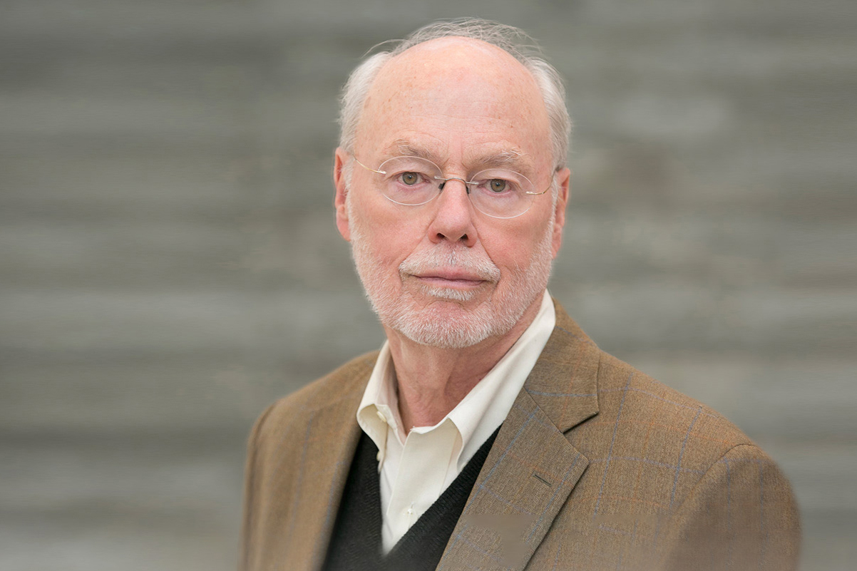
Grossman brought his nascent plans to associate department head and Whitehead Institute for Biomedical Research Member Peter Reddien, and together the two brainstormed individuals who might be willing to lead the class and queue speakers. They reached out to professor of biology and Whitehead Institute Member Richard Young, who served as an advisor to the World Health Organization and National Institutes of Health when a different virus of unknown origins was spreading — HIV. Young was also quick to mount a collaborative research campaign against SARS-CoV-2, the virus that causes Covid-19.
“I give Alan a lot of credit,” Young says. “He thought that it was the responsibility of the department to take the lead in filling the Covid-19 knowledge niche, and asked me if I would take this on and find a partner.”
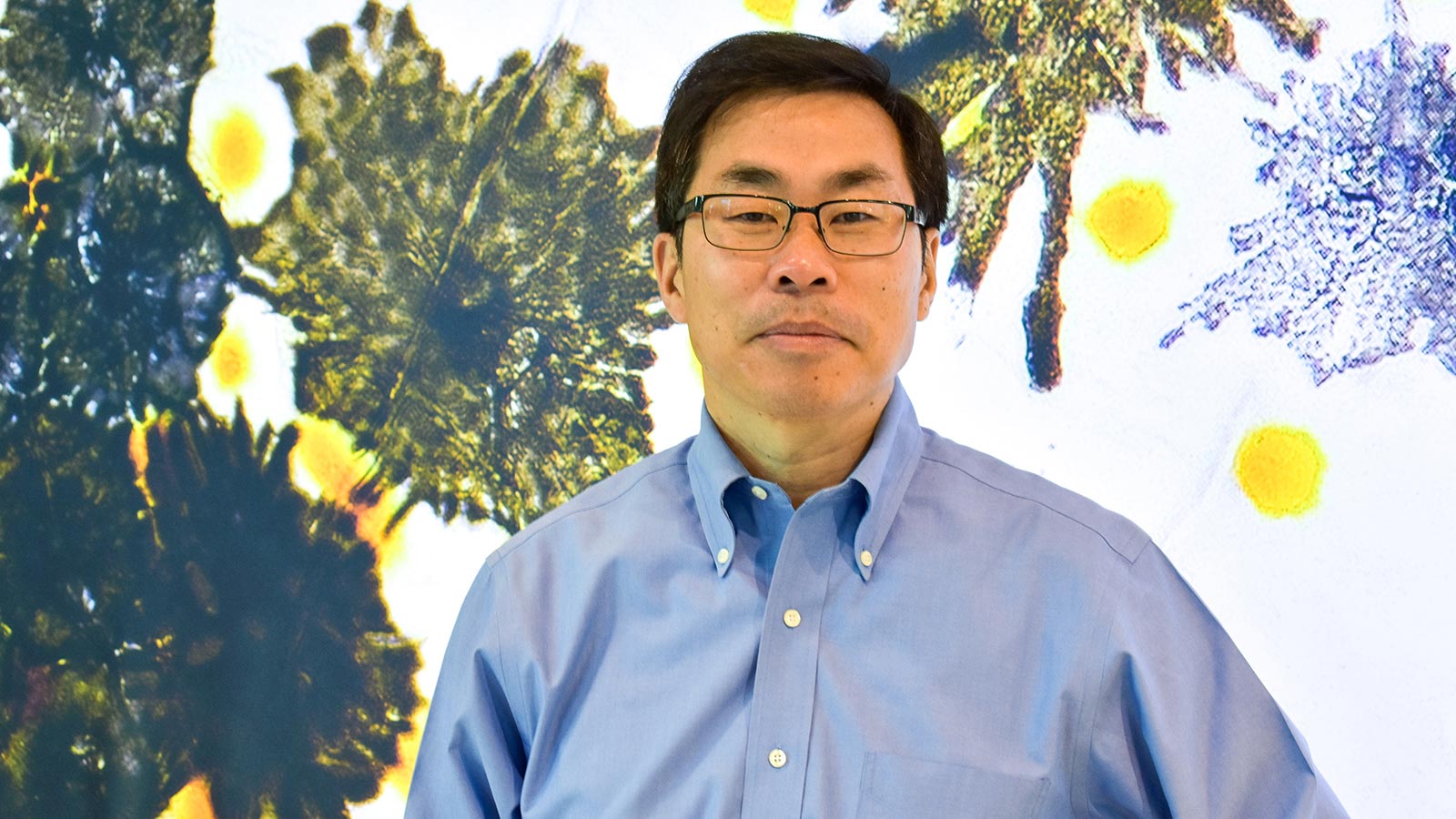
Young contacted Ragon Institute Associate Director Facundo Batista, a world-class expert in immunology and infectious disease. Batista recalls being hesitant at first to co-lead the class; he couldn’t fathom condensing the global emergency into a single course. “But then I realized that the onslaught of information was the very reason we needed to organize this class — to help students and the public avoid misinformation,” he says. “We were filling a gap that the whole world was experiencing.”
Together, Batista and Young generated a list of 14 experts in an array of pandemic-related areas, including Anthony Fauci, director of the National Institute of Allergy and Infectious Diseases, David Baltimore of Caltech, and Kizzmekia Corbett of the National Institutes of Health. Each geared their lecture toward MIT undergraduates with a minimal biology background, and defined key terms and concepts so non-biologists watching the livestream could follow along as well.
Although Batista and Young agree that remote learning pales in comparison to in-person classes, the livestream format opened the talks up to thousands more viewers, and allowed the speakers to present their work without the need for travel. The recordings of each Tuesday lecture were posted on the Department of Biology’s website shortly thereafter, permitting asynchronous viewing for people around the world. The livestream audience regularly exceeded 1,000, and the YouTube views for each recording ranged from 4,000-97,000 and climbing. In many cases, the week’s topic fortuitously coincided with current events. For instance, Corbett spoke about vaccine development just days after the results of Pfizer-BioNTech’s first clinical trial were announced. As one of the NIH scientists who collaborated with Moderna to design another important mRNA-based vaccine, Corbett was able to discuss her reaction to the news and her expectations for Moderna’s imminent clinical trial results.
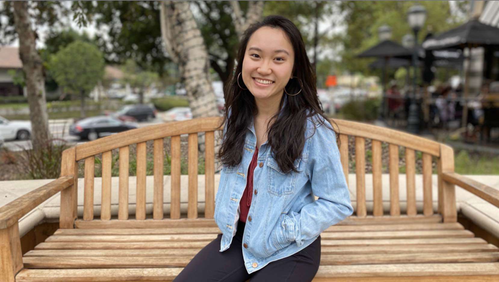
In addition to the livestream audience, each week roughly 300 MIT undergraduates would enter the Zoom room and get the opportunity to ask questions during the Q&A. Participation was unusually high, Young and Batista recall, thanks to the recitation sessions led by graduate student and teaching assistant Lena Afeyan. Afeyan would walk the students through the fundamentals of molecular biology, virology, and any other topics slated to feature heavily in the upcoming lecture. She also invited trainees and medical residents from various institutions to attend, in order to introduce students to the next generation of scientists and health-care professionals. The supplementary reading materials from these sessions are still available online, so biology teachers and other members of the public can access them.
“When I heard that this class was being put together, I hoped I could help make the content more accessible for the students and anyone else joining us,” Afeyan says. “The responses we got were overwhelming. It was incredible to hear from so many teachers, researchers, and alums across the world who watched the course every week.”
Even today, Afeyan, Young, and Batista continue to receive international kudos from scientists and non-scientists alike. At one point, Young was even interviewed by a radio station in Tasmania about the course.
“I learned a lot from 7.00 — not only about Covid-19, but about immunology and biology in general,” says Lucas Marden, a first-year undergraduate who enrolled in the class. “I particularly enjoyed the focus on the real-world response to the pandemic. We learned about everything from designing and developing different tests, treatments, and vaccines, to the scale-up of these technologies. The scientific community’s response to Covid-19 has been incredibly impressive, and I loved learning about it from the people at the forefront of their fields.”
Now, Grossman says, the department is planning to offer the class again this coming fall. “The initial idea stemmed from the need to share clear and reliable information about the pandemic as it began spreading,” he explains. “Although we’ve been living with Covid-19 for over a year now, that need is still present today — perhaps more so as we continue to learn what it will take to tame the virus.”
The next iteration of 7.00 will begin in September, and likely feature some of the same speakers and topics, along with new experts in areas that have recently emerged, such as the evolving viral variants. By arming the MIT community and the public with information from leading experts, Grossman, Batista, Young, and Afeyan hope to help the world navigate this pandemic — and prevent the next one.