Pioneering a deeper understanding of metabolism
Merrill Meadow | Whitehead Institute
March 23, 2022

Metabolism is the sum of life-sustaining chemical reactions occurring in cells and across whole organisms. The human genome codes for thousands of metabolic enzymes, and specific metabolic pathways play significant roles in many biological processes—from breaking down food to release energy, to normal proliferation and differentiation of cells, to pathologies underlying diabetes, cancer, and other diseases.

For decades, Whitehead Institute researchers have helped both to clarify how metabolism works in healthy states and to identify how metabolic processes gone awry contribute to diseases. Among Whitehead Founding Member Harvey Lodish’s wide-ranging accomplishments, for example, are the identification of genes and proteins involved in development of insulin resistance and stress responses in fat cells. His lab explored the hormones controlling fatty acid and glucose metabolism, broadening understanding of obesity and type 2 diabetes. In 1995, the lab cloned adiponectin, a hormone made exclusively by fat cells. A long series of studies has shown that adiponectin causes muscle to burn fatty acids faster – so they are not stored as fat – and increases the metabolism of the sugar glucose. More recently the lab identified and characterized types of RNAs that are specifically expressed in fat cells – including a microRNA unique to brown fat, which burns rather than stores fatty acids. In addition, former Member David Sabatini’s discovery of the mTOR protein and his subsequent work elaborating many ways in which the mTOR pathway affects cells function has proven to be fundamental to understanding the relationship between metabolism and an array of diseases.

Today, Institute researchers continue to pioneer a deeper understanding of how metabolic processes contribute to health and disease – with long-term implications that could range from new treatments for obesity and type 2 diabetes to methods for slowing the aging process. Here are a few examples of Whitehead Institute scientists’ creative and pioneering work in the field of metabolism.

Understanding hibernation and torpor 

Research inspiration comes in many forms. For example, Whitehead Institute Member Siniša Hrvatin – who joined the faculty in January 2022 from Harvard Medical School (HMS) – was inspired to pursue his current research by science-fiction tales about suspended animation for long-term space travel. And during graduate school, he realized that the ability of some mammals to enter a state of greatly reduced metabolism – such as occurs in hibernation –  was a mild but real-world form of suspended animation.

Hrvatin’s doctoral research in Doug Melton’s lab at Harvard University focused primarily on stem cell biology. But his subsequent postdoctoral research positions at Massachusetts Institute of Technology (MIT) and HMS enabled him to begin exploring the mechanisms and impact of reduced metabolic states in mammals. The timing was serendipitous, too, because he was able to use the growing array of genetic tools that were becoming available – and create some new tools of his own as well.

“To survive extreme environments, many animals have evolved the ability to profoundly decrease metabolic rate and body temperature and enter states of dormancy, such as hibernation and torpor,” Hrvatin says. Hibernating animals enter repeated states of significantly reduced metabolic activity, each lasting days to weeks. By comparison, daily torpor is shorter, with animals entering repeated periods of lower-than-normal metabolic activity lasting several hours.

Hrvatin’s lab studies the mysteries of how animals and their cells initiate, regulate, and survive these adaptations. “Our long-term goal is to determine if these adaptations can be harnessed to create therapeutic applications for humans.” He and his team are focusing on three broad questions regarding the mechanisms underlying torpor in mice and hibernation in hamsters.

First: How do the animals’ brains initiate and regulate the metabolic processes involved in this process? During his postdoctoral research,  Hrvatin published details of his discovery of neurons involved in the regulation of mouse torpor. “Now we are investigating how these torpor-regulating neurons receive information about the body’s energy-state,” he explains, “and studying how these neurons then drive a decrease of  metabolic rate and body temperature throughout the body.”

Second: How do individual cells – and their genomes – adapt to extreme or changing environments; and how do these adaptations differ between types of organisms?

“Cells from hibernating organisms ranging from rodents to bears have evolved the ability to survive extreme cold temperatures for many weeks to months,” Hrvatin notes. “We are using genetic screens to identify species-specific mechanisms of tolerance to extreme cold. Then we will explore whether these mechanisms can be induced in non-hibernating organisms – potentially to provide health benefits.”

Third: Can we deliberately and specifically slow down tissue damage, disease progression, and or aging in cells and whole organisms by inducing torpor or hibernation – or facets of those states? It has long been known that hibernating animals live longer than closely related non-hibernators; that cancer cells do not replicate during hibernation; and that cold can help protect neurons from the effects of loss of oxygen. However, the cellular mechanisms underlying these phenomena remain largely unknown. Hrvatin’s lab will induce a long-term hibernation-like state in mice and natural hibernation in hamsters, and study how those states affect processes such as tissue repair, cancer progression, and aging.

“In the lab, if you take many human cell types and put them in a cold environment they die, but cells from hibernators survive,” Hrvatin notes. “We’re fascinated by the cellular processes underlying those survival capacities. As a starting place, we are using novel CRISPR screening approaches to help us identify the genomic mechanisms involved.”

And then? “Ultimately, we hope to take on the biggest question: Is it possible to transfer some of those survival abilities to humans?

Solving a mitochondrial conundrum

When Whitehead Institute postdoctoral researcher Jessica Spinelli was studying cancer metabolism in graduate school, she became interested in what seemed to be a scientific paradox regarding mitochondria, the cell’s energy-producing organelles: Mitochondria are believed to be important for tumor growth; but they generally need oxygen to function, and substantial portions of tumors have very low oxygen levels. Pursuing research in the lab of former Whitehead Institute Member David Sabatini, Spinelli sought to understand how those facts fit together and whether mitochondria could somehow adapt to function with limited oxygen levels.

Recently, Spinelli and colleagues published an answer to the conundrum – one that could inform research into medical conditions including ischemia, diabetes and cancer. In a Science paper for which Spinelli was first author, the team demonstrated that when cells are deprived of oxygen, a molecule called fumarate can serve as a substitute and enable mitochondria to continue functioning.

As Spinelli explains, humans need oxygen molecules for the cellular respiration process that takes place in our cells’ mitochondria. In this process – called the electron transport chain – electrons are passed along in a sort of cellular relay race that, ultimately, allows the cell to create the energy needed to perform its vital functions. Usually, oxygen is necessary to keep that process operating.

Using mass spectrometry to measure the quantities of molecules produced through cellular respiration in varied conditions, Spinelli and the team found that cells deprived of oxygen had a high level of succinate molecules, which form when electrons are added to a molecule called fumarate. “From this, we hypothesized that the accumulation of succinate in low-oxygen environments is caused by mitochondria using fumarate as a substitute for oxygen’s role in the electron transport chain,” Spinelli explains. “That could explain how mitochondria function with relatively little oxygen.” The next step was to test that hypothesis in mice, and those studies provided several interesting findings: Only mitochondria in kidney, liver, and brain tissues could use fumarate in the electron transport chain. And even in normal conditions, mitochondria in these tissues used both fumarate and oxygen to function – shifting to rely more heavily on fumarate when oxygen was reduced. In contrast, heart and skeletal muscle mitochondria made minimal use of fumarate and did not function well with limited oxygen.

“We foresee some exciting work ahead, learning exactly how this process is regulated in different tissues,” Spinelli says, “and, especially, in solid tumor cancers, where oxygen levels vary between regions.”

Seeking a more accurate model of diabetes

Max Friesen, a postdoctoral researcher in the lab of Whitehead Institute Founding Member Rudolf Jaenisch, studies the role of cell metabolism in type 2 diabetes (T2D). An increasingly prevalent disease that affects millions of people around the world, T2D is hard to study in the lab. This has made it very challenging for scientists to detail the cellular mechanisms through which it develops – and therefore to create effective therapeutics.

“It has always been very hard to model T2D, because metabolism differs greatly between species,” Friesen says. “That fact leads to complications when we use animal models to study this disease. Mice, for example, have much higher metabolism and faster heart rates than humans. As a result, researchers have developed many approaches that cure diabetes in mice but that fail in humans.” Nor do most in vitro culture systems—cells in a dish—effectively recapitulate the disease.

But, building on Jaenisch’s pioneering success in developing disease models derived from human stem cells, Friesen is working to create a much more accurate in vitro system for studying diabetes. His goal is to make human stem cell-derived tissues that function as they would in the human body, closely recapitulating what happens when an individual develops diabetes. Currently, Friesen is differentiating human stem cells into metabolic tissues such as liver and adipocytes (fat). He has improved current differentiation protocols by adapting these cells to a culture medium that is much closer to the environment they see in the human body. Serendipitously, the process also makes the cells responsive to insulin at levels that are present in the human bloodstream. “This serves as a great model of a healthy cell that we can then turn into a disease model by exposing the cell to diabetic hyperinsulinemia,” Friesen says.

These advances should enable him to gain a better understanding of how metabolic pathways – such as the insulin signaling pathway – function in a diabetic model versus a healthy control model. “My hope is that our new models will enable us to figure out how dietary insulin resistance develops, and then identify a therapeutic intervention that blocks that disease-causing process,” he explains. “It would be fantastic to help alleviate this growing global health burden.”

Seeing the whole person

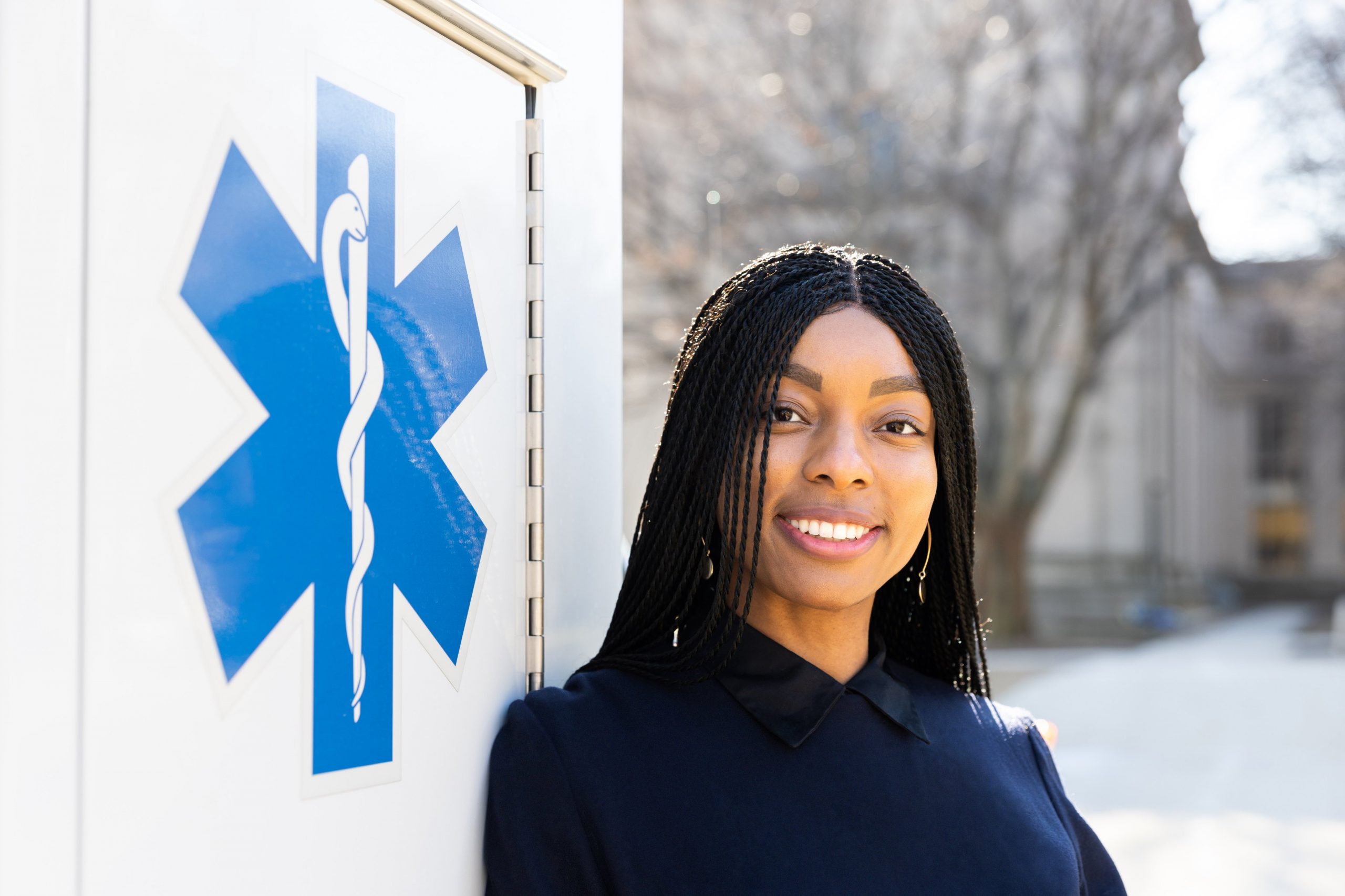
Alumna, Margaret ‘Mo’ Okobi ’16, prepares for a career combining medicine and public health research to improve mental health care services in vulnerable communities

Leah Campbell | School of Science
March 22, 2022

With only one year left of medical school at Harvard, Mo Okobi ’16, took a leave of absence. She didn’t want to take a break as much as a “step back” — a moment to reassess and reframe what she was learning in school within the bigger picture of the U.S. healthcare system. During that year, Okobi earned her master’s in public health from the Harvard School of Public Health.

“It’s so important to keep a broader view of the trends in medicine,” she says, “to look at what medications and therapies we’re using and what is actually working for patients.”

When she applied to the MPH program, Okobi couldn’t have known that her year-long leave to study public health would overlap with a pandemic. Needless to say, it was good timing. Okobi’s joint training in public health and medicine has also uniquely positioned her for a career combining clinical practice and research around chronic mental illness.

Though she says she never expected to find herself on this path, Okobi’s experiences at MIT shaped her approach to medicine and her commitment to providing quality mental healthcare to underserved communities.

Okobi was interested in many aspects of medicine when she enrolled at MIT. She majored in biology, with a minor in chemistry, to explore her broad interests in STEM. She joined MIT Emergency Medical Services, a student-run, volunteer ambulance service, for the same reason.

Being a certified EMT, though, she says, was one of the most formative aspects of her MIT education. She describes it as an “intensive introduction to medicine,” cementing her excitement about her future as a doctor. It was also one of her first exposures to mental health care, and Okobi believes that mental health emergencies made up a large proportion, if not a majority, of their calls.

“MIT EMS was really my first opportunity to actually work with patients very closely and see them in their moments of need,” she says.

While deciding to become a doctor was easy, Okobi’s journey to research wasn’t so smooth. She participated in one official undergraduate research experience at MIT, admitting, “I kind of hated it.” Working in a basic science biology lab, Okobi realized that the bench wasn’t for her.

Fortunately, her mentor, Hazel Sive, a former MIT professor of biology and now Dean of the College of Science at Northeastern University, took the time to talk with Okobi and figure out where she’d thrive. Sive, a South African, connected Okobi with the MIT Africa Program, through which she spent a summer in Johannesburg with the South African National Health Laboratory Services.

That experience, which she describes as a “pivotal framing moment,” helped Okobi understand how a career combining clinical practice and research might look. She worked with scientists studying HIV transmission from mother to child, assessing the quality of testing and resource gaps across different provinces. Near the end of her internship, Okobi was able to go into an HIV clinic and do antibody testing.

“I’m looking at these numbers. I’m making all these graphs and writing this paper, but I’m also seeing the people,” she says. “I loved the clinical work. I loved meeting people and knowing their stories.”

At graduation, Okobi was recognized as a Ronald E. McNair Scholar, an award that goes every year to Black undergraduates who have excelled academically and contributed to the experience of students on campus from underrepresented groups. The award was established in honor of McNair, PhD ’77, an accomplished astronaut who received his doctorate in physics from MIT and tragically perished onboard the Challenger space shuttle in 1986.

Having found a passion for applied, public health research, Okobi spent a year before medical school at a healthcare data analytics startup called Aetion. Aetion uses data from hospitals and insurance companies to analyze healthcare trends like medication usage and clinical outcomes. For a self-described “numbers nerd” like Okobi, it was a great way to learn how healthcare studies are designed and see the big picture behind clinical decisions.

It was her second year of medical school, though, that focused Okobi’s health interests around psychiatry for marginalized populations. During her clinical rotations, she worked at Cambridge Health Alliance (CHA), a public, community-centered hospital. At CHA, she served many non-English speakers and MassHealth recipients and was able to do rotations in outpatient, inpatient, and emergency psychiatric settings.

“I really got to see it from all angles,” Okobi says of her psychiatry rotations. “I loved the practice of creating space for people to talk about their lives…. it’s about the medicine and mental illness, but it’s also about seeing the whole person.”

As for what’s next, Okobi will be heading west for her residency in psychiatry at the University of California San Francisco.

Her work with CHA convinced Okobi to apply for residencies in psychiatry. She’s primarily interested in acute care settings, particularly for those with severe, chronic mental illnesses like schizophrenia. For her, one of the joys of inpatient care is working closely with patients on a daily basis, to, as she describes it, “try to leave a positive, lasting impression on those first encountering psychiatric care.”

Yet, armed with her degree in public health, she’s also committing to combining her clinical practice with ongoing research. At Harvard, she organized a mental health survey for medical and dental students in her class to improve access to mental health resources. Since 2020, she’s been participating in research on psychiatric emergency care utilization with the Boston Emergency Services Team. In 2020, she returned to Aetion as a consultant with their new FDA-backed COVID-19 research group, to lead epidemiological studies examining COVID-19 risk factors.

Looking back, Okobi knows that it may seem like she had a clear professional plan from the get-go. But she stresses that that wasn’t her experience at all, and current students should understand that things have a way of working out if you’re open to trying things.

“My path was very much like ping pong, never knowing where I’m going next,” she says. “The uncertainty never really ends, but I take refuge in those moments of serendipity, when I find something or someone that excites me and challenges me to be a better version of myself.”

Mentorship and medicine

MIT senior Daniel Zhang aims to provide hope for young patients and support to young students.

Celina Zhao | Department of Biology
February 24, 2022

During the virtual spring 2020 semester, Daniel Zhang, a senior majoring in biology, put his time at home to good use. In the garage of his home in San Diego, California, Zhang helped his 13-year-old brother build a lab to study dry eye disease.

This combination of mentorship and medicine feels like second nature to Zhang. When his parents opened a family-run optometry clinic, Zhang was their first patient and then their receptionist. And after a close family member passed away from leukemia, he remembers thinking, “Humans are susceptible to so many diseases — why don’t we have better cures?”

That question propelled him to spend his high school summers studying biomarkers for the early detection of leukemia at the University of California at San Diego. He was invited to present his research at the London International Youth Science Forum, where he spoke to scientists from almost 70 countries. Afterward, he was hooked on the idea of scientific research as a career.

“Research is like standing on the shoulders of giants,” he says. “My experience at the forum was when I knew I loved science and wanted to continue using it to find common ground with others from completely different cultures and backgrounds.”

Exploring the forefront of cancer research

As soon as he arrived at MIT as a first-year undergraduate, Zhang began working under the guidance of postdoc Peter Westcott in professor Tyler Jacks’ lab. The lab focuses on developing better mouse and organoid models to study cancer progression — in Zhang’s case, metastatic colorectal cancer.

One of the ways to model colorectal cancer is by injecting an engineered virus directly into the colons of mice. The viruses, called lentiviral agents, “knock out” tumor suppressor genes and activate the so-called oncogenes that drive cancer forward. However, the imprecise nature of this injection also unintentionally transforms many “off-target” cells into cancer cells, producing a cancer that’s far too widespread and aggressive. Additionally, rare tumors called sarcomas are often initiated rather than adenocarcinomas, the type of tumor found in 95 percent of human cases. As a result, these mouse models are limited in their ability to accurately model colorectal cancer.

To address this problem, Zhang and Westcott designed a method using CRISPR/Cas9 to target a special stem cell called LGR5+, which researchers believe are the types of cells that, when mutated, grow into colorectal cancer. His technique modifies only the LGR5+ cells, which would allow researchers to control the rate at which adenocarcinomas grow. Therefore, it generates a model that is not only much more similar to human colorectal cancer than other models, but also allows researchers to quickly test for other potential cancer driver genes with CRISPR/Cas9. Designing an accurate model is crucial for developing and testing effective new therapies for patients, Zhang says.

During MIT’s virtual spring and fall semesters of 2020, Zhang shifted his focus from benchwork in the lab to computational biology. Using patient data from the Cancer Genome Atlas, Zhang analyzed mutation rates and discovered three genes potentially involved in colorectal cancer tumor suppression. He plans to test their function in his new mouse model to further validate how the dysfunction of these genes drives colorectal cancer progression.

For his work on organoid modeling of colorectal cancer, a third project he’s worked on during his time at the Jacks lab, he also won recognition from the American Association for Cancer Research (AACR). As one of 10 winners of the Undergraduate Scholar Award, he had the opportunity to present his research at the virtual AACR conference in 2021 and again at the next AACR Conference in New Orleans in April 2022.

He credits MIT’s “mens et manus” philosophy, encouraging the hands-on application of knowledge, as a large part of his early success in research.

“I’ve found that, at MIT, a lot of people are pursuing projects and asking questions that have never been thought of before,” Zhang says. “No one has ever been able to develop a late-stage model for colorectal cancer that’s amenable to gene editing. As far as I know, other than us, no one in the world is even working on this.”

Inspiring future generations to pursue STEM

Outside of the lab, Zhang devotes a substantial amount of time to sharing the science he’s so passionate about. Not only has he been awarded the Gene Brown Prize for undergraduate teaching for his time as a teaching assistant for the lab class 7.002 (Fundamentals of Experimental Molecular Biology), but he’s also taken on leadership roles in science outreach activities.

During the 2020-21 academic year, he served as co-director of DynaMIT, an outreach program that organizes a two-week STEM program over the summer for underserved sixth to ninth graders in the greater Boston area. Although the program is traditionally held in-person, in summer 2021 it was held virtually. But Zhang and the rest of the board didn’t let the virtual format deter them from maximizing the fun and interactive nature of the program. They packed and shipped nearly 120 science kits focused on five major topics — astronomy, biology, chemistry, mechanical engineering, and math — allowing the students to explore everything from paper rockets to catapults and trebuchets to homemade ice cream.

“At first, we were worried that most of the students wouldn’t turn on their cameras, since we saw that trend all over MIT classes during the semester,” Zhang says. “But almost everyone had their cameras on the entire time. It was really gratifying to see students come in on Monday really shy, but by Friday be actively participating, making jokes with the mentors, and being really excited about STEM.”

To investigate the long-term impacts of the program, he also helped kick-start a project that followed up with DynaMIT alumni, some of whom have already graduated from college. Zhang says: “We were happy to see that 80-90 percent of DynaMIT alumni enjoyed the program, rating it four or five out of five, and close to 70 percent of them said that DynaMIT had a really positive impact on their trajectory toward a career in STEM.”

Zhang has also served as president of the MIT Pre-medical Society, with the goals of fostering an encouraging environment for premed undergraduates, and providing guidance and resources to first- and second-year students still undecided about the premed path. To achieve these objectives, he pioneered an MIT-hosted mixer with the premedical societies of other Boston colleges, including Wellesley College, Boston University, Tufts University, and Harvard University. At the mixer, students were able to network with each other and listen to guest speakers from the different universities talk about their experiences in medicine. He also started a “big/little” initiative that paired third- and fourth-year mentors with first- and second-year students.

Providing new opportunity and hope

The wealth of activities Zhang has participated in at MIT has inspired his choices for the future. After graduation, he plans to take a gap year and work as a research technician in pediatric oncology before applying to MD/PhD programs.

On the mentorship side, he’s currently working to establish a nonprofit organization called Future African Scientist with his former Ugandan roommate, Martin Lubowa, whom he met at a study abroad program during MIT’s Independent Activities Period in 2020. The organization will teach high schoolers in Africa professional skills and expose them to different STEM topics — a project Zhang plans to work on post-MIT and into the long term.

Ultimately, he hopes to lead his own lab at the intersection of CRISPR-Cas9 technology and cancer biology, and to serve as a mentor to future generations of researchers and physicians.

As he puts it: “All of the experiences I’ve had so far have solidified my goal of conducting research that impacts patients, especially young ones. Being able to provide new opportunity and hope to patients suffering from late-stage metastatic diseases with no current cures is what inspires me every day.”

Investigating pathogens and their life cycles, for the benefit of society

Senior Desmond Edwards has an insatiable curiosity about how the human body works — and how diseases stop it from working.

Leah Campbell | School of Science
November 21, 2021

Desmond Edwards was a little kid when first learned about typhoid fever. Fortunately, he didn’t have the disease. He was looking at a cartoon public health announcement. The cartoon, produced by the Pan American Health Organization, was designed to educate people in his home country of Jamaica about the importance of immunizations for diseases like typhoid. The typhoid character in the cartoon was so unpleasant it gave him nightmares.

Edwards did have his fair share of hospital visits throughout his childhood. But, his own struggles with infection and illness, and those typhoid cartoon nightmares, became his inspiration for pursuing a career studying human disease. At age 6, Edwards was running impromptu baking soda experiments in repurposed glitter containers in his kitchen. Today, he is a senior at MIT, majoring in biology and biological engineering, thanks to a team of dedicated mentors and an insatiable curiosity about how the human body works — or, more accurately, how diseases stop it from working.

Finding a way into research

Edwards knew he wanted to do research but says he assumed that that was something you did after you got your degree. Imagine his surprise, then, upon arriving at MIT in 2018 and meeting classmates who not only had done research, but already had publications. Realizing that he could get a jump-start on his career, he sought out research opportunities and enrolled in the biology class 7.102 (Introduction to Molecular Biology Techniques) for his first-year Independent Activities Period. The class was specifically geared toward first-year students like him with no lab experience.

“It was a great first look at how research is done,” Edwards says of the class. Students took water samples from the Charles River and were expected to identify the strains of bacteria found in those samples using various biological techniques. They looked at the bacteria under a microscope. They examined how the samples metabolized different sources of carbon and determined if they could be stained by different dyes. They even got to try out basic genetic sequencing. “We knew where we were starting. And we knew the end goal,” says Edwards. The in-between was up to them.

Class 7.102 is taught by Mandana Sassanfar, a lecturer in biology and the department’s director of diversity and science outreach. For Sassanfar, the class is also an opportunity to find lab placements for students. In Edwards’ case, she literally led him to the lab of Assistant Professor Becky Lamason, walking up with him one evening to meet a postdoc, Jon McGinn, to talk about the lab and opportunities there. After Edwards expressed his interest to Lamason, she responded within 30 minutes. McGinn even followed up to answer any lingering questions.

“I think that was really what pushed it over the edge,” he says of his decision to take a position in the Lamason lab. “I saw that they were interested not only in having me as someone to help them do research, but also interested in my personal development.”

At the edges of cells and disciplines

The Lamason lab researches the life cycle of two different pathogens, trying to understand how the bacteria move between cells. Edwards has focused on Rickettsia parkeri, a tick-borne pathogen that’s responsible for causing spotted fever. This type of Rickettsia is what biologists call an obligate intracellular pathogen, meaning that it resides within cells and can only survive when it’s in a host. “I like to call it a glorified virus,” Edwards jokes.

Edwards gets excited describing the various ways in which R. parkeri can outsmart its infected host. It’s evolved to escape the phagosome of the cell, the small liquid sac that forms from the cell membrane and engulfs organisms like bacteria that pose a threat. Once it gets past the phagosome and enters the cell, it takes over cellular machinery, just like a virus. At this point of the life cycle, a bacterium will typically replicate so many times that the infected cell will burst, and the pathogen will spread widely. R. parkeri, though, can also spread to uninfected cells directly through the membrane where two cells touch. By not causing a cell to burst, the bacterium can spread without alerting the host to its presence.

“From a disease standpoint, that’s extremely interesting,” says Edwards. “If you’re not leaving the cell or being detected, you don’t see antibodies. You don’t see immune cells. It’s very hard to get that standard immune response.”

In his time in the lab, Edwards has worked on various projects related to Rickettsia, including developing genetic tools to study the pathogen and examining the potential genes that might be important in its life cycle. His projects sit at the intersection of biology and biological engineering.

“For me, I kind of live in between those spaces,” Edwards explains. “I am extremely interested in understanding the mechanisms that underlie all of biology. But I don’t only want to understand those systems. I also want to engineer them and apply them in ways that can be beneficial to society.”

Science for society

Last year, Edwards won the Whitehead Prize from the Department of Biology, recognizing students with “outstanding promise for a career in biological research.” But his extracurricular activities have been driven more by his desire to apply science for tangible social benefits.

“How do you take the science that you’ve done in the lab, in different research contexts, and translate that in a way that the public will actually benefit from it?” he asks.

Science education is particularly important for Edwards, given the educational opportunities he was given to help get to MIT. As a high schooler, Edwards participated in a Caribbean Science Foundation initiative called the Student Programme for Innovation in Science and Engineering. SPISE, as it’s known, is designed to encourage and support Caribbean students interested in careers in STEM fields. The program is modeled on the Minority Introduction to Engineering and Science program (MITES) at MIT. Cardinal Warde, a professor of electrical engineering, is himself from the Caribbean and serves as the faculty director for both MITES and SPISE.

“That experience not only kind of opened my eyes a bit more to what was available, what was in the realm of possibilities, but also provided support to get to MIT,” Edwards says of SPISE. For example, the program helped with college applications and worked with him to secure an internship at a biotech company when he first moved to the United States.

“If education falters, then you don’t replenish the field of science,” Edwards argues. “You don’t get younger generations excited, and the public won’t care.”

Edwards has also taken a leadership role in the MIT Biotechnology Group, a campus-wide student group meant to build connections between the MIT community and thought leaders in industry, business, and academia. For Edwards, the biotech and pharmaceutical industries play a clear role in disease treatment, and he knew he wanted to join the group before he even arrived at MIT. In 2019, he became co-director of the Biotech Group’s Industry Initiative, a program focused on preparing members for industry careers. In 2020, he became undergraduate president, and this year he’s co-president of the entire organization. Edwards speaks proudly of what the Biotech Group has accomplished during his tenure on the executive board, highlighting that they not only have the largest cohort ever this year, but it’s also the first time the group has been majority undergraduate.

Somehow, in between his research and outreach work, Edwards finds time to minor in French, play for the Quidditch team, and serve as co-president on the Course 20 Undergraduate Board, among other activities. It’s a balancing act that Edwards has mastered over his time at MIT because of his genuine excitement and interest in everything that he does.

“I don’t like not understanding things,” he jokes. “That applies to science, but it also extends to people.”

Stem Cell Research Zeroes in on Cancer

Collaborators investigate colon health with novel tools

Deborah Halber | Spectrum
November 9, 2021

In a building at the edge of the Massachusetts General Hospital (MGH) complex, Ömer Yilmaz, MD, and a group of pathology residents gather around a microscope. A resident reads from a chart: a growth was found in the intestine of a patient who had complained of abdominal pain.

Yilmaz, an MIT cancer researcher and a gastrointestinal pathologist, hoped a closer look at the tumor would reveal a noncancerous collection of fat cells or lymphoid cells.

It had taken a couple of days to prepare the biopsy. Somewhere in the hospital, the patient and her family were anxiously awaiting a diagnosis. Yilmaz leaned forward and adjusted the focus on the microscope.

On the tracks of cancer

If the long, twisting tube of the human digestive tract were stretched out straight, it would extend 30 feet, and its absorptive surface area is roughly comparable to the size of a tennis court. A significant chunk of that tube is the large intestine, an intricate place rife with microscopic structures called niches and crypts, evoking an underground cavern or the ocean floor. Besides the skin, the intestines are the body’s primary barrier against external invaders.

Yilmaz, an associate professor of biology at the Koch Institute for Integrative Cancer Research, believes certain cancers and diseases such as inflammatory bowel disease originate with a breakdown of the intestine’s protective barrier. Diet appears to affect intestinal stem cells; these cells can morph into a variety of cell types, and changes in stem cells can lead to cancer, but no one understands exactly how this occurs.

That’s where Yilmaz’s partnership with MIT biomedical engineer and chemist Alex Shalek comes in. Yilmaz and Shalek are both members of the MIT Stem Cell Initiative, which focuses on fundamental biological questions about benign and cancerous adult stem cells.

Shalek, a core member of the Institute for Medical Engineering and Science (IMES), a member of the Koch Institute, and an associate professor of chemistry, develops experimental and computational tools that provide researchers with detailed snapshots of what’s going on inside living cells at a moment in time. Some of these tools, Yilmaz hoped, would enable him to see how intestinal cells react when they encounter an influx of fat or are deprived of food for hours or days.

“In the past, people would have taken a piece of gut that had many different cell types and said, ‘What changes, on average, under different dietary conditions?’” Shalek says. His tools give him and Yilmaz more precise information, providing a window into the discrete molecular responses of individual cells within the colon.

The role of stem cells

Growing up in Battle Creek, Michigan, Yilmaz spent all his free time trailing after his father, a physician who had immigrated from Turkey. He’d make hospital rounds with his dad, visiting the pathology and radiology labs. As Yilmaz grew older, the two would talk about the mechanisms underlying disease.

After completing his MD/PhD at the University of Michigan, Yilmaz did his residency in pathology, the study of disease, at MGH. He began working at the Whitehead Institute with MIT biology professor David M. Sabatini, a pioneer in elucidating the mechanisms under-lying the regulation of growth and metabolism in mammals. Yilmaz had long been fascinated with stem cells’ seemingly miraculous ability to become any kind of cell the body needed. In adults, stem cells are relatively rare, best studied in bone marrow.

When scientists first found stem cells in the intestine in 2007, Yilmaz shifted his research focus. “As soon as intestinal stem cells were identified, I became interested in understanding how they are regulated by diet and aging,” he says.

“We know obesity elevates cancer risk in a wide range of tissues, including the colon, but we don’t know exactly how. And fasting regimens have been known to improve organ and tissue health, but this, too, is not well understood.”

To better study the transition from healthy to diseased cells in the colon, Yilmaz’s team generated colon tumors in mice that closely resemble human tumors. These colon tumors from mice or humans can be grown in culture, creating miniature three-dimensional tumors called organoids.

Subjecting the organoids to different conditions, Yilmaz and Sabatini found that in mice, age-related loss of stem cell function can be reversed by a 24-hour fast. Other studies looked at the type of high-fat diet leading to obesity. Yilmaz determined that a high-fat diet boosted the population of intestinal stem cells and generated even more cells that behaved like stem cells. These stem cells and stem-like cells are more likely to give rise to intestinal tumors.

What’s happening inside

In the microenvironment of the digestive system, the single layer of epithelial cells that line the colon die after only a few days of ferrying nutrients into the bloodstream and lymphatic system.

Stem cells sheltered in protected spaces with fanciful names like the crypts of Lieberkühn generate a hundred grams of new intestinal tissue every day. The source of all the epithelial cells as well as the cells of the villi, a velvety layer of fingerlike projections that line the intestine, stem cells repair and replace tissue continually assaulted by stomach acid, pancreatic enzymes, bile, fats, and bacteria.

Nearby cells guard the stem cells by secreting agents that fight off harmful bacteria, fungi, and viruses and help regulate the composition of the microbiome.

Most of the body’s stem cells, like those deep within bone marrow, are not nearly as prolific as intestinal stem cells, likely because there’s a risk associated with the stem cells’ ability to rapidly replace themselves: mutations.

At the heart of a cell’s behavior is its messenger RNA, or mRNA, the technology used in the Moderna and Pfizer Covid-19 vaccines. These mRNA vaccines teach cells how to make a protein that triggers an immune response to the virus. Each mRNA transcript, a single strand of RNA carrying a specific genetic instruction from the DNA in the nucleus to the cell’s protein-making machinery, determines which protein gets made to help support the cell’s activity.

“From a snapshot of all of the cell’s mRNA, its transcriptome, we can see how it is trying to respond to change,” Shalek says.

Shalek’s tools help him and Yilmaz measure the properties of multiple types of intestinal cells—immune cells, stem cells, and epithelial cells, to name a few—at once to see precisely how these otherwise invisible, minute features collectively orchestrate tissue-wide responses to external signals.

Sequencing a cell’s mRNA makeup requires smashing the cell open and collecting all of its transcripts. Shalek jokingly likened the process to an alien invader beaming human specimens up to a spaceship and investigating what’s happening inside them.

One of the methods Shalek helped develop tags each mRNA within a cell so that it can be traced back to its cell of origin even after it’s been ripped apart. The inexpensive, portable system, called Seq-Well, looks like an ice cube tray. Around the size of a stick of gum, it contains roughly a hundred thousand miniature wells, each approximately 50-by-50-by-50 microns.

Each cell is deposited into its own well, which contains a bead coated with uniquely barcoded DNA molecules; those DNA molecules are designed to latch onto mRNA and ignore the rest of the cell’s components. The wells are sealed and the cells broken apart. The beads are then extracted, processed, and analyzed, providing a record of each cell’s intentions in its last living moments.

The fact that the system can look simultaneously at thousands of individual cells of any type allows Shalek and Yilmaz to check the effect of nutrients on epithelial cells, immune cells, and stem cells all at once.

The Shalek lab is also developing screening tools that are particularly useful for exposing the Yilmaz lab’s organoids to hundreds of nutrients or drugs at one time, potentially reducing the effort needed to identify substances that boost or hinder stem cell function.

Already, Yilmaz and Shalek have used Seq-Well to identify an enzyme that could be a potential future target for a drug that would counter the negative effects of a high-fat diet on intestinal stem cells. More broadly, Yilmaz says, their collaboration is helping develop a very nuanced understanding of a very complex organ.

“Understanding that complexity is what has really driven our collaboration,” Yilmaz says. “Alex has developed the tools that enable us to dissect out individual cell populations and start to understand how environmental factors impact gene expression.”

“Scientists have spent the past 40 years delineating the genetic drivers of colon cancer, and we still have more to learn. But we’ve now entered the era in which we want to understand the impact of environmental and host factors,” Yilmaz says.

Yilmaz hopes to identify nutrients and metabolites that can enhance stem cell function to repair damage after injury, or to identify mechanisms that dampen tumor formation. In addition, biomarkers such as levels of certain substances in the blood could be a key to early intervention, he says.

“Can we identify which obese patients are more prone to developing colon cancer? If so, can we identify therapies that go after weaknesses in their tumors?”

Battling colon cancer

During the time Yilmaz spends at MGH, he looks at slide after slide of biopsied cells. Normal epithelial cells line up in a single, orderly row. After 15 years in medicine, the twisted appearance of diseased cells still shocks him. “You know, in most cases, the number one predictor of how bad a tumor is going to behave isn’t its genetic signature,” he says. “It’s how deep they invade into their organ of origin, whether they have spread to distant organs, and how bad they look under the microscope.” The cells of this patient’s tumor are misshapen, haphazardly stacked on top of each other.

The patient is in her forties. Yilmaz recalled that when he was a resident, colon cancer in a 40-year-old or 30-year-old was a rarity. He now sees such cases almost weekly. Colorectal cancer is among the top three leading causes of cancer-related deaths in the United States, according to the American Cancer Society. It’s expected to cause around 53,000 deaths during 2021. Yilmaz writes up his diagnosis: invasive cancer of the sigmoid colon. The patient’s oncologist will consult with Yilmaz, radiologists, and the surgical team to come up with a treatment plan.

Ultimately, Yilmaz wants to develop strategies to prevent and reduce the growth of tumors in the intestinal tract. The fact that increasingly younger patients are being diagnosed highlights, for him, the importance of diet. “It’s very worrisome,” he says. “We’re at the beginning of a trend where we’re going to see more and more young people afflicted with what can be a fatal disease if not caught early.” Diet could be an important place to start.

He says, “If you can prevent cancer, that’s the best treatment.”

Differences in T cells’ functional state determine resistance to cancer therapy

Researchers decipher when and why immune cells fail to respond to immunotherapy, suggesting that T cells need a different kind of prodding to re-engage the immune response.

Grace van Deelen
October 29, 2021

Non-small cell lung cancer (NSCLC) is the most common type of lung cancer in humans. Some patients with NSCLC receive a therapy called immune checkpoint blockade (ICB) that helps kill cancer cells by reinvigorating a subset of immune cells called T cells, which are “exhausted” and have stopped working. However, only about 35% of NSCLC patients respond to ICB therapy. Stefani Spranger’s lab at the MIT Department of Biology explores the mechanisms behind this resistance, with the goal of inspiring new therapies to better treat NSCLC patients. In a new study published on Oct. 29 in Science Immunology, a team led by Spranger lab postdoc Brendan Horton revealed what causes T cells to be non-responsive to ICB — and suggested a possible solution.

Scientists have long thought that the conditions within a tumor were responsible for determining when T cells stop working and become exhausted after being overstimulated or working for too long to fight a tumor. That’s why physicians prescribe ICB to treat cancer — ICB can invigorate the exhausted T cells within a tumor. However, Horton’s new experiments show that some ICB-resistant T cells stop working before they even enter the tumor. These T cells are not actually exhausted, but rather they become dysfunctional due to changes in gene expression that arise early during the activation of a T cell, which occurs in lymph nodes. Once activated, T cells differentiate into certain functional states, which are distinguishable by their unique gene expression patterns.

In order to determine why some tumors are resistant to ICB, Horton and the research team studied T cells in murine models of NSCLC. The researchers sequenced messenger RNA from the responsive and non-responsive T cells in order to identify any differences between the T cells. Supported in part by the Koch Institute Frontier Research Program, they used a technique called Seq-Well, developed in the lab of fellow Koch Institute member J. Christopher Love, the Raymond A. (1921) and Helen E. St. Laurent Professor of Chemical Engineering and a co-author of the study. The technique allows for the rapid gene expression profiling of single cells, which permitted Spranger and Horton to get a very granular look at the gene expression patterns of the T cells they were studying.

Seq-Well revealed distinct patterns of gene expression between the responsive and non-responsive T cells. These differences, which are determined when the T cells assume their specialized functional states, may be the underlying cause of ICB resistance.

Now that Horton and his colleagues had a possible explanation for why some T cells did not respond to ICB, they decided to see if they could help the ICB-resistant T cells kill the tumor cells. When analyzing the gene expression patterns of the non-responsive T cells, the researchers had noticed that these T cells had a lower expression of receptors for certain cytokines, small proteins that control immune system activity. To counteract this, the researchers treated lung tumors in murine models with extra cytokines. As a result, the previously non-responsive T cells were then able to fight the tumors — meaning that the cytokine therapy prevented, and potentially even reversed, the dysfunctionality.

Administering cytokine therapy to human patients is not currently safe, because cytokines can cause serious side effects as well as a reaction called a “cytokine storm,” which can produce severe fevers, inflammation, fatigue, and nausea. However, there are ongoing efforts to figure out how to safely administer cytokines to specific tumors. In the future, Spranger and Horton suspect that cytokine therapy could be used in combination with ICB.

“This is potentially something that could be translated into a therapeutic that could increase the therapy response rate in non-small cell lung cancer,” Horton says.

Spranger agrees that this work will help researchers develop more innovative cancer therapies, especially because researchers have historically focused on T cell exhaustion rather than the earlier role that T cell functional states might play in cancer.

“If T cells are rendered dysfunctional early on, ICB is not going to be effective, and we need to think outside the box,” she says. “There’s more evidence, and other labs are now showing this as well, that the functional state of the T cell actually matters quite substantially in cancer therapies.” To Spranger, this means that cytokine therapy “might be a therapeutic avenue” for NSCLC patients beyond ICB.

Jeffrey Bluestone, the A.W. and Mary Margaret Clausen Distinguished Professor of Metabolism and Endocrinology at the University of California-San Francisco, who was not involved with the paper, agrees. “The study provides a potential opportunity to ‘rescue’ immunity in the NSCLC non-responder patients with appropriate combination therapies,” he says.

This research was funded by the Pew-Stewart Scholars for Cancer Research, the Ludwig Center for Molecular Oncology, the Koch Institute Frontier Research Program through the Kathy and Curt Mable Cancer Research Fund, and the National Cancer Institute.

Ushering discoveries from bench to bedside

Studying cancer in the Sharp lab helped Courtney JnBaptiste learn strategic thinking skills that he uses as a patent agent, transforming technology into successful biotech businesses.

Raleigh McElvery | Department of Biology
October 17, 2021

Courtney JnBaptiste PhD ’16 spent the first 19 years of his life on the idyllic Caribbean island of Saint Lucia, surrounded by clear waters, sandy beaches, and a robust agricultural community. His family owned their own farm, where they grew bananas and other crops for export. JnBaptiste and his six siblings spent hours each day after school and on the weekends helping to harvest the fruits of their labor. “We were better off than most, but it was a hard existence,” he says. “I had to fight to make something out of life. Where I am today is a big leap.”

He has since moved to the U.S. and completed his PhD at the MIT Department of Biology. Today, he uses the strategic thinking skills he learned during graduate school in his job as a patent agent, helping researchers protect their inventions and start biotech companies.

Despite his exceptional grades, JnBaptiste didn’t enjoy school growing up, and he’d often try to convince his parents that he didn’t need to go to class. His mother, a middle school teacher, was unfazed by his excuses and sent him off to school most days. Despite his protests, JnBaptiste understood his mother’s motto that “education is the key to success.” He knew he’d need good academic standing and self-motivation to attain the financially-stable life he envisioned.

During high school, cable TV became available to his community for the first time, and he was overwhelmed by the deluge of information. He became hooked on Animal Planet and Discovery Channel, and decided that he wanted to be like Jeff Corwin, a biologist, wildlife conservationist, and TV personality. JnBaptiste knew he’d need a college education to reach his career aspirations, and — due to its proximity and promise of opportunity — the U.S. seemed like an ideal destination.

He was accepted to Bethune-Cookman University in Florida and declared a major in biology. He was awarded an environmental research grant, which allowed him to spend several semesters studying the snail populations in Blue Spring State Park. But a summer internship at the University of Kansas Medical Center was what ultimately convinced him to pursue molecular biology rather than environmental sciences. During his junior year, a new biochemistry professor arrived: Christopher Ainsley Davis, a former postdoc in the lab of Cathy Drennan at the MIT Department of Biology.

“When the two of us spoke, he said something that shocked me,” JnBaptiste recalls. “He told me, ‘You’re good enough for MIT and you should apply to their summer research program.’ That just blew my mind. I never thought I was of the MIT caliber — no one had ever challenged me like that before.”

At Davis’ urging, JnBaptiste applied to the MIT Summer Research Program in Biology (MSRP-Bio), and was placed in the lab of Nobel laureate Phil Sharp. In the 1970s, Sharp had co-discovered splicing, a molecular process that happens after DNA has been transcribed into RNA. Segments of the RNA strand are removed, and the remaining parts are stitched back together and translated into proteins that perform vital functions inside the cell.

Two people at a lab bench
JnBaptiste (left) completed his PhD research in the lab of Phil Sharp (right).

Under the supervision of graduate student and postdoc mentors, JnBaptiste spent the summer of 2009 investigating the role that RNA splicing plays in cancer. “It was the best time of my life,” he says. “I just I loved it. I loved the environment. I loved the lab. I loved MIT. And that experience had a profound impact on me.”

He enjoyed campus so much that he returned just one year later to begin his PhD. He was looking forward to returning to the lab bench, but what he didn’t anticipate was that his first semester of classes would be the most rigorous education he’d ever received. He excelled in biochemistry, but found 7.52 (Graduate Genetics) especially difficult. “It was the first time I’d ever failed an exam,” he recalls.

With the help of mentors, classmates, and tutors, JnBaptiste passed genetics and moved on to the stage of his PhD that he was most excited about: lab rotations. After testing out a few different research groups, he ultimately decided to return to the Sharp lab. In his own words: “It was home.”

JnBaptiste’s thesis project focused on a type of RNA known as microRNA (miRNA), which is never translated into a protein. Instead, it remains in its single-stranded RNA form and helps regulate gene expression. The Sharp lab found that removing all the miRNAs from adult cells prompted dramatic activation of embryonic genes. These genes are typically turned off in adult cells, and only expressed during early development when rapid cell division is required. But they can also be hijacked during cancer to create tumors.

JnBaptiste was surprised to find that adding the miRNAs back into the cells didn’t shut down these embryonic genes. In fact, restoring the miRNAs made the cells divide even more rapidly and increased their ability to form tumors — suggesting “global miRNA restoration” would not be a viable approach to treat cancer.

“This model that we developed showed miRNAs control a very important network in the context of both development and cancer,” JnBaptiste explains. “Cancer occurs when normal cellular processes go awry, so understanding those fundamental molecular interactions is critical to fighting the disease.”

By the time he graduated from MIT in 2016, JnBaptiste knew he enjoyed science, but didn’t have ambitions to run his own lab. Instead, he was curious about how lab experiments and research questions engender companies.

When he was still an MSRP-Bio student, JnBaptiste had met an intellectual property lawyer who’d come to speak on campus. He’d been saving her business card ever since, and reached out to her as his time at MIT was drawing to a close. With her assistance, JnBaptiste was offered a job as a scientific advisor at Goodwin Procter, the international law firm where she worked.

JnBaptiste has since transitioned to a similar role as a patent agent at Pabst Patent Group. There, he collaborates with lawyers to write patents protecting new research technology. While scientists are focused on the minutia of their day-to-day lab experiments, JnBaptiste is tasked with understanding the bigger picture, and how those experiments might lay the foundation for successful businesses that could revolutionize therapeutic approaches.

“As a grad student at MIT, I learned a lot about what it takes to be a strategic thinker in science,” JnBaptiste says. “People like my mentor, Phil Sharp, not only recognize a discovery, they look beyond it to envision its future potential as the next biotech company. That’s a skill I’m still honing as I work to develop my business acumen.”

Looking back at his career trajectory thus far, JnBaptiste is struck by the “beauty and diversity” that comes with earning a degree in biology. “Follow your passions,” he advises, “and surround yourself with people who can see the potential and value in you — even when you cannot yet see it yourself.”

Posted: 10.17.21
Top photo: Elisabeth Sherwin/Hello Headshots
Sara Prescott

Education

  • PhD, 2016, Stanford University School of Medicine
  • BA, 2008, Molecular Biology, Princeton University

Research Summary

Our bodies are tuned to detect and respond to cues from the outside world and from within through exquisite collaborations between cells. For example, the cells lining our airways communicate with sensory neurons in response to chemical and mechanical signals, and evoke key reflexes such as coughing. This cellular collaboration protects our airways from damage and stabilizes breathing, but can become dysregulated in disease. Despite their vital importance to human health, fundamental questions about how sensory transduction is accomplished at these sites remain unsolved. We use the mammalian airways as a model system to investigate how physiological insults are detected, encoded, and addressed at essential barrier tissues — with the ultimate goal of providing new ways to treat autonomic dysfunction.

Awards

  • Warren Alpert Distinguished Scholars Award, 2021
  • Life Sciences Research Foundation Fellowship, 2018