CAMBRIDGE, MA–When it comes to gene expression in the endosperm of seeds, gene provenance matters. In this specialized tissue, plants actively strive to keep the expression of genes inherited from the mother versus the father in balance, according to Whitehead Institute scientists.
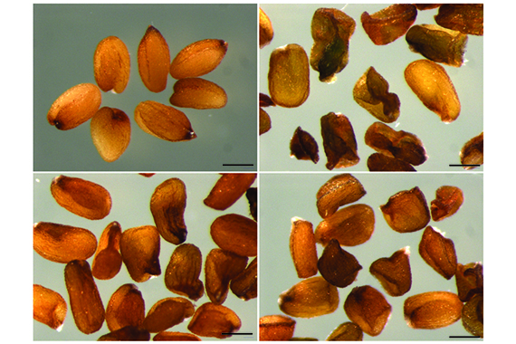
The endosperm, the starchy part of a seed that envelopes and nourishes the developing embryo, comprises two-thirds of the calories in a typical human diet. It is the meat of a coconut and the sweet part of the corn on the cob we eat. In a paper published online December 19 in the journal Cell Reports, Whitehead Member Mary Gehring, first author and former Gehring graduate student Robert Erdmann, and colleagues reveal that the endosperm is also the site where the plant must actively orchestrate a delicate balance between expression of genes inherited from the mother and those of the father. If this critical balance errs toward one parent or the other, seeds can be too small or even abort.
Unlike most plant cells, which have two copies of the genome, cells within the endosperm have three copies: one inherited from the father, and two inherited from the mother. This ratio is established when a sperm cell in the fertilizing pollen grain fuses with the central cell associated with the egg cell in a flower’s ovule. Unlike most cells, the central cell has two nuclei, so when the sperm’s nucleus merges with the central cell, the resulting endosperm is triploid.
The 2-to-1 ratio of maternal to paternal gene expression is crucial, and deviation can have dire consequences: If maternal gene expression is too high, the seeds are too small; if paternal gene expression is too high, the seeds abort. Although plant biologists have known the importance of this ratio for seed viability, the balance was assumed to be passively maintained for the majority of genes. Previously, Gehring determined that a subset of genes expressed in the endosperm are imprinted—their expression is inherited from their parent. But what about the remaining majority of the genome?
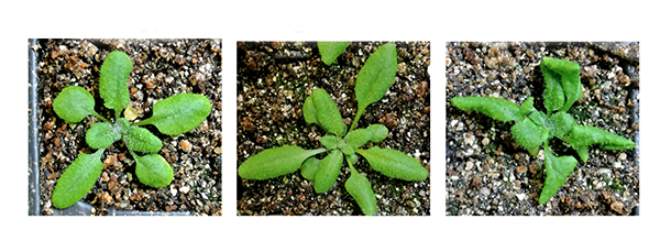
Now Gehring and colleagues have discovered a role for small RNAs—snippets of RNA that interfere with and can reduce gene expression—in actively maintaining this 2-to-1 balance in those genes that are not imprinted. This the first time scientists have documented small RNAs maintaining such a ratio. Using Arabadopsis thaliana and Arabadopsis lyrata plants, Gehring and her lab determined that these small RNAs tamp down the expression of maternally inherited genes. When the enzyme that creates the small RNAs is mutated, fewer small RNAs are produced, and the plant’s carefully balanced gene expression is thrown off. The resulting seeds have excessive maternal gene expression. To understand the significance of this elevated maternal gene expression, Satyaki Rajavasireddy, a postdoctoral researcher in Gehring’s lab and an author of the Cell Reports paper, turned to plants with seeds that abort because they have additional copies of paternal genes. When these plants with extra paternal DNA had their small-RNA-producing enzyme mutated, the outcome was striking: The seeds were rescued and developed to maturity.
Although the research analyzed this phenomenon in A. thaliana and A. lyrata, Gehring expects it to be a widespread manifestation of the tug-of-war between maternal and paternal genetic contributions.
“Maintaining this maternal/paternal balance is crucial for seed development, including in crop plants,” says Gehring, who is also an associate professor of biology at Massachusetts Institute of Technology. “We’ve looked at two species that are separated by 10 million years of evolution, and I anticipate we will find this mechanism in other species as well.”
This work was supported by the National Science Foundation (NSF CAREER grant 1453459).
Written by Nicole Giese Rura
* * *
Mary Gehring’s primary affiliation is with Whitehead Institute for Biomedical Research, where her laboratory is located and all her research is conducted. She is also an associate professor of biology at Massachusetts Institute of Technology.
* * *
Full Citation:
“A small RNA pathway mediates allelic dosage in endosperm”
Cell Reports, online December 19, 2017.
Robert M. Erdmann (1,2), P.R. V. Satyaki (1), Maja Klosinska (1), Mary Gehring (1,2).
1. Whitehead Institute for Biomedical Research, Cambridge, MA 02142 USA
2. Department of Biology, Massachusetts Institute of Technology, Cambridge, MA 02139 USA
December 14, 2017
While our genome contains a vast repertoire of genes that are responsible for virtually all of the cellular and developmental processes life requires, it is the complex dance of regulating their expression that is vital for genetic programs to be executed successfully. Genes must be turned on and off at appropriate times or, in some cases, never turned on or off at all.
Methylation—the addition of chemical tags to DNA—typically reduces the expression of methylated genes. In many cases, DNA methylation can be thought of as roadblocks on a gene. The more methylated a gene is, the less likely it is that it will be active. Such genetic demarcations are critical to ensure that genes involved in particular stages of development are active at the right time, for example. Methylation is essential for proper cellular function, and its dysregulation is associated with diseases, such as cancer in humans. Despite its importance, little is known about how critical methylation patterns are inherited or maintained. Whitehead Institute Member Mary Gehring and her lab have identified a mechanism important for maintaining methylation, that when disrupted, results in the demethylation of large sections of the Arabidopsis plant’s genome. Their work is described this week in the journal Nature Communications.
Using an unusual gene in the plant Arabidopsis, Gehring is teasing apart the mechanisms that underpin methylation. By breaking this unique gene’s “circuit,” Gehring and Ben Williams, a postdoctoral researcher in her lab, have gained important insights into how methylation is maintained, including a surprising finding that previously erased methylation can be restored under certain circumstances.
In order to better understand methylation’s heritability, Gehring and Williams looked closely at an anomaly, the ROS1 gene in Arabidopsis plants, which encodes a protein that removes methylation from its own gene as well as others. Previously, Gehring and Williams had determined that ROS1 methylation actually functions in the complete opposite way from the existing paradigm—unlike most genes, when a short section of this gene is methylated, the gene is actually activated instead of inactivated. Conversely, if it is methylated, the gene is turned on. As a result, ROS1 can act as a rheostat for the Arabidopsis genome: As methylation increases, ROS1 turns on and begins removing methyl groups, and as methylation decreases, ROS1 shuts off and reduces its demethylating activity.
In the current research, Williams altered methylation at ROS1 so that its activity was uncoupled from methylation levels in the genome, in order to see what effects such a change would have on methylation throughout the entire genome. When he analyzed the plants’ methylation, it was haywire. Methylation was lost throughout the genome and progressively decreased in subsequent generations, except in a particular part of the genome called the heterochromatin—genomic areas that are strongly repressed. Interestingly, Williams found that, despite the alteration of the ROS1regulatory circuit, these heterochromatic sections of the genome actually regain their methylation and approach full methylation by the fourth generation— the same time point by which the rest of the genome has lost much of its methylation .
The researchers determined that the ROS1 circuit they uncovered is important for methylation homeostasis because it causes heritable loss of methylation when disrupted. And yet methylation returns at some locations, albeit not immediately, suggesting that Arabidopsis enlists multiple mechanisms to maintain methylation homeostasis. Gehring and Williams are intrigued by that delay in remethylation and are working to identify its cause as well as other mechanisms that may also be at work regulating this critical process.
This work was supported by the National Institute of General Medical Sciences of the National Institutes of Health (R01GM112851).
Written by Nicole Giese Rura
* * *
Mary Gehring’s primary affiliation is with Whitehead Institute for Biomedical Research, where her laboratory is located and all her research is conducted. She is also an associate professor of biology at Massachusetts Institute of Technology.
* * *
Full Citation:
“Stable transgenerational epigenetic inheritance requires a DNA methylation-sensing circuit”
Nature Communications, December 14, 2017.
Ben P. Williams (1) and Mary Gehring (1,2).
1. Whitehead Institute for Biomedical Research, Cambridge, MA 02142
2. Department of Biology, Massachusetts Institute of Technology, Cambridge, MA 02139
Whitehead researchers unravel fundamental molecular machinery that propels chromosome movement
November 16, 2017
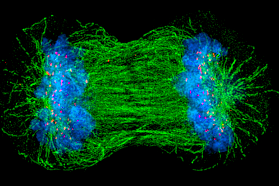
Each day, billions of cells in the human body undergo a vital ritual, wherein one cell divides to form two. This process, known as cell division, is as beautiful as it is essential, undergirding the body’s growth in times of both health and disease. Despite the fact that cell division (or “mitosis”) has been a basic topic in high school biology classes for the past 70 years, the mechanisms by which cells conduct this critical event remain poorly understood. In particular, there are lingering uncertainties about how chromosomes — large units of DNA that include our genes — get properly allocated so that both daughter cells receive intact, complete copies of their genetic blueprint.
“People have been watching chromosomes move, align, and segregate for more than a century — it’s such a fundamental aspect of biology,” says Iain Cheeseman, a member of the Whitehead Institute for Biomedical Research and an associate professor of biology at Massachusetts Institute of Technology. “It’s also much more elegant and complicated than we ever anticipated.”
Cheeseman and members of his Whitehead laboratory have discovered many of the molecular movers and shakers that ensure chromosomes get to the right place at the right time. These components assemble together — like the parts of an engine — to establish robust connections with chromosomes and ultimately power their movement within cells. “The most important unanswered question in the field is how do these components work together? That is, how do you build a machine that is more than the sum of its parts?” he says.
Now, in two recent papers in the journals eLife and Current Biology, Cheeseman and his colleagues help shed light on this central question.
Back to the drawing board
As recently as 15 years ago, scientists assumed that in dividing cells, chromosomes move the way many other cellular objects move — transported by tiny molecular motors. These mini-motors are specifically designed to travel along roads made from rod-like structures called microtubules. Like a car cruising on a highway, they can carry cargo over long distances. Although such microtubule-based vehicles seemed a logical suspect, when scientists inactivate them in human cells, chromosomes can still move and segregate just fine. So something else must contribute the necessary molecular muscle.
“We basically had to throw out the major hypothesis that was out there and go back to the drawing board,” says Cheeseman.
Over the last several years, he and other scientists in the field have helped develop a clearer view of how this process works and who the key players are that enable a very different type of movement. Consider a car sitting motionless on a highway. Rather than revving its own engine to generate motion, the highway itself moves, shrinking or growing while the car hangs on. “It is a radically different way of imagining this movement process,” says Cheeseman. “An important part of my lab’s mission has been to figure out how do you build a motor like that? What are the factors required and how do they act?”
Of course, the highway — or more precisely, the microtubule — must grow and shrink as needed. But even more important, there must also be an apparatus that can enable chromosomes to hold on to such a dynamic structure. As Cheeseman and his colleagues have uncovered, this coupling requires a suite of highly sophisticated molecular players.
Building an unusual machine
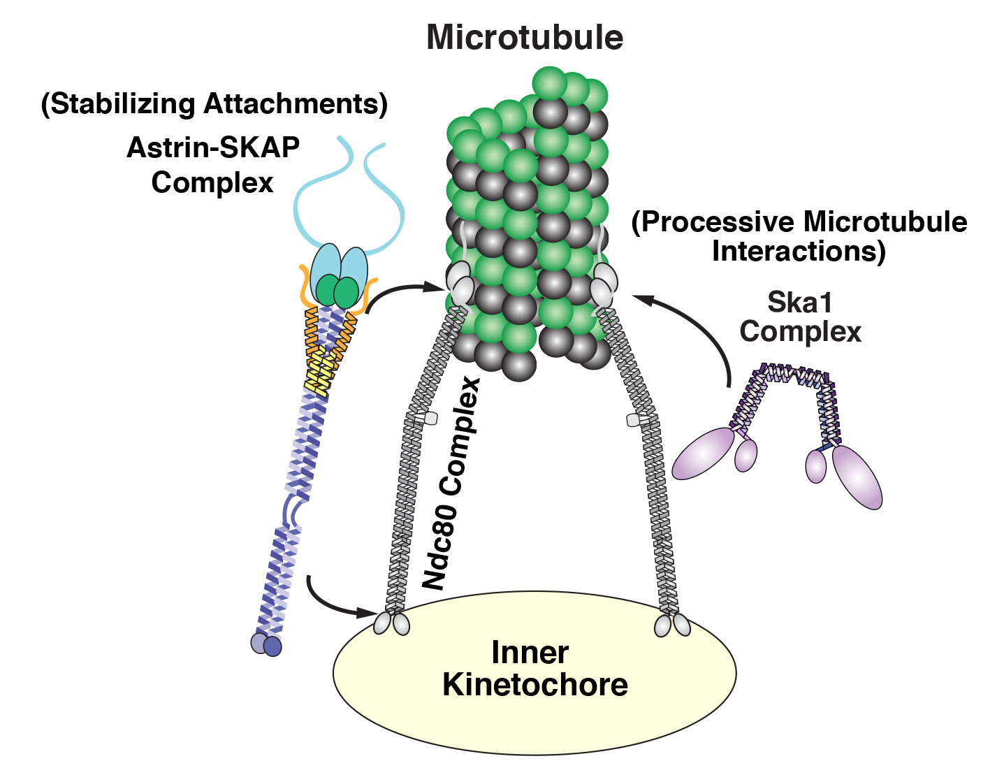
Cheeseman and his laboratory have focused on three key groups or complexes of proteins that play essential roles in chromosome segregation in human cells. These components assemble together to form a kind of molecular tether point on chromosomes (called the kinetochore) where microtubules attach.
Diagram of the kinetochore/microtube interface Courtesy: David Kern/Whitehead Institute
Among this trio of parts, the most critical is the Ndc80 complex. “It is the major connection between the kinetochore and the microtubule,” says Cheeseman. As a postdoc, he discovered the biochemical properties that enable this Ndc80 complex to grab on to microtubules, research that sparked his lab’s quest to study the various pieces of the kinetochore machinery and how they work.
While Ndc80 forms a critical linkage, it lacks some key capabilities, like processivity — the ability to keep ahold of something while it moves. In a series of papers, one published in 2009, another in 2012, and a new one in Current Biology, Cheeseman’s team revealed that Ska1 can perform this crucial function. That is, it has the biochemical capacity to enable chromosomes to hang onto microtubules while they grow and as they shrink, an activity that it can impart to the Ndc80 complex. “These are pretty powerful properties,” says Cheeseman.
Diving even deeper into Ska1’s bag of tricks, Julie Monda and Ian Whitney, lead authors of the Current Biology paper, went on to decipher the precise molecular features that enable the complex’s dynamic capabilities, uncovering multiple surfaces that associate with microtubules and enable Ska1 to undergo something akin to molecular somersaults. These somersaults are what allow it to maintain its association with microtubules.
The third complex, Astrin-SKAP, also plays a unique role. As Cheeseman’s team described in their recent eLife paper, led by first author David Kern, it serves as a master stabilizer — like a final drop of superglue to secure everything in place. “It’s the last thing that comes in and helps lock down these interactions, so you can stabilize and maintain them,” says Cheeseman.
Uncovering its role was no easy feat. Astrin-SKAP proved to be rather temperamental biochemically, complicating Kern’s efforts to purify and manipulate it in the laboratory. Also, as he and his colleagues discovered, a tiny piece of the structure had previously gone undetected; it works alongside the rest of the complex and is required for its normal function. Perhaps the most important revelation was that Astrin-SKAP doesn’t just work alone — it also coordinates with Ndc80. “This is an important finding for how we think about these components as a whole,” saysCheeseman.
Although questions remain about how all of these parts work together and how other pieces may come into play, Cheeseman believes these studies provide an exciting start. “The first human kinetochore component wasn’t identified until 1987, when many of the other key processes in the cell had already been intensively studied,” he says. “There are so many exciting questions that are accessible now that we have these tools and knowledge.”
Now, he and his colleagues will continue to meld approaches in cell biology and biochemistry to decode the inner workings of the kinetochore. That includes understanding how the various components operate not only in individual cells, but also in multicellular organisms.
“We are currently thinking a lot about the physiological context— that is, what matters to cells and to an organism,” says Cheeseman. “The work that our lab and others have conducted over the past two decades has given us a molecular handle on this problem. I’m excited to be able to apply these finding to understanding the ways that cell division is altered in development and in disease states.”
Written by Nicole Davis
* * *
Iain Cheeseman’s primary affiliation is with Whitehead Institute for Biomedical Research, where his laboratory is located and all his research is conducted. He is also an associate professor of biology at Massachusetts Institute of Technology.
* * *
Full citations:
“Astrin-SKAP complex reconstitution reveals its kinetochore interaction with microtubule-bound Ndc80”
eLife 2017;6:e26866 August 25, 2017. DOI: 10.7554/eLife.26866
David M Kern (1,2), Julie K Monda (1,2), Kuan-Chung Su (1), Elizabeth M Wilson-Kubalek (3), and Iain M Cheeseman (1,2).
1. Whitehead Institute for Biomedical Research, 455 Main Street, Cambridge, MA 02142, USA
2. Department of Biology, Massachusetts Institute of Technology, Cambridge, MA 02142, USA
3. Department of Cell Biology, The Scripps Research Institute, La Jolla, CA 92037, USA
“Microtubule tip tracking by the spindle and kinetochore protein Ska1 requires diverse tubulin-interacting surfaces”
Current Biology, online November 16, 2017. DOI: 10.1016/j.cub.2017.10.018
Julie K. Monda (1,2,6), Ian P. Whitney (1,6), Ekaterina V. Tarasovetc (3,4), Elizabeth Wilson-Kubalek (5), Ronald A. Milligan (5), Ekaterina L. Grishchuk (3), and Iain M. Cheeseman (1,2).
1. Whitehead Institute for Biomedical Research, 455 Main Street, Cambridge, MA 02142, USA
2. Department of Biology, Massachusetts Institute of Technology, Cambridge, MA 02142, USA
3. Department of Physiology, Perelman School of Medicine, University of Pennsylvania, Philadelphia, PA 19104, USA
4. Center for Theoretical Problems of Physicochemical Pharmacology, Russian Academy of Sciences, Moscow, Russia
5. Department of Cell Biology, The Scripps Research Institute, La Jolla, CA 92037, USA
6. These authors contributed equally
November 7, 2017
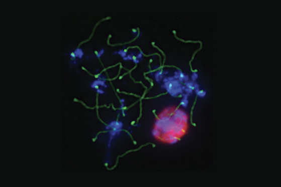
CAMBRIDGE, MA – Sperm production requires progression through a well-orchestrated series of transitions in the testes that move diploid spermatogonia cells, with two complete sets of chromosomes, through a series of transitions to produce haploid sperm, with one copy of each chromosome, poised to swim and fertilize an available egg. There are four major transitions in sperm production, or spermatogenesis. The first is spermatogonial differentiation, during which spermatogonia differentiate, losing their stem-cell like qualities. The resulting spermatocytes then initiate meiosis and undergo two rounds of cell division to generate haploid spermatids. The spermatids undergo elongation and then the resulting sperm are released.
The signals that control progression through these transitions were poorly understood until 2015, when David Page, Member and Director of Whitehead Institute, professor of biology at Massachusetts Institute of Technology, and investigator with Howard Hughes Medical Institute and colleagues determined that retinoic acid (RA), a derivative of vitamin A that has been shown to play a key role in a number of developmental processes, was responsible for coordinating the first two stages of spermatogenesis-differentiation and meiosis. Now, in a paper published this week in the journal Proceedings of the National Academy of Sciences, Page, first author Tsutomu Endo, and colleagues extend those findings to show that RA signaling in mice coordinates the second two transitions as well.
The researchers used chemical manipulation of RA levels to determine that RA controlled the second two transitions, spermatid elongation and sperm release, in addition to the first two. With this knowledge in hand, the researchers were then able to drill down and get a better picture of how RA regulates male gamete production. One outstanding question has been how males are able to continually produce sperm throughout their lifetime, in contrast with females whose egg production and maturation is limited. Page and colleagues measured RA levels in the testes and discovered that it is cyclically produced, driving production of sperm during the male lifetime. In addition to the timing of RA production, the researchers also examined its source. From which cells was the RA signal coming? During the first two transitions, they determined that the RA was coming from the somatic Sertoli cells, the support cells of the testes, and in the second two transitions they determined that it was being released by the germ cells themselves-the meiotic (pachytene-stage) spermatocytes were found to be secreting RA to other germ cells in the testes.
These findings not only contribute to our fundamental understanding of male gamete formation, they also provide important clues for the field of reproductive technology. For years, scientists have been working on making gametes in the laboratory, but have had difficulty making functional sperm. This discovery of the role of RA in spermatogenesis adds important tools to the toolbox of assisted reproduction. The work shows that RA is required in both the early and late transitions of spermatogenesis and sheds light on an important component of laboratory efforts for sperm production.
Other researchers involved include Elizaveta Freinkman and Dirk G. de Rooij.
This research was supported by Howard Hughes Medical Institute (HHMI) and the United States Department of Defense (DoD W81XWH-15-1-0337)
Written by Lisa Girard
****
David Page’s primary affiliation is with Whitehead Institute for Biomedical Research, where his laboratory is located and all his research is conducted. He is also a Howard Hughes Medical Institute Investigator and a Professor of Biology at the Massachusetts Institute of Technology.
****
Paper cited: Endo, T et al. Periodic production of retinoic acid by meiotic and somatic cells coordinates four transitions in mouse spermatogenesis. Proc Natl Acad Sci. DOI: 10.1073/pnas.1710837114. Epub 2017 Nov 6.
Other work cited: Endo T et al. Periodic retinoic acid-STRA8 signaling intersects with periodic germ-cell competencies to regulate spermatogenesis. Proc Natl Acad Sci. DOI: 10.1073/pnas.1505683112. Epub 2015 Apr 20.
October 30, 2017
CAMBRIDGE, Mass. – A few years ago while paddling off the coast near La Jolla, California, avid surfer Roland Kersten noticed a piece of red algae (Laurenica pacifica) bobbing alongside his surfboard. Kersten, whose background is in natural product chemistry, was intrigued.
Natural products—chemicals from living organisms such as plants and algae—represent a rich source of potential therapeutics. A majority of anti-cancer drugs are natural product-based or inspired. One such well-known natural product—the potent anti-cancer drug Taxol—was identified in the bark of a yew tree.
Marine algae, like the red algae Kersten found, are often rich in compounds called sesquiterpenes, some of which have been shown to have potential medicinal attributes. Since the 1970s, scientists identified many sesquiterpenes produced by Laurencia species with anti-cancer properties. The identification techniques usually required about tens of milligrams of purified compounds, which were obtained from more than a kilogram of algae. Because Laurencia and the reef ecosystems in which it thrives are protected, and such large-scale harvesting for scientific or medicinal purposes is no longer tenable, Kersten had to devise a different approach to analyze its sesquiterpenes.
Kersten received a collection permit to clamber over the rocky shore at deep low tide to collect a hand-sized sample of the red algae. Now a postdoctoral fellow in the lab of Whitehead Member and Massachusetts Institute of Technology assistant professor of biology Jing-Ke Weng, Kersten’s first task was to search the RNA sequences of all genes expressed in his red algae sample to find those whose product seemed likely to be enzymes that make sesquiterpenes. In order to determine the product generated by these enzymes, he engineered them in yeast and isolated its sesquiterpene products.
In order to define the first step in the biogenesis of sesquiterpenes in red algae, Kersten wanted to see the precise 3-D structure of the isolated sesquiterpene. But the small handful of algae he had obtained produced only a fraction of the amount required for x-ray crystallography, the established method for determining a compound’s absolute structure. So Kersten tried a method recently developed by collaborator Makoto Fujita at the University of Tokyo that requires only a few nanograms of material: soaking extracted compounds into a special crystalline sponge, which supports the sample’s molecular shape while it is bombarded with x-rays to accurately determine the 3-D conformation of a molecule. A new combination of the crystalline sponge method and nuclear magnetic resonance spectroscopy by the Fujita group revealed the structure of prespatane.
With the compound’s structure in hand, Kersten is closer to understanding how Laurenciabiosynthesizes its sesquiterpenes and how to engineer yeast to produce the same molecules for medicinal research at scale—without touching the red algae flourishing on protected reefs. And the novel workflow—spanning genomics, metabolomics, synthetic biology, and x-ray crystallography with crystalline sponges—established by Weng, Kersten, and their collaborators may expedite the identification of other promising compounds produced by organisms from both land and sea.
Other contributors to this work include Shoukou Lee of Tokyo University, Daishi Fujita of Tokyo University and Whitehead Institute, Tomáš Pluskal of Whitehead Institute. The team also collaborated with researchers from Scripps Institution of Oceanography and Salk Institute of Biological Sciences.
This work was supported by Howard Hughes Medical Institute, the Simons Foundation, the Helen Hay Whitney Foundation, the Pew Scholars Program in the Biomedical Sciences, the Searle Scholars Program, and the Japan Science and Technology Agency.
Written by Nicole Giese Rura
* * *
Jing-Ke Weng’s primary affiliation is with Whitehead Institute for Biomedical Research, where his laboratory is located and all his research is conducted. He is also an assistant professor of biology at Massachusetts Institute of Technology.
* * *
Full Citation:
“A Red Algal Bourbonane Sesquiterpene Synthase Defined by Microgram-scale NMR-coupled Crystalline Sponge XRD Analysis”
Journal of the American Chemical Society, online October 30, 2017.
Roland D. Kersten (1,6), Shoukou Lee (2,6) , Daishi Fujita (1,2) , Tomáš Pluskal (1) , Susan Kram (3), Jennifer E. Smith (3) , Takahiro Iwai (2) , Joseph P. Noel (4) , Makoto Fujita (2), Jing-Ke Weng (1,5).
1. Whitehead Institute for Biomedical Research, 455 Main Street, Cambridge, MA, United States
2. Graduate School of Engineering, The University of Tokyo, JST-ACCEL, Tokyo, Japan
3. Scripps Institution of Oceanography, University of California San Diego, La Jolla, CA, United States
4. Howard Hughes Medical Institute, Jack H. Skirball Center for Chemical Biology and Proteomics, The Salk Institute for Biological Studies, La Jolla, CA, United States
5. Department of Biology, Massachusetts Institute of Technology, Cambridge, MA, United States
6. These authors contributed equally
A pro-metastatic transcription factor’s journey from anonymity to a promising target for breast cancer therapy
October 20, 2017
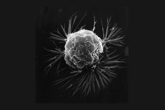
An overwhelming majority of deaths from cancer are associated not with the primary tumor, but instead with its metastases to other sites in the body. For this reason, understanding the properties of cancer cells that give them a high metastatic potential, and identifying molecular strategies to intervene, is critical for improving clinical outcomes.
One of the hallmarks of cancer cells with high metastatic potential is an epithelial to mesenchymal transition (EMT). This shift in their gene expression landscape is a harbinger for both invasive behavior and anti-cancer drug resistance. One signaling pathway active in cells that have undergone EMT transition, the PERK pathway, has been of particular interest to Whitehead Institute Member and Massachusetts Institute of Technology associate professor of biology Piyush Gupta and postdoctoral researchers in Gupta’s lab, Yu-Xiong Feng and Dexter Jin. The PERK signaling pathway has been a sought-after target for a number of types of cancer, including breast cancer. Drug companies had largely given up on the PERK signaling pathway as a target, however, because when it is inhibited, it also has the unintended consequence of affecting glucose regulation to the degree that mice given PERK inhibitors typically develop diabetes within a few weeks. Gupta and colleagues hypothesized that downstream elements of the pathway could include targets with more specific effects on metastatic behavior, potentially enabling the development of therapies that do not result in the unintended consequences associated with inhibiting PERK.
In a recent article in Nature Communications, Gupta, Feng, Jin, and colleagues describe CREB3L1, a factor downstream of the PERK pathway that is active in the subset of triple negative breast cancer cells and tumor cells that have undergone an EMT transition. CREB3L1 expression is associated with distant metastasis and is important for the transformed cell’s invasive and drug resistant properties. While factors like CREB3L1, called transcription factors, are usually difficult to target with small molecules, Gupta and his team zeroed in on a unique property it shares with only a small handful of other factors-it is normally stuck to the membrane of a cellular compartment called the endoplasmic reticulum and, in order for it to be active, it need to be cut free by factors called proteases. Gupta and colleagues show that certain protease inhibitors can actually stop the activation of CREB3L1 in its tracks, along with the invasive and drug resistance properties its activation confers.
While the PERK signaling pathway has been an attractive target for anticancer therapy, its more general cellular role made it an intractable target. The downstream factor of the pathway CREB3L1 is a potential new target for breast cancer therapy whose specificity of action makes it an attractive option for targeting metastatic behavior.
By Lisa Girard
Citation:
Feng Y-X, Jin DX, et al. “Cancer-specific PERK signaling drives invasion and metastasis through CREB3L1.” Nature Communications DOI:10.1038/s41467-017-01052-y
October 6, 2017
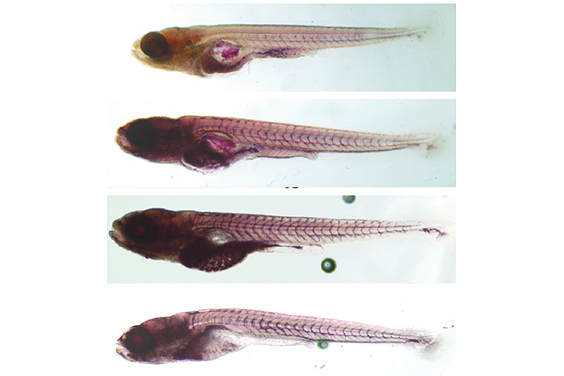
CAMBRIDGE, Mass. – For the first time, Whitehead Institute scientists have documented a direct link between deletions in two genes—fam57ba and doc2a—in zebrafish and certain brain and body traits, such as seizures, hyperactivity, enlarged head size, and obesity.
“Finding the molecular connections between a brain and a body phenotype is indeed really paradigm shifting,” says Whitehead Institute Member Hazel Sive, who is also a professor of biology at MIT. “It lets us think about the common control of these two aspects of phenotype, which is very interesting and could be useful for developing therapies for these phenotypes.”
Both genes reside in the 16p11.2 region of human chromosome 16. About 1 in 2000, or around 4 million people worldwide, have deletions in this region, and these deletions are associated with multiple brain and body symptoms, including autism spectrum disorders, developmental delay, intellectual disability, seizures, and obesity.
Scientists have had difficulty teasing apart the relationship between specific traits and deletions in this region, because it includes at least 25 genes, and because there is not a one-to-one mapping of gene to phenotype. Instead, multiple genes seem to create a web of interactions that produce a variety of characteristics.
To solve such a complex puzzle, Jasmine McCammon, a postdoctoral researcher in Sive’s lab, enlisted the zebrafish as a “living test tube”. The Sive group uses zebrafish to study the genetic/phenotype connections associated with human disorders. Like the human genome, the zebrafish genome has two copies of each gene, and scientists can remove the function of multiple genes to produce phenotypes that are reminiscent of human symptoms.
The results from McCammon’s initial screen with zebrafish indicate that two genes in the 16p11.2 region could be key for brain development: fam57ba and doc2a. (fam57bencodes a ‘ceramide synthase’ that makes a kind of lipid, and doc2a encodes a regulator of secretion.) McCammon investigated further by deleting one copy of fam57ba and doc2a individually; the effect was minimal. However, simultaneously removing a copy of both genes revealed significant synergy between them. Compared with controls, fish with only one copy of each gene exhibit hyperactivity, increased propensity for seizures, increased body and head size, and fat content. When both copies of only fam57ba are removed, the fish are much larger and with a higher fat content. All of the study’s results are published in the journal Human Molecular Genetics.
Although her findings use zebrafish and are far from the clinic, McCammon was struck by how much people affected by deletions in this genome identified with her results.
“When I spoke with the parents of some kids with neurodevelopmental disorders, I was surprised how much the brain/body connection that we described resonated with them,” she says. “They said that yes, their child has autism, but he also has really weak muscle tone. Or she has a gastrointestinal problem and that’s been more problematic than her behavior issues. For me, it’s been really revealing to talk to people who’ve actually experienced this as opposed to reading about statistics in journals.”
The mechanisms underlying this brain/body connection are still not well understood. One of the identified genes, fam57ba, provides some intriguing hints as to how metabolism and brain function could be intertwined, because it produces an enzyme that plays a role in lipid production and is believed to be a metabolic regulator. The lipid type, ceramide, also has a functional role in various signaling pathways and affects synaptic function, although its primary role is not in the synapse, but providing structure in cell membranes.
For Sive, the two identified genes could be just the beginning. “Our data suggest that there may be metabolic genes involved in human neurodevelopmental disorders,” she says. “This is a nascent field, that we’re very interested in going forward.”
This work was supported by Jim and Pat Poitras, Len and Ellen Polaner, and the Markell-Balkin-Weinberg Postdoctoral Fellowship.
Written by Nicole Giese Rura
* * *
Hazel Sive’s primary affiliation is with Whitehead Institute for Biomedical Research, where her laboratory is located and all her research is conducted. She is also a professor of biology at Massachusetts Institute of Technology.
* * *
Full Citation:
“The 16p11.2 homologs fam57ba and doc2a generate certain brain and body phenotypes”
Human Molecular Genetics, Volume 26, Issue 19, 1 October 2017.
Jasmine M. McCammon(1), Alicia Blaker-Lee(1), Xiao Chen(2), and Hazel Sive (1,2).
1. Whitehead Institute for Biomedical Research, Cambridge, MA 02142, USA
2. Department of Biology, Massachusetts Institute of Technology, Cambridge, MA 02139, USA
Justin Chen
March 13, 2017
The biology department welcomes Eliezer Calo back to MIT
By Justin Chen
As the newest faculty member of the MIT biology department, Eliezer Calo is working in Building 68, the same building where he was first inspired to become a scientist. Professor Calo’s relationship with MIT began eleven years ago when he was a chemistry major at the University of Puerto Rico with hazy career aspirations. Encouraged by his instructors to attend a Minority Access to Research Careers (MARC) conference, Calo came across a booth advertising the MIT Summer Research Program (MSRP). Even though Calo initially associated MIT with engineering and math, he applied for and received a summer internship position in Professor Stephen Bell’s lab studying DNA replication. “Experiencing the scope of MIT’s biological research and seeing how collaborative and enthusiastic people were about biology was eye opening,” Calo says. “That was the summer I decided to do a PhD.”
MSRP launched Calo’s scientific career and cemented his love for MIT. After graduating from the University of Puerto Rico, he returned to MIT’s biology department for graduate school and earned a PhD under the mentorship of Professor Jackie Lees. He then moved to Stanford University for postdoctoral training with Professor Joanna Wysocka. He began his faculty position at MIT in January and became an extramural member of the Koch Institute in March.
“We are thrilled to welcome Eliezer back to MIT as a faculty member,” says Biology Department head Alan Grossman. “He and the two other new faculty members, Professors Stefani Spranger and Sebastian Lourido, exemplify the energy and cutting edge research in the department. We eagerly anticipate many years of exciting discoveries from their labs.”
Now leading his own lab, Calo seeks to understand how cells assemble ribosomes and the roles they play in development and in disease. Ribosomes, intricate molecular machines, create building blocks of the body by translating the genome into proteins. In order to sustain growth, human cells assemble millions of ribosomes. When defects in ribosome assembly occur during embryonic development, cells are unable to grow and divide, leading to developmental disorders.
One such disorder is Treacher Collins syndrome, which arises from a genetic alteration that impairs the expression of a gene named Treacle,whose protein product assists in ribosome assembly. Surprisingly, although Treacle is expressed in most cells during early embryo development, the mutation affects only the nascent face: individuals have smaller facial bones making up their cheeks and jaws.
“Treacher Collins and other syndromes caused by abnormal ribosome assembly and function challenge our understanding of the ribosome,” Calo explains. “We think of ribosomes as constitutively expressed molecular machines required only for protein synthesis. These diseases, however, suggest that ribosomes might unexpectedly have very specific developmental roles as well.”
Describing how a single genetic mutation warps cell biology and triggers disease is a difficult task. In the case of Treacher Collins Syndrome, the precise mechanism remains unknown but scientists have identified two potential factors. First, cells destined to become facial bones grow quickly during development and may be especially sensitive to reduced protein production. Second, new research suggests that Treacher Collins may also be caused by defective ribosomes activating cancer suppressor pathways, leading to slower cell division and cell death.
To design a simplified model of Treacher Collins syndrome, Calo has used CRISPR gene editing technology to introduce disease-relevant mutations into human embryonic stem cells in culture. The cells are then grown and differentiated into the specific facial tissues affected by Treacher Collins syndrome. These in vitro cell communities allow Calo to closely observe abnormalities as they arise during development and better understand how decreased protein levels, tumor suppressor pathways, or other factors yet to be discovered contribute to cell death.
To determine whether the results in cultured cells apply to whole organisms, Calo plans to validate his findings in zebrafish. Mutant zebrafish, like humans, have craniofacial defects and allow researchers to screen chemicals that may lessen facial anomalies. By working with human embryonic stem cells in culture and then testing the findings in zebrafish, Calo has created a powerful two-pronged approach to understand Treacher Collins and address fundamental questions of ribosome biology and disease.
As Calo establishes his new laboratory, he is also reprising a familiar role of instructor and mentor. While performing graduate research, he served as a teaching assistant for MIT’s introductory biology course (7.01) and as a program assistant for MSRP. Calo, who still runs into former students in New York, Boston, and Stanford, enjoys learning about their accomplishments and future goals. Now a professor, Calo will inspire the next generation of biologists by advising graduate students and MSRP researchers. “My MSRP experience shaped the course of my scientific career, so I look forward to having MSRP students working in my lab,” Calo says. “I want them to experience what it is like to do research at MIT.”
Posted: 12.5.17
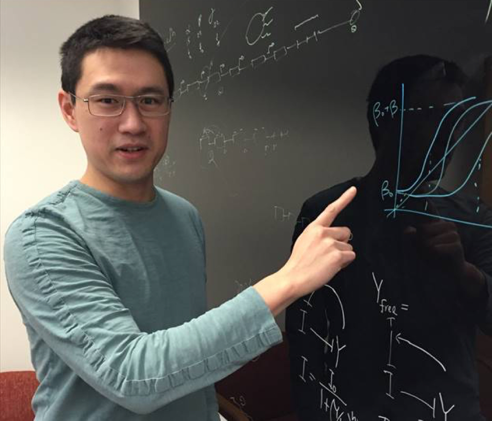
May 4, 2016
Gene-Wei Li is the newest member of the MIT Department of Biology. He opened his lab on the second floor in building 68 about one year ago. But who is Gene? Born in California and raised in Taiwan, Gene fell in love with math and physics and a boyhood dream to figure out quantum teleportation. It was not until he arrived at Harvard that he discovered the field of biophysics. “As a physicist I like thinking about numbers and when I came to Harvard I suddenly realized there was so much biophysics going on in a diversity of labs,” Gene says of his years in graduate school.
In his thesis project, Gene was looking at how transcription factors find their target through single molecule imaging in bacterial cells. He became focused on protein dynamics. Do transcription factors diffuse through cytosol and randomly land on DNA or do they scan through in a directional manner? He discovered they do a bit of both. “As a cell you would optimize the amount of transcription factors searching at any given time and the number of sites. You would not want to crowd the DNA,” Gene explains smiling.
Despite his work in a biological system, Gene admits he still saw himself primarily as a physicist at the beginning of his postdoc. “When I started my postdoc at the Weissman lab at UCSF, I did not even know what ubiquitin was,” he laughs. That was soon to change. At UCSF, Gene utilized a novel method called ribosome profiling which enables the study of protein synthesis rates by looking at ribosome density. “In my postdoc, I was lucky to get a paper published early on and so I had an opportunity to explore what I enjoyed. Quantification is always hard so I decided to see whether there is a good metric to measure, and found a striking result that density corresponds to stoichiometry really well. All the subunits are made in proportion to their stoichiometry. While this makes intuitive sense it was not necessarily obvious before,” Gene describes his postdoctoral experience. What about single subunit proteins? “No protein acts alone,” Gene replies, “we need to look at a whole system — enzymes could be diffusing but receive substrates and the amount of enzyme matters. Make enough but not too much because that would be wasteful.”
Are there physical and quantitative principles behind the precise control of transcription and translation? How do cells fine-tune their RNA and protein production to result in correct stoichiometric complexes? And importantly, if a cell is engineered in the lab to express exogenous proteins are there detrimental effects? Gene’s growing team at MIT (currently, two graduate students, a technician, an undergraduate, and a joint postdoc) are focused on cracking precisely these key questions. “As a mentor, my philosophy is to be supportive but leave freedom for students and postdocs to explore on their own too. In graduate school, I was stuck on a project for two years but was also allowed to follow side stories that both eventually went to fruition.”
Gene’s lab uses bacteria (E.coli and B. subtilis) in their experimental work. “Their operons are surprisingly conserved despite a billion years’ separation. The power is in comparison though – even though the gene order and protein stoichiometry are conserved, these bugs use different tricks of post-transcriptional controls to get the same amount of proteins,” Gene says of his model organisms. Being a young faculty in the MIT Department of Biology is a very humbling experience because it has so much history, he adds. “Boris Magasanik from this department was one of the pioneers of bacterial physiology — we know the system much better now, we can quantitate it better too but he laid the foundation. My lab space is formerly Alexander Rich’s who discovered polysomes — now we are stretching polysomes individually and looking at the actual distribution of ribosomes along the mRNA.”
In his free time, Gene enjoys traveling with his wife though it has become more difficult with the recent birth of their son (congratulations!) and his three-year old brother. He loves meeting people of different backgrounds and thinking about science from different perspectives. “It takes a while to adjust from postdoc to faculty – becoming a manager, accountant, grant writer, colleague, mentor – leaving less time for research,” he says. “The nice thing about the MIT Biology Department is that I can knock on any door and ask for advice on things big and small.”
 The researchers used chemical manipulation of RA levels to determine that RA controlled the second two transitions, spermatid elongation and sperm release, in addition to the first two. With this knowledge in hand, the researchers were then able to drill down and get a better picture of how RA regulates male gamete production. One outstanding question has been how males are able to continually produce sperm throughout their lifetime, in contrast with females whose egg production and maturation is limited. Page and colleagues measured RA levels in the testes and discovered that it is cyclically produced, driving production of sperm during the male lifetime. In addition to the timing of RA production, the researchers also examined its source. From which cells was the RA signal coming? During the first two transitions, they determined that the RA was coming from the somatic Sertoli cells, the support cells of the testes, and in the second two transitions they determined that it was being released by the germ cells themselves-the meiotic (pachytene-stage) spermatocytes were found to be secreting RA to other germ cells in the testes.