Whitehead Institute team develops new method to study human brain cells
Nicole Davis | Whitehead
November 25, 2019

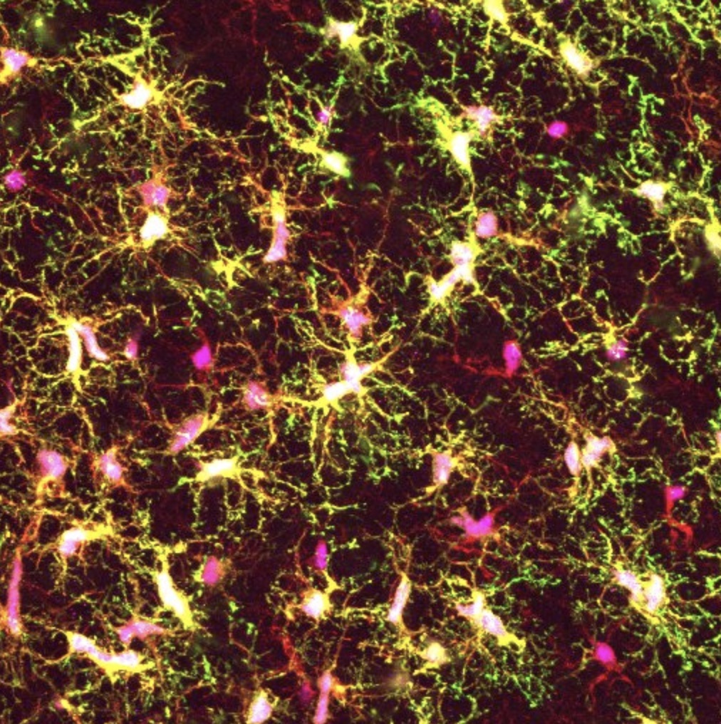
A groundswell of evidence connects defects in the function of microglia, the brain’s resident immune cells, to neurodegenerative diseases, yet the tools for studying these cells in the laboratory have been limited. Now, a team of Whitehead Institute scientists has developed a new experimental platform for generating microglia from human stem cells that includes transplantation into newborn mice. As described online November 26 in the Proceedings of the National Academy of Sciences (PNAS), this new method yields microglial cells that resemble those in the human brain more closely than previous approaches, which could help enable future studies aimed at unravelling the role of microglia in neurodegeneration and other brain disorders.

“The dysfunction of microglia is implicated in a wide variety of brain conditions, and yet our knowledge of them, especially in humans, is really quite limited,” says senior author Rudolph Jaenisch, a Founding Member of the Whitehead Institute and professor of biology at the Massachusetts Institute of Technology. “This new approach will help us lift the hood on these important yet enigmatic brain cells.”

Microglia are increasingly recognized as key players in brain health and disease, but the majority of what is known about them comes from studies of mice, not humans. Yet human and mouse microglia are quite distinct — in humans, the cells are much larger, and have a more branched appearance, suggesting significant differences in their biology.

To address this gap in knowledge, multiple research teams have recently devised methods to generate microglia using human stem cells and grow them under laboratory conditions that mimic their natural environment. However, this approach has a fundamental drawback: the cultured cells do not look like microglia nor do they behave much like them, even though they display the appropriate molecular hallmarks.

“That really suggests to us that this is not the optimal approach to study how microglia are behaving in healthy and diseased brains,” says first author Devon Svoboda, a postdoctoral fellow in the Jaenisch lab. “We set out to create a new method in which the stem-cell derived microglial cells can reside in the brains of mice — one of the best models of the human brain that we have.”

Transplanting human cells into mice — creating “chimeras” — is a well-established technique. However, Svoboda and her colleagues discovered they needed to use special strains of mice that carry human genes for certain growth factors, called cytokines, which are required for microglial development and survival. The researchers utilized mice that carry human genes for four crucial cytokines: CSF1, IL3, SCF, and GM-CSF.

“What is special about these chimeras is really the mice we are using,” says Svoboda. “They express the human alleles of these cytokines which is key because the mouse versions are not able to communicate with receptors on human microglia, so the cells die.”

After transplanting the stem-cell derived microglia into these mice, the research team examined the cells’ morphology and their molecular characteristics. They found that the transplanted cells closely resembled those found in the human brain.

Further analyses revealed some striking differences between the team’s “chimera-grown” microglia and those grown in the laboratory using conventional cell culture methods. Surprisingly, Svoboda and her colleagues found that the cultured microglia showed strong similarities to the diseased microglia from patients with multiple sclerosis, another brain condition in which the cells are implicated.

“If you want to learn more about the role of microglia in disease, then studying them in culture is probably not the best way,” says Svoboda. “The chimeras and the in vitro methods really complement each other, and we think there is a place for both systems in microglia research going forward.”

The Whitehead-led team plans to extend their initial studies in several ways. One is to identify which cytokines and other growth factors are most crucial to microglial development. That knowledge could help improve existing cell culture methods and enable them to more closely mirror the cells’ natural environment. Another key direction is to use the new chimera-based system to create models of neurodegenerative diseases, including Alzheimer’s and Parkinson’s disease, to understand how microglia respond to diseased neurons and, in turn, how diseased microglia can impair neuron function.

Our chimera-based method will give us a good handle to begin to stringently test the role of microglia in brain health and disease,” says Jaenisch. “This is an important step forward for the field.”

Support for this work was provided by the Cure Alzheimer’s Foundation, MassCATS, and NIH Grants R01 AG058002-01, R01 MH104610, R37 CA084198, and U19 AI131135 (to R.J.). L.D.S. is supported by NIH Grants R24 OD26440, AI32963, and CA034196. J.S. is supported by the National Institute of Child Health and Human Development (K99HD096049).

Written by Nicole Davis

***

Rudolf Jaenisch’s primary affiliation is with Whitehead Institute for Biomedical Research, where his laboratory is located and all his research is conducted. He is also a professor of biology at Massachusetts Institute of Technology.

***

Paper cited:

Human iPSC-derived microglia assumer a primary microglia-like state after transplantation into the neonatal mouse brain.

PNAS, online November 26, 2019. DOI: 

Devon S. Svoboda (1)M. Inmaculada Barrasa (1)Jian Shu (1,3)Rosalie Rietjens (1)Shupei Zhang (1)Maya Mitalipova (1)Peter Berube (3)Dongdong Fu (1)Leonard D. Shultz (4)George W. Bell (1), and Rudolf Jaenisch (1,2)


1. Whitehead Institute, Cambridge, MA 02142, USA

2. Department of Biology, Massachusetts Institute of Technology, Cambridge, MA 02139, USA

3. Broad Institute of MIT and Harvard, Cambridge, MA 02142

4. The Jackson Laboratory Cancer Center, The Jackson Laboratory, Bar Harbor, ME 04609

Building a roadmap for salicylic acid
Nicole Giese Rura | Whitehead Institute
November 25, 2019

Salicylic acid, which may be best known as a treatment for skin conditions such as acne and warts and in its modified form as aspirin, is a critical plant hormone involved in growth and development as well as regulating plants’ immune defenses. Unable to move and evade physical damage or attacks by bacteria and other pathogens, plants respond to these assaults through the biosynthesis of salicylic acid, which in turn controls cascades of other defense responses. Consequently, control of salicylic acid production in agricultural plants could boost crops’ resilience to pathogens and insects, thereby reducing the overuse of potentially toxic pesticides that can lead to pathogen resistance. Yet scientists have been missing a key tool necessary for manipulating salicylic acid levels in plants: a full description of the pathway necessary to synthesize the hormone. Now Whitehead Institute Member Jing-Ke Weng, along with Weng lab postdoc Michael Torrens-Spence, have uncovered the last missing steps in the Arabidopsis plant’s salicylic acid pathway and solved a puzzle that has dogged Weng and his field for decades.

The quest to define the salicylic acid biosynthesis pathway started about 50 years ago when researchers determined that salicylic acid is principally formed downstream from a ubiquitous compound called chorismate. In 2001 another step was resolved: Chorismate is converted to isochorismate before eventually becoming salicylic acid. Encouraged by this progress, many in the fields of plant biology and biochemistry thought that the rest of the biosynthesis pathway in plants would be quickly defined by looking for enzymes similar to those that comprise the bacterial version of the pathway, rather an almost two decade-long drought in discoveries followed instead.

Weng and Torrens-Spence tried a different tack using genetic and biochemical methods to break the dry spell in the identification of the pathway’s missing links. Their work is described online this week in the journal Molecular Plant. From previous research, Torrens-Spence knew that the enzymes encoded by two genes – PBS3 and EPS1 – play roles in salicylic acid accumulation after pathogen attacks. In order to determine the role of these enzymes in salicylic acid biosynthesis pathway, Torrens-Spence generated plants lacking in S3H and DMR6, two genes known to breakdown salicylic acid and keep its production in check. With those genes disrupted, plants overproduce salicylic acid to an extreme extent, resulting in a severely stunted growth and other physical traits associated with surplus salicylic acid. Using these transgenic plants, Torrens-Spence had a model in which he could see if a particular gene affects salicylic acid production: If Torrens-Spence mutates genes responsible for salicylic acid biosynthesis, salicylic acid production should be abolished along with the associated visible plant characteristics. Mutations in PBS3 and EPS1 did just that – they rescued the stunted phenotypes associated with salicylic acid overproduction, and the plants accumulated less salicylic acid in their leaves than plants without the PBS3 or EPS1 mutations.

Next Torrens-Spence analyzed and compared the metabolites – the compounds created by cellular processes – in the leaves of plants without mutations and plants with PBS3 or EPS1 mutations. The results identified the probable products of the PBS3 protein’s enzymatic activity and also determined that the EPS1 protein likely acts downstream of PBS3. In order to confirm PBS3 and EPS1’s roles in salicylic acid biosynthesis, Torrens-Spence recreated the pathway in the test tube and in a relative of the tobacco plant. In both models, the reconstructed pathway efficiently converts isochorismate into salicylic acid. Interestingly, Torrens-Spence found that the intermediate produced by PBS3 could be spontaneously converted to salicylic acid in plants, but EPS1 greatly increased this step’s efficiency.

A recent evolutionary study indicates that PBS3 and variations of this gene are found throughout flowering plants, and Torrens-Spence’s work uncovered that PBS3 is an essential enzyme in the production of salicylic acid likely across all flowering plants as well. EPS1 is found only within the mustard family, which includes broccoli, Brussel sprouts, and turnips. According to Torrens-Spence and Weng, other enzymes may fulfill a role similar in plants that lack EPS1. Though the EPS1 aspect of the biosynthesis pathway described by Torrens-Spence and Weng are specific to Arabidopsis, their work provides a roadmap that researchers could follow to explore salicylic acid production in other organisms.

Weng, who has been trying to solve salicylic acid’s biosynthesis pathway in plants since he was in graduate school, says that he’s proud to have finally identified the remaining steps in Arabidopsis. With the complete salicylic acid biosynthesis pathway in Arabidopsis now known, agricultural scientists can use it to try to precisely manipulate salicylic acid’s immunological benefits in crop plants without the stunted growth associated with its excessive production.


This work was supported by the Pew Scholar Program in the Biomedical Sciences, the Searle Scholars Program, and the National Science Foundation (CHE-1709616).


Written by Nicole Giese Rura


***

Jing-Ke Weng’s primary affiliation is with Whitehead Institute for Biomedical Research, where his laboratory is located and all his research is conducted. He is also an associate professor of biology at Massachusetts Institute of Technology.

***

Citation:

“PBS3 and EPS1 complete salicylic acid biosynthesis from isochorismate in Arabidopsis”

Molecular Plant, online November 21, 2019 [online] DOI:10.1016/j.molp.2019.11.005

1. Whitehead Institute, Cambridge, MA 02142, USA

2. Department of Biology, Massachusetts Institute of Technology, Cambridge, MA 02139, USA

Committed to reproduction
Greta Friar | Whitehead Institute
November 21, 2019

Cambridge, MA – Early in mammalian embryonic development, long before the organism’s ultimate form has taken shape, a precious subset of its cells are set aside for future use in creating offspring. This task bestows on that subset of cells a special kind of immortality. While the majority of the embryo’s cells go on to construct the growing body, and their journey begins and ends in that body, the cells that are set aside, called primordial germ cells (PGCs), will eventually produce sperm and eggs, which will in turn produce a new body—and so the circle of life continues.

An embryo’s earliest cells are pluripotent, meaning they have the potential to develop into many different cell types—for example, heart, brain, blood—but the descendants of these cells eventually become committed to a specific identity, after which each can only produce one type of cell. Scientists have long believed that when PGCs are set aside, they are immediately committed to the path of producing egg and sperm cells. However, new research from Whitehead Institute Director David Page, also a professor of biology at the Massachusetts Institute of Technology (MIT) and a Howard Hughes Medical Institute investigator, and postdoctoral researcher Peter Nicholls, suggests that instead, the primordial germ cells’ fate remains flexible for much longer: until much closer to the end of embryonic development. In most species, PGCs are set aside long before the gonads—the testes or ovaries—form, and then later travel to these developing gonads where they will ultimately produce sex cells. Page and Nicholls have found evidence that the fate of these PGCs remains flexible until shortly after they reach the gonads. Their findings, which appear in the journal PNAS on November 21, deepen our understanding of the process of reproduction.

“A fundamental question in biology is how we get from one generation to the next,” Page says. “And the cells that are tasked with producing the next generation are an important part of that story.”

Establishing a new timeline for when PGCs become committed could also shed light on the origins of some reproductive tract cancers, including testicular cancer, the incidence of which is on the rise, and which is already the most commonly diagnosed cancer in young men.

Although PGCs are precursors of sperm and eggs, they also share many features with pluripotent cells, like embryonic stem cells. If migrating PGCs are isolated and cultured like embryonic stem cells, the PGCs show indicators of pluripotency, and are able to spontaneously form tumors containing multiple cell types—a trademark of pluripotent cells. Page and Nicholls found evidence confirming that shortly after the PGCs reach the gonads, they lose this capacity to produce pluripotent cell lines, and their ability for tumor formation. From that point on, the PGCs can only develop into eggs and sperm, no matter their environment.

The researchers then set out to identify the gene that prompts PGCs to become committed to produce only eggs or sperm. First, Nicholls identified a set of genes that are activated around the time that PGCs enter the gonads in mice and humans, and of those, focused on the genes that appeared to have equivalents involved in sex cell commitment across a variety of animals, not just in mammals. He then narrowed in on one of these genes, Dazl, as the single gene necessary for PGCs to become irrevocably committed to their path as sex cells. Nicholls found that when the Dazl gene is deleted from mice, PGCs travel to the gonads but don’t develop into committed precursors of egg and sperm, suggesting that Dazl is the key ingredient in the recipe for sex cell commitment.

In the absence of Dazl, PGCs remain uncommitted, and in some cases, will form gonadal tumors. The researchers argue, based on their findings, that testicular cancer and other gonadal cancers may develop from PGCs that have travelled to the gonads, but have not properly committed to becoming sex cells and so are prone to forming tumors. In Dazl-deficient mice, which had large amounts of uncommitted PGCs, more than one out of four males developed testicular tumors at a young age. The early onset of the tumors is consistent with that seen in children and men with testicular cancer, most of whom are under 45 years old.

The researchers also found that female Dazl-deficient mice developed gonadal tumors, though at a lower rate than males. Further research demonstrated that the testis environment is particularly favorable for tumor formation from uncommitted PGCs.

“Testicular cancer is on the rise for reasons not yet known, and our findings suggest that the cancer has embryonic origins,” Page says. “Understanding the nature of primordial germ cells will be important for investigating and addressing this disease.”

The researchers hope that, along with providing insights into gonadal cancers, their work could help improve the derivation of eggs and sperm from stem cells in the lab. Figuring out the specifics of the process for sex cell commitment should allow researchers recreate it in a dish. Nicholls is also excited about the evolutionary implications of the work: he found evidence that a similar process of sex cell commitment occurs across a wide variety of species. In particular, research with DAZL-deficient pigs—whose last common ancestor with mice and humans lived 95 million years ago—provides strong evidence that this DAZL-dependent process has been in play since the early days of modern mammals.

“This work completely shifts the timing for when sex cells become committed in mammals,” Nicholls says. “Furthermore, our data suggest that a common set of factors might operate in sex cell commitment not only in mammals, but perhaps across all vertebrates, regardless of how the primordial germ cells are first established.”

This work was supported by the Howard Hughes Medical Institute; a Hope Funds for Cancer Research Fellowship; an Early Career Fellowship; a DFG grant; a research grant from Biogen, Inc.; the National Natural Science Foundation of China; and a National Institutes of Health SBIR award.

Written by Greta Friar

***

David Page’s primary affiliation is with Whitehead Institute for Biomedical Research, where his laboratory is located and all his research is conducted. He is also a Howard Hughes Medical Institute Investigator and a Professor of Biology at the Massachusetts Institute of Technology.

***

Citation:

Mammalian germ cells are determined after PGC colonization of the nascent gonad

PNAS, online, Nov 21, 2019, DOI: 10.1073/pnas.1910733116

Peter K. Nicholls (1), Hubert Schorle (1,2),  Sahin Naqvi (1,3), Yueh-Chiang Hu (1,4), Fan Yuting (1,5), Michelle A. Carmell (1), Ina Dobrinski (6), Adrienne L. Watson (7), Daniel F. Carlson (7), Scott C. Fahrenkrug (7) and David C. Page (1,3,8)

1. Whitehead Institute, Cambridge, MA 02142, USA

2. Department of Developmental Pathology, Institute of Pathology, University of Bonn Medical

School, Bonn 53127, Germany

3. Department of Biology, Massachusetts Institute of Technology, Cambridge, MA 02139, USA

4. Divisions of Developmental Biology and Reproductive Sciences, Cincinnati Children’s

Hospital Medical Center, Cincinnati, OH 45229, USA

5. Reproductive Medicine Center, Sixth Affiliated Hospital, Sun Yat-sen University, Guangzhou,

510655, China

6. Department of Comparative Biology & Experimental Medicine, Faculty of Veterinary

Medicine, University of Calgary, Alberta, T2N 4N1, Canada

7. Recombinetics, Inc., Saint Paul, MN 55104, USA

8. Howard Hughes Medical Institute, Whitehead Institute, Cambridge, MA 02142, USA

Creating my niche in grad school

How diversity and outreach initiatives helped me find my place in MIT

YamilexA.-S.
November 15, 2019

Imagine being in a roller coaster that’s on fire, adrift, going full speed. That was my first year at MIT. Coming straight from an undergraduate institution in Puerto Rico, it was difficult for me to get used to the fast pace in which topics were taught in a different language and to the amount of work we had to constantly do. Recognizing these struggles, I convinced myself that I had to work even harder. However, towards the end of my first year, I couldn’t help but feel like something was missing. While struggling with that inner voice, I stumbled one day upon my personal statement for graduate school applications. I remember thinking, “who wrote this?”. One sentence in particular felt completely foreign to me: “I wish to provide a voice and an example that encourages minority students to pursue a career in science.” How was it that one year into my PhD program, I had completely lost the drive to start paving the path for people that looked and felt like me?

Being in STEM, it is quite easy to feel that if science isn’t the most important thing in your life, you are probably doing something wrong. For me, however, although science is definitely an important part of my life, it surely isn’t my entire life. I realize this isn’t a popular view among my peers, especially at a place like MIT, and it took some time for me to even embrace this mentality. I knew I appreciated my science more when I started doing things that fulfilled me outside the lab. This was clear in my mind, but I didn’t know where to start. How could I begin creating my niche in grad school?

While talking to a friend of mine about my interest in getting involved in diversity initiatives, I learn about a graduate student group called the Biology Diversity Community (BDC). The main mission of this group was to help foster networking amongst underrepresented graduate students in the biology community and connect students to resources that may be helpful. As it turned out, they were looking for volunteers to help plan out activities for the upcoming academic year. I reached out to the organizers, who listened to my ideas on how to create a healthy environment for students with a diverse array of backgrounds. They must have liked those ideas, since they then allowed me to carry out some activities for the semester including one for the MIT Summer Research Program (MSRP).

The MSRP is a summer program that enables undergraduate students from all over the country to conduct cutting-edge research at MIT, especially those from disadvantaged or underrepresented groups. My idea was to host a BDC-MSRP mixer to encourage summer students to interact with the greater MIT Biology community. A few days into the planning of the event, I started having doubts and worrying that very few people would actually show up. In between sending emails, ordering food, and publicizing the event, this thought kept haunting me. I wanted the mixer to be a success, not only because this was my first time planning an event of this magnitude, but also because this was part of fulfilling my major goal at MIT. I wanted to give back to the community that allowed me to be where I am today.

When the day of the event came, I was happy and reassured to see roughly 50 people show up, including post-docs, graduate students, and faculty! Many interns thanked me for the event, saying it made them feel like they were part of the community. I had a chance to speak with people from my program that I didn’t even know, and learned more about their lives and their experience in the department. Overall, I was genuinely excited my event was helpful for both the interns and the department.  

Enabling the bonding between upper year grad students and prospective students led me to become an organizer for BioPals. BioPals’ main goal is to increase communication amongst biology graduate students from all levels. First-year graduate students get paired with upper-year students (aka, “Pals”) to meet on a monthly basis and interact with other pairs during social events. I was part of the kickoff year as a BioPals mentee, which made my first semester here more bearable. My biopal was a fifth-year student that gave me all the pointers I needed to survive my first year. She went the extra mile to ensure I was ok, from giving me a gift after I passed my first round of exams, to staying with me for over an hour when I was having an anxiety attack. She really inspired me to become a resource for incoming first-years. I currently work on organizing BioPals along with 4 other students from my year. BioPals is now starting its second year, with more social events and roughly an 80% participation rate from first-year students.

With all these activities, I feel a sense of purpose by doing something that matters to me. That sentence in my personal statement doesn’t feel that foreign to me anymore. I can safely say that I have “provided a voice and an example that encourages minority students” to pursue a career in science. I feel happy I am able to do the two things I love during my graduate school trajectory: helping others and doing science!

I could continue talking about my niche forever, but I want to take some time to address yours! I have found that diversity and outreach are some of the things that keep me sane and happy in graduate school. If your niche is mentoring, policy, or startups, (or anything), make some time for that! Graduate school is long. You can run your experiment the next day or troubleshoot that equipment piece later. Make your graduate experience one that is worth looking back on. And I hope in no time, you’ll create your own niche in grad school!

Scholarships Open Up Learning Opportunities at MIT
MIT Better World
November 18, 2019

When Muskaan Aggarwal ’20 was considering colleges, she was looking for undergraduate research opportunities and a strong humanities program. “Choosing MIT was a convergence of factors,” she says. “I knew that there’s no better place for biological research than Cambridge, but I did not know that MIT students are required to take eight humanities classes over their four years. It was so surprising to learn that it’s built into the degree!”

And there was another important draw to the Institute: “Scholarship support was a big factor in me coming to MIT because I probably would not have been able to afford it otherwise,” says Aggarwal, who is a recipient of the Malcolm E. and Donna M. Wheeler Scholarship. As one of five universities in the country with need-blind admissions for both US and international students, MIT is committed to meeting the full financial need of every accepted undergraduate through scholarships. “My scholarship has made it possible for me to pursue extracurriculars based on my passions,” she continues. “It would be much more difficult to participate in those experiences if I had to support myself by working multiple jobs.”

In addition to majoring in biology, Aggarwal minors in ancient and medieval studies and participates in the Burchard Scholars Program, which facilitates monthly faculty-led humanities seminars. “To be a good scientist, you need to be able to communicate your work very effectively, and you cannot do that without a humanities background,” she says, noting that her minor and major intersect in interesting ways. “With ancient and medieval studies, we have very little evidence with which to reconstruct the past, so imagination is key. It’s similar with biology—we’ve learned so much but there’s still so much we don’t know; we have to combine existing knowledge with imagination to construct the future.”

Since her first year at MIT, Aggarwal has been working in the lab of Angelika Amon, who is the Kathleen and Curtis Marble Professor in Cancer Research, through the Undergraduate Research Opportunities Program. “In Professor Amon’s lab, I’ve been fortunate to be able to work with Marianna Trakala [postdoc researcher], an incredible mentor, since the infancy of the project. Our project explores how deviation from the normal chromosome number can lead to tumorigenesis,” Aggarwal says.

Aggarwal is planning to become a physician-scientist to pursue both patient care and research—her “true love”—but she is also looking for ways to integrate her other passions into her future profession. She sees MIT as the ideal place to explore a wide range of interests—and the scholarship support she receives is a vital component of her education. “MIT is an extraordinary place. In high school, I never imagined that I would be minoring in ancient and medieval studies, or dancing with middle school girls on Monday afternoons as a SHINE mentor, or writing a review of a Dutch film about a famous Swedish author for The Tech,” she says. “I could have done research at other schools, but would I be working in the lab of someone like Professor Amon, who won nearly every single big prize in science in the past year? I’m immensely grateful that the scholarship has given me the opportunity to explore all of my interests during college.”

Researchers discover a new toxin that impedes bacterial growth
Raleigh McElvery
November 6, 2019

An international research collaboration has discovered a new toxin, which bacteria inject into their neighboring cells to hinder growth and compete for limited resources. Their findings were published on November 6 in Nature.

At McMaster University in Ontario, Canada, co-senior author John Whitney and his team were studying a secretion system that allows bacteria to deliver these deleterious molecules, when they came across a new toxin. This toxin was an enzyme, and one they had never seen before. Based on their structural analyses, it looked a lot like the enzymes that synthesize guanosine tetra- and penta-phosphate, collectively known as “(p)ppGpp.” (p)ppGpp is a signaling molecule that helps bacteria safely dial down their growth rate in response to starvation. Suspecting the toxin might produce (p)ppGpp in recipient cells and ultimately impact their growth, the McMaster team shared their findings with Michael Laub, a professor of biology at MIT and a Howard Hughes Medical Institute investigator.

Researchers identified Tas1 in Pseudomonas aeruginosa bacteria. Credit: U.S. Centers for Disease Control and Prevention – Medical Illustrator.

Boyuan Wang, a postdoc in the Laub lab who specializes in (p)ppGpp synthesis, examined the unknown enzyme’s activity to determine its product. He soon realized that, rather than making (p)ppGpp, this enzyme was instead producing related molecules, adenosine tetraphosphate and adenosine pentaphosphate, collectively referred to as (p)ppApp. Somehow, (p)ppApp production was hindering growth.

“Scientists have known about (p)ppApp for decades, but it hadn’t been shown to have a physiological role in organisms until now,” says Wang, a co-first author. Researchers had previously speculated that (p)ppApp was merely a non-specific product generated during (p)ppGpp synthesis, so it was surprising to find an enzyme that made it specifically.

The researchers named their enzyme Tas1, and determined that it uses the cell’s main energy currency, ATP, and its precursor, ADP, to produce (p)ppApp. In fact, one molecule of Tas1 was enough to consume 180,000 molecules of ATP per minute — two orders of magnitude faster than the fastest known (p)ppGpp synthetases work to make (p)ppGpp. Using metabolomic analyses, the MIT group showed that this exceptional rate of (p)ppApp production requires so much energy that there’s not enough left to carry out essential cellular processes, effectively killing the bacterium.

“Bacteria can inject only one Tas1 molecule at a time, and yet the toxin has such a powerful impact on its target, depleting the ATP supply in a matter of minutes,” Wang says. “The secretion system is kind of like a miniaturized intercontinental ballistic missile in terms of its structure and impact, except it functions ‘intercompartmentally’ between two bacteria.”

“It’s amazing that the first (p)ppApp synthase ever discovered actually serves as a novel, and quite clever, means of killing another cell,” says Laub, a co-senior author. “Findings like these really highlight the diversity of mechanisms that bacteria use to inhibit each other’s growth.”

Tas1, the researchers believe, may augment other known toxins that bacteria inject into one another to hinder cell growth, including those that work in the cytoplasm or target the cell envelope.

As a biochemist, Wang is excited by the prospect of using Tas1 as a tool in future experiments to deplete ATP, and probe the networks of metabolic regulation within bacteria and higher organisms.

“It’s fascinating to uncover the strategies nature uses to repurpose proteins,” Wang says. “Before this study, we wouldn’t have considered the possibility that a member of this protein family could be used as a deadly toxin.”

Image: Tas1, a newly discovered enzyme, has a similar structure to the widespread bacterial Rel proteins that produce (p)ppGpp to promote survival during starvation. Tas1 alters its specificity to quickly produce large amounts of (p)ppApp, serving as a toxin in Pseudomonas aeruginosa and killing competing bacteria. Credit: Boyuan Wang.

Citation:
“An interbacterial toxin inhibits target cell growth by synthesizing (p)ppApp”
Nature, online November, 6, DOI: 10.1038/s41586-019-1735-9
Shehryar Ahmad, Boyuan Wang, Matthew D. Walker, Hiu-Ki R. Tran, Peter J. Stogios, Alexei Savchenko, Robert A. Grant, Andrew G. McArthur, Michael T.  Laub, and John C. Whitney.

MicroRNAs work together to tune gene expression in the brain
Raleigh McElvery
November 4, 2019

A new study from the MIT Department of Biology suggests we may need to re-think how certain RNAs operate to impact development and disease.

According to the “central dogma” of biology, DNA is converted into messenger RNA (mRNA) before being expressed as a protein. However, not all RNAs are destined to become proteins. MicroRNAs (miRNAs) are small, non-coding RNAs, which regulate a variety of cellular processes by binding to mRNAs and destabilizing them to reduce their expression.

A single miRNA can target hundreds of different mRNAs. And yet, on its own, an individual miRNA only represses the expression of each mRNA target by about 10-20%. Given that the effects of a single miRNA are so mild, researchers couldn’t understand how they could exert such powerful control over so many processes. One theory is that, rather than acting alone, perhaps multiple miRNAs bind to the same target mRNA in concert to exert enhanced repression. However, few studies have explored this idea in-depth, or identified examples of such co-regulation.

In a new study published in Genome Research on October 24, MIT biologists were able to pinpoint specific miRNAs that collaborate with one another to repress mRNA expression in the brain — adding credence to the notion that miRNAs often collaborate with one another.

“The idea that miRNAs may work by co-targeting sets of transcripts together has been around for a while,” says Jennifer Cherone, the study’s lead author. “But it’s only recently that certain key advances — like better annotations of where transcripts end and more accurate predictions of miRNA target sites — have allowed us to uncover these relationships and rigorously test them in the lab.”

Using powerful computational analyses to compare target sets of different miRNAs, Cherone was able to identify hundreds of distinct miRNAs, which — despite their sequence differences — bound many of the same mRNAs. Of all the tissues she examined, the brain appeared to have the most co-targeting. So she narrowed her focus to explore the overlapping functions of just two miRNAs that worked together there: miR-138 and miR-137.

“That was a really interesting observation and a functional demonstration of the overlap between these two miRNAs,” she says. “One miRNA can rescue the loss of a completely different miRNA if they share targets.”If she deleted miR-138 from her cells, they could no longer differentiate and become neurons. However, when she added miR-138’s co-targeting partner, miR-137, the cells were once again able to differentiate.

Cherone went on to identify an entire group of miRNAs within the brain, nine in total, that also shared similar targets. She selected several genes targeted by three or more of these miRNAs, and mutated every possible combination of the miRNA sites to determine their individual contributions. She ultimately established that subsets of the miRNAs could repress gene expression between five- and tenfold if they were expressed at the same time and bound close together.

According to Cherone, “seeing a tenfold repression by miRNAs is unheard of.” Such strong repression can have serious phenotypic consequences. She attributes this finding to the lab’s advanced computational strategies, which allowed them to systematically and unbiasedly identify the miRNAs that work together and their gene targets.

Why might a single gene be regulated by so many different miRNAs? There are more evolutionary paths to acquire sites for many different miRNAs than paths to acquire sites for the same miRNA. And, the authors explain, this arrangement may allow more precise control of cell type-specific expression.

Given that their miRNAs of interest primarily worked in the brain, the researchers wondered why this tissue might require so much co-targeting. One idea is that mRNAs in the brain tend to have longer regions where more miRNAs can bind to exert their effects. Another possibility is that mRNA expression in the brain must be especially fine-tuned, because too much or too little expression could have severe ramifications for neuronal function and development. For instance, fragile X-associated tremor/ataxia syndrome (FXTAS) can result from fairly subtle changes in proteins levels.

“Co-targeting appears to be widespread in many tissues, not just the brain,” says senior author Christopher Burge, a professor of biology at MIT. “This means that strategies to modulate the activity of a miRNA in a genetic or therapeutic context will be most effective when they take into account the levels of the other miRNAs that frequently partner with the miRNA of interest.”

“It’s time to start thinking of miRNAs as working together in networks, rather than functioning as individual units,” Cherone says. “If you want to know the function of a given miRNA, you have to understand the group it’s collaborating with, and explore its function within that group.”

Top image: Graphical illustration of co-targeting by miRNAs. Credit: Jennifer Cherone.

Citation:
“Cotargeting among microRNAs in the brain.”
Genome Research, online October 24, 2019, DOI: 10.1101/gr.249201.119
Jennifer M. Cherone, Vjola Jorgji, and Christopher B. Burge.

Golden Anniversary for Luria’s Gold Medal
Koch Institute
October 22, 2019

Fifty years ago, on the heels of an extraordinary summer—the Cuyahoga River Fire, Stonewall Riots, Apollo 11, Manson and Woodstock—a microbiologist in Cambridge, Massachusetts received one more heady piece news. Originally from Torino, Italy, Salvador E. Luria had joined the faculty of MIT ten years earlier. It was here in the US, where he immigrated in 1940, that he conducted the research that had just won him the Nobel Prize.

Luria, with collaborators Max Delbrück and Alfred Hershey, won the Nobel in Medicine for discoveries about the replication mechanism and genetic structure of viruses called bacteriophages. Luria also showed that bacterial resistance to these viruses is genetically inherited, uncovering mutations that permit them to overcome immunological barriers.The scientists’ work illuminated key unanswered questions in virology and genetics, pioneered biological materials and techniques, and is regarded as being primarily responsible for modern advances in the control of viral diseases and for advances in molecular biology.

By time of his award, however, Luria was looking for new challenges, and shortly after the passage of the National Cancer Act of 1971 he successfully applied for funds to build a cancer research facility at MIT.  As founder and first director of the fledgling MIT Center for Cancer Research (CCR), he oversaw the conversion of a former chocolate factory abutting campus into a research laboratory and National Cancer Institute-designated basic cancer research center; he also sought out and recruited scientists with expertise in genetics, immunology, and cell biology. Luria and his founding faculty opened the CCR in 1974, setting in motion an unprecedented era of progress in cancer research.  Under his leadership, the Center set the standard for investigating the fundamental nature of cancer.  Among their accomplishments, faculty members isolated the first human oncogene, discovered RNA splicing, and made numerous other seminal contributions to cancer biology and genetics.  Beyond their importance in understanding the disease, these advances laid the groundwork for new methods to treat and diagnose cancer.  At institutions around the world, generations of CCR-trained scientists have shaped the evolution of cancer research in their own labs.  Here at MIT, the legacy of Luria and the CCR continues to flourish through the work of the Koch Institute for Integrative Cancer Research, where cancer scientists and engineers collaborate to create the next generation of cancer solutions.

As a tribute to the individual who spearheaded the formation of MIT’s first dedicated cancer research effort the Koch Institute is working, with friends and the MIT administration, to name the Koch Institute’s main meeting space the Salvador E. Luria Auditorium.  A hub of daily life in the Koch Institute, the Luria Auditorium will host scientific and community meetings and programs, visiting presenters, K-12 educational workshops, and special and public events. The Luria Auditorium will also include an installation describing the history of the CCR and its many contributions to cancer science.

Contributions to the campaign to name the Salvador E. Luria Auditorium can be made online or by mail to Lisa Marks Schwarz, Managing Director of Development, Koch Institute at MIT, 77 Massachusetts Avenue (76-158), Cambridge, MA 02139. Your support will help to ensure that the pioneering work of Luria and the CCR remain a living legacy within the heart of the Koch Institute.

Researchers discover new source of drug resistance in pancreatic cancer
Lucy Jackub
October 17, 2019

The best available treatments for pancreatic cancer are highly toxic, and, as chemotherapies go, not very effective. The drug gemcitabine has been used for decades to extend the life of patients, but very high doses are required to combat the tumor, which grows in the pancreas surrounded by stiff, fibrous, noncancerous tissue called stroma. This hallmark of pancreatic cancer makes it unusually difficult to treat: the more stromal tissue accumulates, the less the drug works, while patients still endure brutal side effects. Only 8.5 percent of pancreatic cancer patients survive five years beyond their diagnosis, so there’s an urgent need to figure out why existing treatments are failing.

Scientists have known for a long time that gemcitabine fights cancer by killing cells during replication, though why it works for pancreatic cancer in particular is a bit of a mystery. The drug is a small molecule that masquerades as the nucleoside deoxycytidine, one unit in the nucleic acids that make up DNA. Once gemcitabine is integrated into a replicating strand of DNA, additional nucleosides can’t be joined to it. The new DNA strand can’t be completed, and the cell dies. Now, researchers from MIT have discovered that non-cancer cells in the pancreatic stromal tissue secrete astonishing quantities of deoxycytidine. They found that competition with deoxycytidine makes its imposter, gemcitabine, less effective, explaining why higher doses of the drug are needed as more stromal tissue grows around the tumor.

“That was an answer we were looking for — what is making pancreatic tumors resistant to gemcitabine?” says Michael Hemann, associate professor of biology, a member of MIT’s Koch Institute for Integrative Cancer Research, and co-senior author of the study. “Understanding the basic mechanisms of these drugs allows us to return to the clinic with improved strategies to treat patients with cancer.”

Douglas Lauffenburger, a professor of biological engineering, is also a co-senior author of the study, which represents a collaboration between the Hemann lab, the Lauffenburger lab, and the Vander Heiden lab, and appeared online in Cancer Research on September 4. Hemann lab graduate student Simona Dalin is the lead author.

The mystery ingredient

For years, researchers at MIT have been investigating different sources of chemotherapy resistance in stromal tissue. When Dalin took up the study two years ago, she was building on the findings of a former postdoc in the Hemann lab, Emanuel Kreidl. Kreidl had found that stellate cells, one type of cell in the pancreatic stromal tissue surrounding the tumor, were releasing something into the microenvironment of the pancreas that disrupted the function of gemcitabine.

Cells secrete all sorts of things — micro RNAs, fatty acids, proteins — that may be taken up and used by neighboring cells. Biologists call these ambient materials around the cell its “media.”  Kreidl had tried boiling, digesting, and filtering the stellate cell media, but nothing he did made gemcitabine any more effective against the cancer cells. The usual suspects commonly implicated in drug resistance caused by neighboring cells, like proteins, would break down under such tests. “That’s when we knew there was something new here,” says Dalin. Her challenge was to figure out what that mystery ingredient was.

Mark Sullivan PhD ‘19, then a graduate student and biochemist in Vander Heiden lab, was enlisted to help separate the stellate cell media into its molecular components and identify them. After doing so, Dalin says, “it was fairly obvious that deoxycytidine was the thing that we were looking for.” Because gemcitabine works by taking deoxycytidine’s place in DNA replication, it made sense that the presence of a lot of deoxycytidine could make it difficult for gemcitabine to fulfill its function.

Molecules pass in and out of cells through gates in the cell membrane, called transporters. Using a drug that blocks certain transporters, Dalin was able to shut the gate in the stellate cells through which deoxycytidine is released. With less deoxycytidine around, the gemcitabine was effective at lower doses, confirming her hypothesis. Now, the researchers just needed to figure out how and where deoxycytidine was getting in the way of the drug.

Once inside the cell, a nucleoside must have one or more phosphate groups added to it by several enzymes in order to become a nucleotide that can be used to build DNA. Gemcitabine goes through the same process. The researchers determined that gemcitabine was competing with deoxycytidine for the first of those enzymes, deoxycytidine kinase. When they flooded the cell with that enzyme, gemcitabine didn’t have to wait in line for its phosphate groups — and could get into the DNA to work its fatal subterfuge.

Upending Assumptions

Going forward, the Hemann lab aims to identify drugs that could inhibit the production of deoxycytidine and restore the tumor’s sensitivity to gemcitabine. Senthil Muthuswamy, an associate professor of medicine at Beth Israel Deaconess Medical Center who was not involved in the research, says this study provides “new and important insights” into how and why tumors develop resistance to gemcitabine. The findings, he adds, are “likely to have important implications for developing ways to overcome gemcitabine resistance in pancreatic cancer.”

The study’s findings may shed light on other cancer treatments that work similarly to gemcitabine. For every nucleoside, there are look-alike molecules, or analogs, that are used in cancer therapies. For example, the purine analog fludarabine is used to treat acute myeloid leukemia, another tenacious carcinoma. These generic drugs have been adopted through trial and error in the clinic, but scientists don’t fully understand why they are effective at the molecular level.

In theory, nucleoside analog drugs should work interchangeably; every nucleoside is necessary in either the replication of DNA or RNA. In practice, though, these drugs are only effective for certain cancers. The MIT researchers speculate that the sheer amount of deoxycytidine being produced in the pancreas could suggest that pancreatic cells have a particular need for deoxycytidine that also makes them more responsive to its analogs — perhaps explaining why gemcitabine targets pancreatic cancer cells effectively.

“Understanding more about nucleoside biology, and more about which organs have high levels of which nucleosides, might help us understand when to use which chemotherapies,” Dalin says.

This study leaves the researchers with many questions about how and why nucleosides are produced in the body, a realm of basic biology that is still poorly understood. It’s generally assumed that cells only make nucleosides for their own internal use in DNA replication. But pancreatic stellate cells produce a lot of deoxycytidine, far more than they need for themselves, suggesting the excess nucleosides may serve some unknown purpose in neighboring cells. Although more experiments are needed to determine this mysterious purpose, the MIT researchers have some ideas.

“These extra nucleosides introduce a possibility that perhaps making deoxycytidine is a normal function of stellate cells in the pancreas, in order to provide building blocks for the cells around them,” says Hemann. “And that’s a real surprise.”

This work was funded in part by a David H. Koch Fellowship and the MIT Center for Precision Cancer Medicine.

Image: Deoxycytidine and gemcitabine, its look-alike molecule, enter a cancer cell through the same gate in the cell membrane and are altered by the same enzyme (dCK) before they are integrated into DNA. Credit: Courtesy of the researchers.

Citation:
“Deoxycytidine Release from Pancreatic Stellate Cells Promotes Gemcitabine Resistance.”
Cancer Research, online Sept. 4, 2019, DOI: 10.1158/0008-5472.CAN-19-0960.
Dalin, S., Sullivan, M.R., Lau, A.N., Grauman-Boss, B., Mueller, H.S., Kreidl, E., Fenoglio, S., Luengo, A., Lees, J.A., Vander Heiden, M.G. and Lauffenburger, D.A.