New research from the Yilmaz Lab suggests liver cells exposed to too much fat revert to an immature state that is more susceptible to cancer-causing mutations.
Anne Trafton | MIT News
December 22, 2025
One of the biggest risk factors for developing liver cancer is a high-fat diet. A new study from MIT reveals how a fatty diet rewires liver cells and makes them more prone to becoming cancerous.
The researchers found that in response to a high-fat diet, mature hepatocytes in the liver revert to an immature, stem-cell-like state. This helps them to survive the stressful conditions created by the high-fat diet, but in the long term, it makes them more likely to become cancerous.
“If cells are forced to deal with a stressor, such as a high-fat diet, over and over again, they will do things that will help them survive, but at the risk of increased susceptibility to tumorigenesis,” says Alex K. Shalek, director of the Institute for Medical Engineering and Sciences (IMES), the J. W. Kieckhefer Professor in IMES and the Department of Chemistry, and a member of the Koch Institute for Integrative Cancer Research at MIT, the Ragon Institute of MGH, MIT, and Harvard, and the Broad Institute of MIT and Harvard.
The researchers also identified several transcription factors that appear to control this reversion, which they believe could make good targets for drugs to help prevent tumor development in high-risk patients.
Shalek; Ömer Yilmaz, an MIT associate professor of biology and a member of the Koch Institute; and Wolfram Goessling, co-director of the Harvard-MIT Program in Health Sciences and Technology, are the senior authors of the study, which appears today in Cell. MIT graduate student Constantine Tzouanas, former MIT postdoc Jessica Shay, and Massachusetts General Brigham postdoc Marc Sherman are the co-first authors of the paper.
Cell reversion
A high-fat diet can lead to inflammation and buildup of fat in the liver, a condition known as steatotic liver disease. This disease, which can also be caused by a wide variety of long-term metabolic stresses such as high alcohol consumption, may lead to liver cirrhosis, liver failure, and eventually cancer.
In the new study, the researchers wanted to figure out just what happens in cells of the liver when exposed to a high-fat diet — in particular, which genes get turned on or off as the liver responds to this long-term stress.
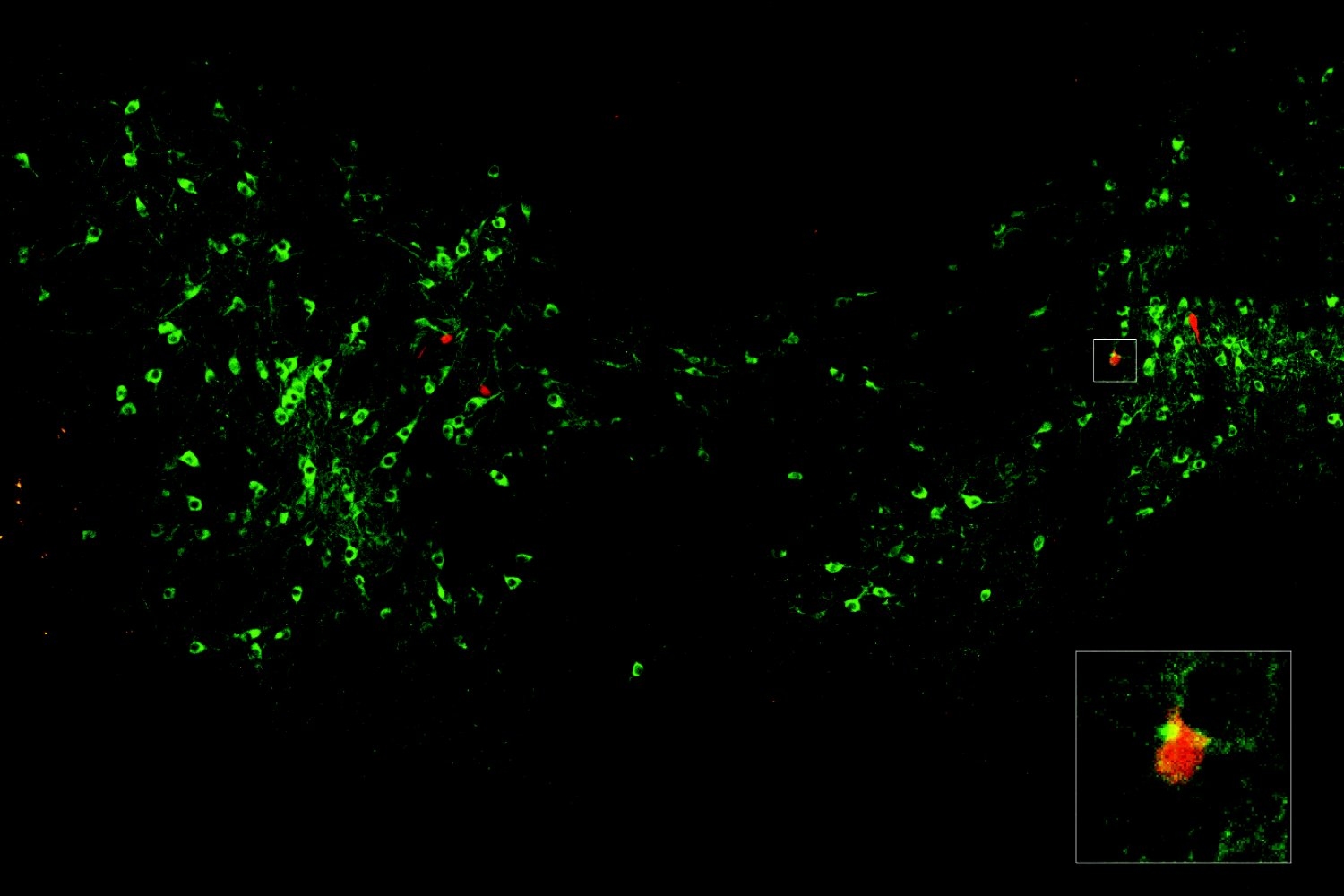
To do that, the researchers fed mice a high-fat diet and performed single-cell RNA-sequencing of their liver cells at key timepoints as liver disease progressed. This allowed them to monitor gene expression changes that occurred as the mice advanced through liver inflammation, to tissue scarring and eventually cancer.
In the early stages of this progression, the researchers found that the high-fat diet prompted hepatocytes, the most abundant cell type in the liver, to turn on genes that help them survive the stressful environment. These include genes that make them more resistant to apoptosis and more likely to proliferate.
At the same time, those cells began to turn off some of the genes that are critical for normal hepatocyte function, including metabolic enzymes and secreted proteins.
“This really looks like a trade-off, prioritizing what’s good for the individual cell to stay alive in a stressful environment, at the expense of what the collective tissue should be doing,” Tzouanas says.
Some of these changes happened right away, while others, including a decline in metabolic enzyme production, shifted more gradually over a longer period. Nearly all of the mice on a high-fat diet ended up developing liver cancer by the end of the study.
When cells are in a more immature state, it appears that they are more likely to become cancerous if a mutation occurs later on, the researchers say.
“These cells have already turned on the same genes that they’re going to need to become cancerous. They’ve already shifted away from the mature identity that would otherwise drag down their ability to proliferate,” Tzouanas says. “Once a cell picks up the wrong mutation, then it’s really off to the races and they’ve already gotten a head start on some of those hallmarks of cancer.”
The researchers also identified several genes that appear to orchestrate the changes that revert hepatocytes to an immature state. While this study was going on, a drug targeting one of these genes (thyroid hormone receptor) was approved to treat a severe form of steatotic liver disease called MASH fibrosis. And, a drug activating an enzyme that they identified (HMGCS2) is now in clinical trials to treat steatotic liver disease.
Another possible target that the new study revealed is a transcription factor called SOX4, which is normally only active during fetal development and in a small number of adult tissues (but not the liver).
Cancer progression
After the researchers identified these changes in mice, they sought to discover if something similar might be happening in human patients with liver disease. To do that, they analyzed data from liver tissue samples removed from patients at different stages of the disease. They also looked at tissue from people who had liver disease but had not yet developed cancer.
Those studies revealed a similar pattern to what the researchers had seen in mice: The expression of genes needed for normal liver function decreased over time, while genes associated with immature states went up. Additionally, the researchers found that they could accurately predict patients’ survival outcomes based on an analysis of their gene expression patterns.
“Patients who had higher expression of these pro-cell-survival genes that are turned on with high-fat diet survived for less time after tumors developed,” Tzouanas says. “And if a patient has lower expression of genes that support the functions that the liver normally performs, they also survive for less time.”
While the mice in this study developed cancer within a year or so, the researchers estimate that in humans, the process likely extends over a longer span, possibly around 20 years. That will vary between individuals depending on their diet and other risk factors such as alcohol consumption or viral infections, which can also promote liver cells’ reversion to an immature state.
The researchers now plan to investigate whether any of the changes that occur in response to a high-fat diet can be reversed by going back to a normal diet, or by taking weight-loss drugs such as GLP-1 agonists. They also hope to study whether any of the transcription factors they identified could make good targets for drugs that could help prevent diseased liver tissue from becoming cancerous.
“We now have all these new molecular targets and a better understanding of what is underlying the biology, which could give us new angles to improve outcomes for patients,” Shalek says.
The research was funded, in part, by a Fannie and John Hertz Foundation Fellowship, a National Science Foundation Graduate Research Fellowship, the National Institutes of Health, and the MIT Stem Cell Initiative through Foundation MIT.