MIT biologists find highly concentrated droplets can help cells keep enzymes organized and control growth signals.
Anne Trafton | MIT News
June 1, 2026
Within the past decade, biologists have discovered that one strategy cells use to keep their contents organized is a phenomenon known as phase separation.
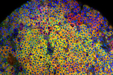
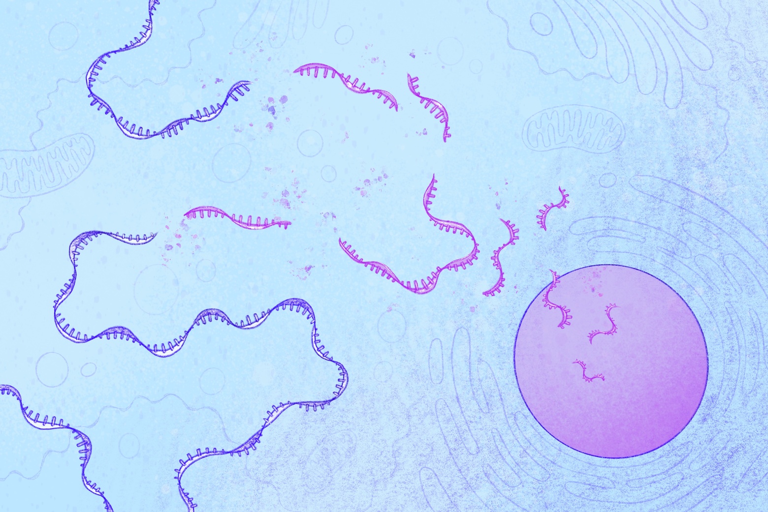
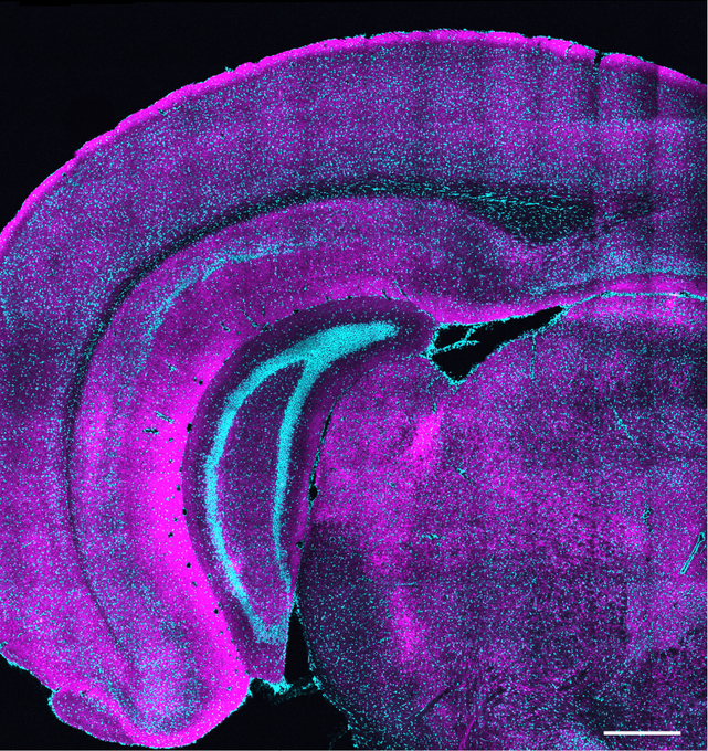
Similar to the way oil forms droplets that float in a vinegar solution, proteins inside cells can phase separate to form highly concentrated droplets that keep them organized within the cell. In a new study, MIT researchers have now shown that this droplet formation is critical for controlling the function of a class of enzymes called kinases.
The researchers found that condensing into droplets optimizes the biochemical conditions needed for kinases to catalyze reactions, allowing them to more rapidly activate cell signaling pathways. In some cases, droplet formation can even change which reactions the kinases perform.
“Many biological molecules have this propensity to spontaneously separate. We were really interested in asking, if we have these kinases forming droplets, what is the consequence of that in the context of signaling?” says Lindsay Case, an assistant professor of biology at MIT and the senior author of the study.
Learning more about how these droplets form could help researchers design drugs that target kinases, some of which can be overactive in cancer cells.
“Understanding the chemistry of these compartments, and what molecules go into them and what molecules don’t go into them, could help us design drugs that better localize to their target of interest,” Case says.
Nicholas Lea, an MIT graduate student, is the lead author of the paper, which appears today in Cell Reports.
Forming droplets
Since her days as a graduate student, Case has been studying how the physical organization of molecules inside cells affects their function. As a postdoc, she began studying how phase separation might affect a signaling pathway that allows cells to sense when they’re attached to their environment, so they can respond appropriately.
Some of the proteins in this pathway are kinases, which activate other proteins by adding phosphate groups to them. Kinases can also activate themselves through a process called autophosphorylation.
“Inside of the cell, you have these kinase molecules that are responsible for carrying a signal through the cell, and we know that the organization of these molecules changes. When the information is present, they’re organized in a different way than when the information is not present,” Case says. “We think that having the right molecules in the right place is incredibly important for the right biochemistry to occur.”
Phase separation is one of the methods that cells appear to use for this organization. The most familiar example of phase separation can be seen in a salad dressing, where oil forms droplets to minimize contact with water-based vinegar. Proteins can phase separate when they are highly concentrated, leading them to self-assemble into dense droplets floating in the cell’s cytoplasm.
Case hypothesized that this phase separation, which brings kinases together at a high density, might help cells to boost the enzymes’ activity because they are more likely to bump into and phosphorylate each other.
In this study, Case and Lea set out to test that hypothesis, focusing on an enzyme called focal adhesion kinase (FAK). This kinase, which becomes activated when cells attach to their surrounding environment, activates pro-growth and pro-survival signals. In cancer cells, this signaling pathway can go awry, allowing cells to proliferate even when they detach from their original locations.
Scientists already knew that when cells are properly attached to their environment, that adhesion signal causes FAK to accumulate at the cell membrane. In the new study, the MIT team mimicked that effect by overexpressing FAK in cells. These cells were floating freely in a solution, not attached to any surface. Even so, the high concentration of FAK caused the kinase to phase separate into droplets, which turned on the pro-growth signal.
“It was surprising that just by condensing this protein into a droplet, you can actually turn on a signaling pathway that should be turned off,” Case says. “If FAK concentration is too high, you’re always getting these droplets and you’re always signaling, regardless of what the receptors that are supposed to be controlling this are doing.”
The findings suggest that in cancer cells, overexpression of FAK may lead to phase separation, which then helps to drive cancer progression and metastasis.
“It may be that for some kinases, you’re not supposed to form these droplets in the cytoplasm because it leads to this always-on signal, and then the cells no longer listen to the information coming from the environment,” Case says.
Interfering with FAK’s ability to form droplets could offer a new strategy for cancer drug development, she says.
Controlling reactions
The researchers also studied two other kinases, Mst2 and Abl. They found that these enzymes could also phase separate at high concentrations, and that this increased their activity. While phase separation of FAK in the cytoplasm may occur only in cancerous cells, for Mst2, it appears to be a strategy that healthy cells use to control a signaling pathway called Hippo, which promotes cell growth and survival.
Additionally, for both Mst2 and Abl, the researchers discovered that phase separation can lead the enzymes to phosphorylate additional targets, which may lead them to activate different signaling pathways.
“It’s not just that you’re getting faster phosphorylation, but in those cases, the patterns of what is actually getting phosphorylated were very different inside of the droplet compared to what might be happening in a non-droplet context,” Case says. “The kinase is able to phosphorylate amino acid residues beyond the set of canonical sites that have been described before.”
The researchers also found that when these droplets form, they attract high concentrations of ATP, the molecule that kinases use as a source of phosphate. This occurs because kinases tend to contain floppy sections containing many positively charged amino acids, which attract negatively charged ATP.
Using a machine-learning model, the researchers predicted that about 45 percent of the 500 kinases found in human cells would have the ability to form droplets like those seen in this study. Those kinases were also more likely to be highly positively charged, which could help them to recruit ATP into the droplets.
In future work, Case hopes to explore the possibility of designing drugs that could mimic ATP’s ability to be attracted into droplets within a cell, which could help reduce negative side effects of the drugs.
“By localizing drugs to the compartment where your target localizes, that could reduce off-target effects by concentrating the drug with the target of interest and reducing interactions with other molecules,” Case says.
The research was funded by a Searle Scholars Program Award, the U.S. Air Force Office of Scientific Research, the National Institutes of Health, the Royal G. and Mae H. Westaway Family Memorial Fund, and a David H. Koch Graduate Fellowship.