Biologists identify new targets for cancer vaccines

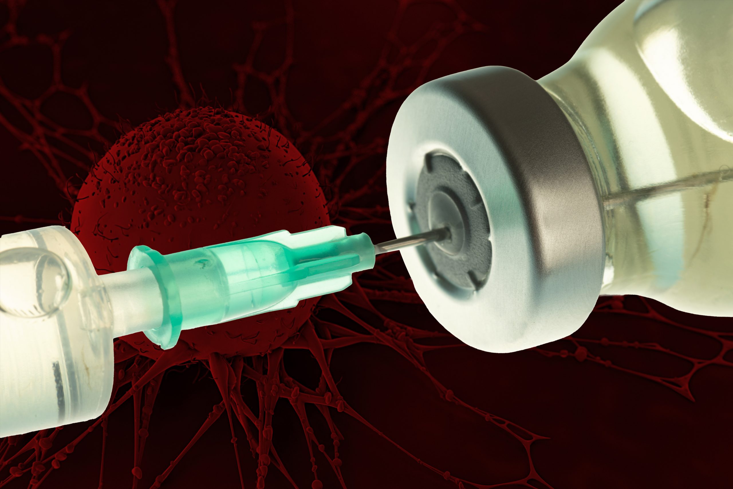
Vaccinating against certain proteins found on cancer cells could help to enhance the T cell response to tumors.

Anne Trafton | MIT News Office
September 16, 2021

Over the past decade, scientists have been exploring vaccination as a way to help fight cancer. These experimental cancer vaccines are designed to stimulate the body’s own immune system to destroy a tumor, by injecting fragments of cancer proteins found on the tumor.

So far, none of these vaccines have been approved by the FDA, but some have shown promise in clinical trials to treat melanoma and some types of lung cancer. In a new finding that may help researchers decide what proteins to include in cancer vaccines, MIT researchers have found that vaccinating against certain cancer proteins can boost the overall T cell response and help to shrink tumors in mice.

The research team found that vaccinating against the types of proteins they identified can help to reawaken dormant T cell populations that target those proteins, strengthening the overall immune response.

“This study highlights the importance of exploring the details of immune responses against cancer deeply. We can now see that not all anticancer immune responses are created equal, and that vaccination can unleash a potent response against a target that was otherwise effectively ignored,” says Tyler Jacks, the David H. Koch Professor of Biology, a member of the Koch Institute for Integrative Cancer Research, and the senior author of the study.

MIT postdoc Megan Burger is the lead author of the new study, which appears today in Cell.

T cell competition

When cells begin to turn cancerous, they start producing mutated proteins not seen in healthy cells. These cancerous proteins, also called neoantigens, can alert the body’s immune system that something has gone wrong, and T cells that recognize those neoantigens start destroying the cancerous cells.

Eventually, these T cells experience a phenomenon known as “T cell exhaustion,” which occurs when the tumor creates an immunosuppressive environment that disables the T cells, allowing the tumor to grow unchecked.

Scientists hope that cancer vaccines could help to rejuvenate those T cells and help them to attack tumors. In recent years, they have worked to develop methods for identifying neoantigens in patient tumors to incorporate into personalized cancer vaccines. Some of these vaccines have shown promise in clinical trials to treat melanoma and non-small cell lung cancer.

“These therapies work amazingly in a subset of patients, but the vast majority still don’t respond very well,” Burger says. “A lot of the research in our lab is aimed at trying to understand why that is and what we can do therapeutically to get more of those patients responding.”

Previous studies have shown that of the hundreds of neoantigens found in most tumors, only a small number generate a T cell response.

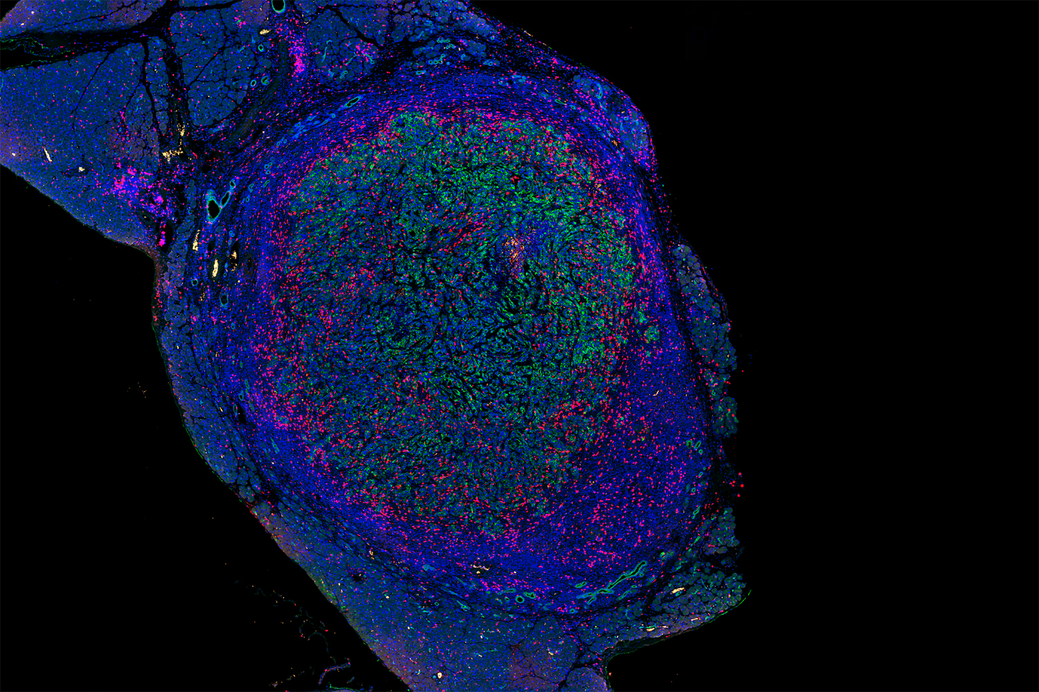
The new MIT study helps to shed light on why that is. In studies of mice with lung tumors, the researchers found that as tumor-targeting T cells arise, subsets of T cells that target different cancerous proteins compete with each other, eventually leading to the emergence of one dominant population of T cells. After these T cells become exhausted, they still remain in the environment and suppress any competing T cell populations that target different proteins found on the tumor.

However, Burger found that if she vaccinated these mice with one of the neoantigens targeted by the suppressed T cells, she could rejuvenate those T cell populations.

“If you vaccinate against antigens that have suppressed responses, you can unleash those T cell responses,” she says. “Trying to identify these suppressed responses and specifically targeting them might improve patient responses to vaccine therapies.”

Shrinking tumors

In this study, the researchers found that they had the most success when vaccinating with neoantigens that bind weakly to immune cells that are responsible for presenting the antigen to T cells. When they used one of those neoantigens to vaccinate mice with lung tumors, they found the tumors shrank by an average of 27 percent.

“The T cells proliferate more, they target the tumors better, and we see an overall decrease in lung tumor burden in our mouse model as a result of the therapy,” Burger says.

After vaccination, the T cell population included a type of cells that have the potential to continuously refuel the response, which could allow for long-term control of a tumor.

In future work, the researchers hope to test therapeutic approaches that would combine this vaccination strategy with cancer drugs called checkpoint inhibitors, which can take the brakes off exhausted T cells, stimulating them to attack tumors. Supporting that approach, the results published today also indicate that vaccination boosts the number of a specific type of T cells that have been shown to respond well to checkpoint therapies.

The research was funded by the Howard Hughes Medical Institute, the Ludwig Center at Harvard University, the National Institutes of Health, the Koch Institute Support (core) Grant from the National Cancer Institute, the Bridge Project of the Koch Institute and Dana-Farber/Harvard Cancer Center, and fellowship awards from the Jane Coffin Childs Memorial Fund for Medical Research and the Ludwig Center for Molecular Oncology at MIT.

New drug combo shows early potential for treating pancreatic cancer

Researchers find three immunotherapy drugs given together can eliminate pancreatic tumors in mice.

Anne Trafton | MIT News Office
August 5, 2021

Pancreatic cancer, which affects about 60,000 Americans every year, is one of the deadliest forms of cancer. After diagnosis, fewer than 10 percent of patients survive for five years.

While some chemotherapies are initially effective, pancreatic tumors often become resistant to them. The disease has also proven difficult to treat with newer approaches such as immunotherapy. However, a team of MIT researchers has now developed an immunotherapy strategy and shown that it can eliminate pancreatic tumors in mice.

The new therapy, which is a combination of three drugs that help boost the body’s own immune defenses against tumors, is expected to enter clinical trials later this year.

“We don’t have a lot of good options for treating pancreatic cancer. It’s a devastating disease clinically,” says William Freed-Pastor, a senior postdoc at MIT’s Koch Institute for Integrative Cancer Research. “If this approach led to durable responses in patients, it would make a big impact in at least a subset of patients’ lives, but we need to see how it will actually perform in trials.”

Freed-Pastor, who is also a medical oncologist at Dana-Farber Cancer Institute, is the lead author of the new study, which appears today in Cancer Cell. Tyler Jacks, the David H. Koch Professor of Biology and a member of the Koch Institute, is the paper’s senior author.

Immune attack

The body’s immune system contains T cells that can recognize and destroy cells that express cancerous proteins, but most tumors create a highly immunosuppressive environment that disables these T cells, helping the tumor to survive.

Immune checkpoint therapy (the most common form of immunotherapy currently being used clinically) works by removing the brakes on these T cells, rejuvenating them so they can destroy tumors. One class of immunotherapy drug that has shown success in treating many types of cancer targets the interactions between PD-L1, a cancer-linked protein that turns off T cells, and PD-1, the T cell protein that PD-L1 binds to. Drugs that block PD-L1 or PD-1, also called checkpoint inhibitors, have been approved to treat cancers such as melanoma and lung cancer, but they have very little effect on pancreatic tumors.

Some researchers had hypothesized that this failure could be due to the possibility that pancreatic tumors don’t express as many cancerous proteins, known as neoantigens. This would give T cells fewer targets to attack, so that even when T cells were stimulated by checkpoint inhibitors, they wouldn’t be able to identify and destroy tumor cells.

However, some recent studies had shown, and the new MIT study confirmed, that many pancreatic tumors do in fact express cancer-specific neoantigens. This finding led the researchers to suspect that perhaps a different type of brake, other than the PD-1/PD-L1 system, was disabling T cells in pancreatic cancer patients.

In a study using mouse models of pancreatic cancer, the researchers found that in fact, PD-L1 is not highly expressed on pancreatic cancer cells. Instead, most pancreatic cancer cells express a protein called CD155, which activates a receptor on T cells known as TIGIT.

When TIGIT is activated, the T cells enter a state known as “T cell exhaustion,” in which they are unable to mount an attack on pancreatic tumor cells. In an analysis of tumors removed from pancreatic cancer patients, the researchers observed TIGIT expression and T cell exhaustion from about 60 percent of patients, and they also found high levels of CD155 on tumor cells from patients.

“The CD155/TIGIT axis functions in a very similar way to the more established PD-L1/PD-1 axis. TIGIT is expressed on T cells and serves as a brake to those T cells,” Freed-Pastor says. “When a TIGIT-positive T cell encounters any cell expressing high levels of CD155, it can essentially shut that T cell down.”

Drug combination

The researchers then set out to see if they could use this knowledge to rejuvenate exhausted T cells and stimulate them to attack pancreatic tumor cells. They tested a variety of combinations of experimental drugs that inhibit PD-1 and TIGIT, along with another type of drug called a CD40 agonist antibody.

CD40 agonist antibodies, some of which are currently being clinically evaluated to treat pancreatic cancer, are drugs that activate T cells and drive them into tumors. In tests in mice, the MIT team found that drugs against PD-1 had little effect on their own, as has previously been shown for pancreatic cancer. They also found that a CD40 agonist antibody combined with either a PD-1 inhibitor or a TIGIT inhibitor was able to halt tumor growth in some animals, but did not substantially shrink tumors.

However, when they combined CD40 agonist antibodies with both a PD-1 inhibitor and a TIGIT inhibitor, they found a dramatic effect. Pancreatic tumors shrank in about half of the animals given this treatment, and in 25 percent of the mice, the tumors disappeared completely. Furthermore, the tumors did not regrow after the treatment was stopped. “We were obviously quite excited about that,” Freed-Pastor says.

Working with the Lustgarten Foundation for Pancreatic Cancer Research, which helped to fund this study, the MIT team sought out two pharmaceutical companies who between them have a PD-1 inhibitor, TIGIT inhibitor, and CD40 agonist antibody in development. None of these drugs are FDA-approved yet, but they have each reached phase 2 clinical trials. A clinical trial on the triple combination is expected to begin later this year.

“This work uses highly sophisticated, genetically engineered mouse models to investigate the details of immune suppression in pancreas cancer, and the results have pointed to potential new therapies for this devastating disease,” Jacks says. “We are pushing as quickly as possible to test these therapies in patients and are grateful for the Lustgarten Foundation and Stand Up to Cancer for their help in supporting the research.”

Alongside the clinical trial, the MIT team plans to analyze which types of pancreatic tumors might respond best to this drug combination. They are also doing further animal studies to see if they can boost the treatment’s effectiveness beyond the 50 percent that they saw in this study.

In addition to the Lustgarten Foundation, the research was funded by Stand Up To Cancer, the Howard Hughes Medical Institute, Dana-Farber/Harvard Cancer Center, the Damon Runyon Cancer Research Foundation, and the National Institutes of Health.

Alison E. Ringel

Education

  • PhD, 2015, Johns Hopkins University School of Medicine
  • BA, 2009, Molecular Biology & Biochemistry/Physics, Wesleyan University

Research Summary

We investigate crosstalk between CD8+ T cells and their environment at a molecular level, by dissecting the biological and metabolic programs engaged under conditions of stress. Using an array of approaches to model and perturb the local microenvironment, our research aims to reveal both the adaptive molecular changes as well as intrinsic vulnerabilities in T cells that arise within the tumor niche. Our goal is to understand how disease states remodel the fundamental mechanisms that regulate immune cell function and contribute to pathogenesis.

Awards

  • Forbeck Scholar, 2021
Yadira Soto-Feliciano

Education

  • PhD, 2016, MIT
  • BS, 2008, Chemistry, University of Puerto Rico-Mayagüez 

Research Summary

We study chromatin — the complex of DNA and proteins that make up our chromosomes. We aim to understand how post-translational modifications to these building-blocks, as well as the factors that regulate these events, play essential roles in maintaining the integrity of cells, tissues, and ultimately entire organisms. We implement a combination of functional genomics, biochemical, genetic, and epigenomic approaches to study how chromatin and epigenetic factors decode the chemical language of chromatin, and how these are dysregulated in diseases such as cancer.

Awards

  • AACR Gertrude B. Elion Cancer Research Award, 2023
  • V Foundation Award, 2022
  • NIH MOSAIC K99/R00 Postdoctoral Career Transition Award, 2021
  • Eddie Méndez Scholar Award, Fred Hutchinson Cancer Research Center, 2020
  • Damon Runyon-Sohn Pediatric Cancer Fellowship, Damon Runyon Cancer Research Foundation, 2017
Francisco J. Sánchez-Rivera

Education

  • PhD, 2016, Biology, MIT
  • BS, 2008, Microbiology, University of Puerto Rico at Mayagüez

Research Summary

The overarching goal of the Sánchez-Rivera laboratory is to elucidate the cellular and molecular mechanisms by which genetic variation shapes normal physiology and disease, particularly in the context of cancer. To do so, we develop and apply genome engineering technologies, genetically-engineered mouse models (GEMMs), and single cell lineage tracing and omics approaches to obtain comprehensive biological pictures of disease evolution at single cell resolution. By doing so, we hope to produce actionable discoveries that could pave the way for better therapeutic strategies to treat cancer and other diseases.

Awards

  • V Foundation Award, 2022
  • Hanna H. Gray Fellowship, Howard Hughes Medical Institute, 2018-2026
  • GMTEC Postdoctoral Researcher Innovation Grant, Memorial Sloan Kettering Cancer Center, 2020-2022
  • 100 inspiring Hispanic/Latinx scientists in America, Cell Mentor/Cell Press, 2020
Biologists discover a trigger for cell extrusion

Study suggests this process for eliminating unneeded cells may also protect against cancer.

Anne Trafton | MIT News Office
May 5, 2021

For all animals, eliminating some cells is a necessary part of embryonic development. Living cells are also naturally sloughed off in mature tissues; for example, the lining of the intestine turns over every few days.

One way that organisms get rid of unneeded cells is through a process called extrusion, which allows cells to be squeezed out of a layer of tissue without disrupting the layer of cells left behind. MIT biologists have now discovered that this process is triggered when cells are unable to replicate their DNA during cell division.

The researchers discovered this mechanism in the worm C. elegans, and they showed that the same process can be driven by mammalian cells; they believe extrusion may serve as a way for the body to eliminate cancerous or precancerous cells.

“Cell extrusion is a mechanism of cell elimination used by organisms as diverse as sponges, insects, and humans,” says H. Robert Horvitz, the David H. Koch Professor of Biology at MIT, a member of the McGovern Institute for Brain Research and the Koch Institute for Integrative Cancer Research, a Howard Hughes Medical Institute investigator, and the senior author of the study. “The discovery that extrusion is driven by a failure in DNA replication was unexpected and offers a new way to think about and possibly intervene in certain diseases, particularly cancer.”

MIT postdoc Vivek Dwivedi is the lead author of the paper, which appears today in Nature. Other authors of the paper are King’s College London research fellow Carlos Pardo-Pastor, MIT research specialist Rita Droste, MIT postdoc Ji Na Kong, MIT graduate student Nolan Tucker, Novartis scientist and former MIT postdoc Daniel Denning, and King’s College London professor of biology Jody Rosenblatt.

Stuck in the cell cycle

In the 1980s, Horvitz was one of the first scientists to analyze a type of programmed cell suicide called apoptosis, which organisms use to eliminate cells that are no longer needed. He made his discoveries using C. elegans, a tiny nematode that contains exactly 959 cells. The developmental lineage of each cell is known, and embryonic development follows the same pattern every time. Throughout this developmental process, 1,090 cells are generated, and 131 cells undergo programmed cell suicide by apoptosis.

Horvitz’s lab later showed that if the worms were genetically mutated so that they could not eliminate cells by apoptosis, a few of those 131 cells would instead be eliminated by cell extrusion, which appears to be able to serve as a backup mechanism to apoptosis. How this extrusion process gets triggered, however, remained a mystery.

To unravel this mystery, Dwivedi performed a large-scale screen of more than 11,000 C. elegans genes. One by one, he and his colleagues knocked down the expression of each gene in worms that could not perform apoptosis. This screen allowed them to identify genes that are critical for turning on cell extrusion during development.

To the researchers’ surprise, many of the genes that turned up as necessary for extrusion were involved in the cell division cycle. These genes were primarily active during first steps of the cell cycle, which involve initiating the cell division cycle and copying the cell’s DNA.

Further experiments revealed that cells that are eventually extruded do initially enter the cell cycle and begin to replicate their DNA. However, they appear to get stuck in this phase, leading them to be extruded.

Most of the cells that end up getting extruded are unusually small, and are produced from an unequal cell division that results in one large daughter cell and one much smaller one. The researchers showed that if they interfered with the genes that control this process, so that the two daughter cells were closer to the same size, the cells that normally would have been extruded were able to successfully complete the cell cycle and were not extruded.

The researchers also showed that the failure of the very small cells to complete the cell cycle stems from a shortage of the proteins and DNA building blocks needed to copy DNA. Among other key proteins, the cells likely don’t have enough of an enzyme called LRR-1, which is critical for DNA replication. When DNA replication stalls, proteins that are responsible for detecting replication stress quickly halt cell division by inactivating a protein called CDK1. CDK1 also controls cell adhesion, so the researchers hypothesize that when CDK1 is turned off, cells lose their stickiness and detach, leading to extrusion.

Cancer protection

Horvitz’s lab then teamed up with researchers at King’s College London, led by Rosenblatt, to investigate whether the same mechanism might be used by mammalian cells. In mammals, cell extrusion plays an important role in replacing the lining of the intestines, lungs, and other organs.

The researchers used a chemical called hydroxyurea to induce DNA replication stress in canine kidney cells grown in cell culture. The treatment quadrupled the rate of extrusion, and the researchers found that the extruded cells made it into the phase of the cell cycle where DNA is replicated before being extruded. They also showed that in mammalian cells, the well-known cancer suppressor p53 is involved in initiating extrusion of cells experiencing replication stress.

That suggests that in addition to its other cancer-protective roles, p53 may help to eliminate cancerous or precancerous cells by forcing them to extrude, Dwivedi says.

“Replication stress is one of the characteristic features of cells that are precancerous or cancerous. And what this finding suggests is that the extrusion of cells that are experiencing replication stress is potentially a tumor suppressor mechanism,” he says.

The fact that cell extrusion is seen in so many animals, from sponges to mammals, led the researchers to hypothesize that it may have evolved as a very early form of cell elimination that was later supplanted by programmed cell suicide involving apoptosis.

“This cell elimination mechanism depends only on the cell cycle,” Dwivedi says. “It doesn’t require any specialized machinery like that needed for apoptosis to eliminate these cells, so what we’ve proposed is that this could be a primordial form of cell elimination. This means it may have been one of the first ways of cell elimination to come into existence, because it depends on the same process that an organism uses to generate many more cells.”

Dwivedi, who earned his PhD at MIT, was a Khorana scholar before entering MIT for graduate school. This research was supported by the Howard Hughes Medical Institute and the National Institutes of Health.

Catching key moments of cancer progression
Whitehead Institute
February 9, 2021

Important moments of cancer — mutation, tumor formation, metastasis —  are fleeting, easy-to-miss events. Even with modern medical technologies and methods, they often happen unobserved, and are only realized later when these cells spawn life-threatening conditions.

In recent years, however, new methods of tracking individual cells through time have allowed researchers to get closer to the origin of these events, and Whitehead Institute scientists are turning the power of these technologies to study cells involved in several different types of cancer. “With [these technologies], you can track down rare events in the past, identify all the offspring of an event, and see how they’ve changed their behavior,” said Whitehead Institute Member Jonathan Weissman.  “You can ask, when a cell picks up an oncogene, how does it mutate, and further evolve, and proliferate and metastasize?”

Read on to learn how three Whitehead Institute Members are using specially engineered mice, CRISPR-based technologies, and other methods to track cells at different stages of cancer development, pushing the boundaries of what we understand about how the disease starts and proliferates. From the initial beginnings of a tumor, sometimes in the darkness of a still-forming embryo, to a tumor’s growth and eventual metastasis to other sites in the body, Whitehead Institute scientists Rudolf Jaenisch, Jonathan Weissman, and Robert Weinberg study the pivotal points in a cancer’s growth and spread.
The birth of a tumor

For around 800 children each year in the United States, the seeds are sown during fetal development for a rare and unpredictable childhood cancer called neuroblastoma. To understand how the disease develops, scientists need to study what happens to these cancerous “seeds” as the embryo  matures. But they can hardly study a living human embryo, and fetal development is such a complex process that it is near impossible to simulate it in the lab.

To make matters more complex, the cancer grows and then may shrink unpredictably after the children are born and as they age. Sometimes the tumors disappear on their own; other times they grow uncontrollably.

In 2020, Institute Founding Member Rudolf Jaenisch’s lab introduced a new way of tracking the cells involved in the disease using his tried and true method for modeling such complex conditions: chimeric mice. A chimera is a conglomeration of two species — in this case, mostly mouse, with a few strategically placed human stem cells.

To create chimeras to study neuroblastoma, Jaenisch, who is also a Professor of Biology at Massachusetts Institute of Technology (MIT), along with collaborators at the Koch Institute for integrative Cancer Research at MIT and the Dana-Farber Cancer Institute, engineered human stem cells with glowing proteins to make them easy to see under the microscope. The cells contained a special genetic-switch that allowed the researchers to induce tumors by adding a certain chemical. These human stem cells were then induced to form a more specialized cell type – a neural crest stem cell.

Neural crest cells are a group of developing stem cells that go on to form the peripheral nervous system as well as other parts of the body such as the facial bones. It is neural crest cells that mutate into neuroblastoma tumors in humans, so the researchers hoped that by using these cells, they could create “human” tumors in mice. The researchers injected these human neural crest cells into mice so that they could readily incorporate with the host’s cells.

After the mice were born, the researchers were then able to take samples from the mice over the course of their development to see whether these implanted cells would form neural crest-derived tumors and, if so, what happened as they grew — something they would never have been able to do with neuroblastoma tumors in human infants and children.

The tumors in the chimeric mice pups developed in similar forms to human neuroblastomas — specifically, they formed characteristic rosette shapes — very similar to those seen in patient’s samples. With the help of Stefani Spranger, an assistant professor of biology at MIT and a Member of the Koch Institute for Integrative Cancer Research at MIT, they were able to track the cells’ interactions with the mice’s immune systems, and learn how the cancer “tricks” the immune cells into letting it stick around.

Now that they are able to model the formation of neuroblastoma tumors, the researchers hope to find a way to eliminate the tumors in the mice. “This is a model that will allow us to approach how to get rid of the tumors,” said Malkiel Cohen, a former postdoc in Jaenisch’s lab and first author of the paper, published in the journal Cell Stem Cell describing the work.

Cancer genealogy

A recent project from the lab of Whitehead Institute Member Jonathan Weissman focuses on another essential moment of cancer progression: metastasis. Metastasis happens when cancer spreads from a primary tumor to distant places in the body. Weissman, an expert in genome editing, created a CRISPR-based method to track the lineage of individual cancer cells in real time as they proliferate and metastasize.

Weissman and his collaborators, including graduate student Matthew Jones and then postdoctoral researcher Jeffrey Quinn, adapted the technology from a similar tool designed by Michelle Chan, a former postdoc in Weissman’s former lab at the University of California, San Francisco (UCSF) who is now an assistant professor at Princeton University. Chan designed a CRISPR mechanism to track the lineages of embryonic cells as they developed into specialized tissues.

“What Michelle Chan was able to do was uncover how tissues that look very similar to one another, actually come from disparate sources,” said Matthew Jones, a graduate student in Weissman’s lab and a co-first author on the paper describing the new method. “That has rapid implications about how tissues organize themselves, and we wanted to apply it to cancer.”

To create this system, the researchers engineered cancer cells with added genes: one for Cas9, the gene that codes for CRISPR’s “molecular scissors,” others for fluorescing proteins for microscopy, and a few sequences that would serve as targets for the CRISPR technology.

They then implanted thousands of these cells into the lungs of mice to simulate a tumor, using a model designed by Trever Bivona, a cancer biologist at UCSF. As the cells in the model tumors began to divide, the Cas9 protein began making small snips in the target sites in the cells’ DNA. When the cells fixed these snips, they patched in or deleted a few random nucleotides, leading to a unique “barcode” in each cell. Because these barcodes were added to each cell’s DNA, they were heritable and able to be passed on through generations of cells.

With the help of Nir Yosef, a computer scientist at the University of California, Berkeley, the researchers organized the data into “family trees” of cancer cells spanning multiple generations. By taking samples from different areas of the body, the researchers were able to resolve exactly when a cell jumped from where it started, in the lungs, to a distant tissue.

When they compared the trees, the researchers noticed that some cells were highly metastatic, jumping around multiple times over the course of the experiment, while others stayed put throughout. “We were excited to uncover some of these really rare, but consequential events that happened in the past that you would never be able to observe, and rarely be able to infer from a static snapshot,” Jones said.

By comparing highly metastatic and non-metastatic cells, they were able to identify metastasis-associated genes and answer questions about how the tumors were evolving and adapting. “It’s an entirely new way to look at the behavior and evolution of a tumor,” Weissman said. “We think it can be applied to many different problems in cancer biology.”

The next steps

A natural tumor begins with a single cell, mutated in a way that leads it to “go rogue,” so to speak. To mimic this in a model system, Dian Yang, another post-doc in the Weissman Lab, is collaborating with researchers in the lab of MIT Professor of Biology and Koch Institute Director Tyler Jacks’ to create a mouse model with the CRISPR lineage recording tool embedded in its DNA.

The model is based on a mouse model created by Jacks’ lab to study lung cancer. The lab has created genetically engineered mice that when left alone, live completely normal lives. But upon adding a trigger (an enzyme called “Cre”) — in Jacks’ and Yang’s case, they deliver the trigger to the lung using a virus — oncogenic signals are activated and lead to spontaneous tumors in the mice’s lungs.

Being able to “switch on” the mouse model in this way has a number of advantages.  “Each tumor will start from one single transformed cell, which we can then watch in its native environment as it evolves,” Yang said. “Then, we can look back later at how the cells metastasize.”

Adding the CRISPR system to Jacks’ existing cancer model will also allow researchers to study cancerous cells on a broader time scale. “Usually, when we harvest samples from the mice, it is like a snapshot, just one sample at one stage,” Yang said. “You can see what it looks like, you can analyze gene expression at the time of sampling, you can even take a time series, but you don’t know what happened in the past.”

In a sense, the lineage recording technology embedded in the genome of the mice now makes it possible to look back in time. “When you have a million cells in a tumor, you can use the lineage network of these cells to find out how they’re related, and connect the current state with the past evolutionary lineage history,” he said. “I think this will provide a new dimension of biological information for us to understand biology that is not just limited to cancer biology.”The making of metastasis

Weissman’s lab’s method for tracking the lineage of cancer cells can illuminate the nature of the cells that leave the primary tumor and scatter throughout the body. But what actually happens to these cells to cause them to metastasize?

That’s where Whitehead Institute Founding Member Robert Weinberg’s research comes in. Weinberg has been studying cancer for decades. His early work helped to answer the question of how cells that form a primary tumor become cancerous. Weinberg identified the first human oncogene, a gene that causes otherwise-normal cells to form tumors. This finding, combined with others, demonstrated that cancer is a disease driven by damaged genes, at least in its origins. Weinberg has since turned his attention to the question of how cancer cells acquire the ability to spread.

Around 90 percent of cancer deaths are caused not by the primary tumor, but by its metastasis. Based on previous work, there is no single genetic switch that can be flipped to equip a cancer cell for metastasis. Instead, cells must go through a series of changes over time. Most cancer cells fail to acquire all of the necessary traits, and so may, for example, spread to new tissues but rarely form tumors there. Weinberg’s lab tracks cancer cells to help fill in the “road map” that cancer cells follow on the way to metastasis, in the hopes that their insights can be used to prevent or treat metastatic cancers.

One important change that cancer cells undergo is called the epithelial to mesenchymal transition (EMT), a cellular process that causes the cancer cells to express different genes and go from being immobile to mobile and invasive. Cancer cells undergoing this transition to be able to spread are called “quasi-mesenchymal.” The ability to spread does not fully explain metastasis, however.

“There’s two aspects of the metastasis problem,” Weinberg said. “The first aspect is how cancer cells physically leave the primary tumor and get seeded into a distant tissue. In other words, the physical translocation of the cells. The second step represents the subsequent ability of the already seeded cells to figure out how to make a living in a distant tissue.”

In other words, how do those transplanted cells adapt to a new tissue environment, which might offer them only inhospitable conditions? “That represents the major unsolved problem of metastasis,” he said.

Weinberg hopes to study this more in the future; for now though, his lab has found that studying quasi-mesenchymal cells can serve another purpose. Anushka Dongre, a postdoc in Weinberg’s lab, found that these cells are resistant to a common type of cancer treatment, known as immune checkpoint therapy, and can even protect the other cancer cells around them from that therapy. If as little as ten percent of a tumor consists of quasi-mesenchymal cells, then the whole tumor may become resistant.

By using a tumor’s epithelial/mesenchymal profile, Dongre demonstrated that she could predict how likely a breast cancer tumor was to respond to a particular immune checkpoint therapy. This finding could help physicians match patients to the best treatment plan, by indicating ahead of time whether the treatment will work. She also identified a way to eliminate the quasi-mesenchymal cells’ protective effect by suppressing a certain enzyme that they employ to defend themselves.

Weinberg’s lab continues to study pivotal changes in the lives of cancer cells, such as the EMT, so that they can better understand metastasis and, they hope, help find effective treatments for patients with metastatic cancers.Tracking cells into the future

Scientists have been tracking cells for more than a century, and Whitehead Institute scientists will be tracking cancer cells for decades to come. In the coming years, Weinberg plans to continue to investigate the mysteries of metastasis. For Weissman’s part, he hopes to continue refining his CRISPR technique, with the end goal of eventually being able to predict the behavior of cancer cells. “We want to be able to measure where they are, where they’re going at any time, and then predict where they’re going to be in the future,” he said.

With new technologies and ever-expanding fields, there is limitless potential in these various methods. “That’s what is so exciting about the cell tracking field right now,” said Matt Jones. “It’s really pushing the boundaries on what we can capture from our measurements.”

Catching cancer in the act
Eva Frederick | Whitehead Institute
January 21, 2021

When cancer is confined to one spot in the body, doctors can often treat it with surgery or other therapies. Much of the mortality associated with cancer, however, is due to its tendency to metastasize, sending out seeds of itself that may take root throughout the body. The exact moment of metastasis is fleeting, lost in the millions of divisions that take place in a tumor. “These events are typically impossible to monitor in real time,” said Whitehead Institute Member Jonathan Weissman.

Now, researchers led by Weissman, who is also a professor of biology at Massachusetts Institute of Technology and an investigator with the Howard Hughes Medical Institute, have turned a CRISPR tool into a way to do just that. In a paper published January 21 in Science, Weissman’s lab, in collaboration with Nir Yosef, a computer scientist at the University of California, Berkeley, and Trever Bivona, a cancer biologist at the University of California, San Francisco (UCSF), treats cancer cells the way evolutionary biologists might look at species, mapping out an intricately detailed family tree. By examining the branches, they can track the cell’s lineage to find when a single tumor cell went rogue, spreading its progeny to the rest of the body.

“With this method, you can ask questions like, ‘How frequently is this tumor metastasizing? Where did the metastases come from? Where do they go?’” Weissman said. “By being able to follow the history of the tumor in vivo, you reveal differences in the biology of the tumor that were otherwise invisible.”

Scratch paper cells

Scientists have tracked the lineages of cancer cells in the past by comparing shared mutations and other variations in their DNA blueprints. These methods, however, depend to a certain extent on there being enough naturally occurring mutations or other markers to accurately show relationships between cells. That’s where Weissman and co-first authors Jeffrey Quinn, then a postdoctoral researcher in Weissman’s lab, and Matthew Jones, a graduate student in Weissman’s lab, saw an opportunity to use CRISPR technology — specifically, a method developed by Weissman Lab member Michelle Chan to track embryo development — to facilitate tracking. Instead of simply hoping that a cancer lineage contained enough lineage-specific markers to track, the researchers decided to use Chan’s method to add in markers themselves. “Basically, the idea is to engineer a cell that has a genomic scratchpad of DNA, that then can be ‘written’ on using CRISPR,” Weissman said. This ‘writing’ in the genome is done in such a way that it becomes heritable, meaning a cell’s grand-offspring would have the ‘writing’ of its parent cells and grandparent cells recorded in its genome.

Where did you come from, where did you go? 
To create these special “scratchpad” cells, Weissman engineered human cancer cells with added genes: one for the bacterial protein Cas9 — the famed “molecular scissors” used in CRISPR genome editing methods — others for glowing proteins for microscopy, and a few sequences that would serve as targets for the CRISPR technology. They then implanted thousands of the modified human cancer cells into mice, mimicking a lung tumor (a model developed by collaborator Bivona).  Mice with human lung tumors often exhibit aggressive metastases, so the researchers reasoned they would provide a good model for tracking cancer progression in real time. As the cells began to divide, Cas9 made small cuts at these target sites. When the cell repaired the cuts, it patched in or deleted a few random nucleotides, leading to a unique repair sequence called an indel. This cutting and repairing happened randomly in nearly every generation, creating a map of cell divisions that Weissman and the team could then track using special computer models that they created by working with Yosef, a computer scientist.
Revealing the invisible
Tracking cells this way yielded some interesting results. For one thing, individual tumor cells were much different from each other than the researchers expected. The cells the researchers used were from an established human lung cancer cell line called A549. “You’d think they would be relatively homogeneous,” Weissman said. “But in fact, we saw dramatic differences in the propensity of different tumors to metastasize — even in the same mouse. Some had a very small number of metastatic events, and others were really rapidly jumping around.”To find out where this heterogeneity was coming from, the team implanted two clones of the same cell in different mice. As the cells proliferated, the researchers found that their descendents metastasized at a remarkably similar rate. This was not the case with the offspring of different cells from the same cell line — the original cells had apparently evolved different metastatic potentials as the cell line was maintained over many generations. The scientists next wondered what genes were responsible for this variability between cancer cells from the same cell line. So they began to look for genes that were expressed differently between nonmetastatic, weakly metastatic and highly metastatic tumors. Many genes stood out, some of which were previously known to be associated with metastasis — although it was not clear whether they were driving the metastasis or simply a side effect of it. One of them, the gene that codes for the protein Keratin 17, is much more strongly expressed in low metastatic tumors than in highly metastatic tumors. “When we knocked down or overexpressed Keratin 17, we showed that this gene was actually controlling the tumors’ invasiveness,” Weissman said. Being able to identify metastasis-associated genes this way could help researchers answer questions about how tumors evolve and adapt. “It’s an entirely new way to look at the behavior and evolution of a tumor,” Weissman said. “We think it can be applied to many different problems in cancer biology.”
Where did you come from, where did you go? 
Weissman’s CRISPR method also allowed the researchers to track with more detail where metastasizing cells went in the body, and when. For example, the progeny of one implanted cancer cell underwent metastasis five separate times, spreading each time from the left lung to other tissues such as the right lung and liver. Other cells made a jump to a different area, and then metastasized again from there. These movements can be mapped neatly in phylogenetic trees (see image), where each color represents a different location in the body. A very colorful tree shows a highly metastatic phenotype, where a cell’s descendents jumped many times between different tissues. A tree that is primarily one color represents a less metastatic cell. Mapping tumor progression in this way allowed Weissman and his team to make a few interesting observations about the mechanics of metastasis. For example, some clones seeded in a textbook way, traveling from the left lung, where they started, to distinct areas of the body. Others seeded more erratically, moving first to other tissues before metastasizing again from there.One such tissue, the mediastinal lymph tissue which sits between the lungs, appears to be a hub of sorts, said co-first author Jeffrey Quinn. “It serves as a waystation that connects the cancer cells to all of this fertile ground that they can then go and colonize,” he said. Therapeutically, the discovery of metastasis “hubs” like this could be extremely useful. “If you focus cancer therapies on those places, you could then slow down metastasis or prevent it in the first place,” Weissman said.In the future, Weissman hopes to move beyond simply observing the cells and begin to predict their behavior. “It’s like with Newtonian mechanics — if you know the velocity and position and all the forces acting on a ball, you can figure out where the ball is going to go at any time in the future,” Weissman said. “We’re hoping to do the same thing with cells. We want to construct essentially a function of what is driving differentiation of a tumor, and then be able to measure where they are at any given time, and predict where they’re going to be in the future.”The researchers are optimistic that being able to track the family trees of individual cells in real time will prove useful in other settings as well. “I think that it’s going to unlock a whole new dimension to what we think about as a measurable quantity in biology,” said co-first author Matthew Jones. “That’s what’s really cool about this field in general is that we’re redefining what’s invisible and what is visible.”

Notes

Jeffrey J. Quinn, Matthew G. Jones, Ross A. Okimoto, Shigeki Nanjo, Michelle M. Chan, Nir Yosef, Trever G. Bivona, Jonathan S. Weissman. “Single-cell lineages reveal the rates, routes, and drivers of metastasis in cancer xenografts.” Science, Jan. 21, 2021.

Why cancer cells waste so much energy

MIT study sheds light on the longstanding question of why cancer cells get their energy from fermentation.

Anne Trafton | MIT News Office
January 19, 2021

In the 1920s, German chemist Otto Warburg discovered that cancer cells don’t metabolize sugar the same way that healthy cells usually do. Since then, scientists have tried to figure out why cancer cells use this alternative pathway, which is much less efficient.

MIT biologists have now found a possible answer to this longstanding question. In a study appearing in Molecular Cell, they showed that this metabolic pathway, known as fermentation, helps cells to regenerate large quantities of a molecule called NAD+, which they need to synthesize DNA and other important molecules. Their findings also account for why other types of rapidly proliferating cells, such as immune cells, switch over to fermentation.

“This has really been a hundred-year-old paradox that many people have tried to explain in different ways,” says Matthew Vander Heiden, an associate professor of biology at MIT and associate director of MIT’s Koch Institute for Integrative Cancer Research. “What we found is that under certain circumstances, cells need to do more of these electron transfer reactions, which require NAD+, in order to make molecules such as DNA.”

Vander Heiden is the senior author of the new study, and the lead authors are former MIT graduate student and postdoc Alba Luengo PhD ’18 and graduate student Zhaoqi Li.

Inefficient metabolism

Fermentation is one way that cells can convert the energy found in sugar to ATP, a chemical that cells use to store energy for all of their needs. However, mammalian cells usually break down sugar using a process called aerobic respiration, which yields much more ATP. Cells typically switch over to fermentation only when they don’t have enough oxygen available to perform aerobic respiration.

Since Warburg’s discovery, scientists have put forth many theories for why cancer cells switch to the inefficient fermentation pathway. Warburg originally proposed that cancer cells’ mitochondria, where aerobic respiration occurs, might be damaged, but this turned out not to be the case. Other explanations have focused on the possible benefits of producing ATP in a different way, but none of these theories have gained widespread support.

In this study, the MIT team decided to try to come up with a solution by asking what would happen if they suppressed cancer cells’ ability to perform fermentation. To do that, they treated the cells with a drug that forces them to divert a molecule called pyruvate from the fermentation pathway into the aerobic respiration pathway.

They saw, as others have previously shown, that blocking fermentation slows down cancer cells’ growth. Then, they tried to figure out how to restore the cells’ ability to proliferate, while still blocking fermentation. One approach they tried was to stimulate the cells to produce NAD+, a molecule that helps cells to dispose of the extra electrons that are stripped out when cells make molecules such as DNA and proteins.

When the researchers treated the cells with a drug that stimulates NAD+ production, they found that the cells started rapidly proliferating again, even though they still couldn’t perform fermentation. This led the researchers to theorize that when cells are growing rapidly, they need NAD+ more than they need ATP. During aerobic respiration, cells produce a great deal of ATP and some NAD+. If cells accumulate more ATP than they can use, respiration slows and production of NAD+ also slows.

“We hypothesized that when you make both NAD+ and ATP together, if you can’t get rid of ATP, it’s going to back up the whole system such that you also cannot make NAD+,” Li says.

Therefore, switching to a less efficient method of producing ATP, which allows the cells to generate more NAD+, actually helps them to grow faster. “If you step back and look at the pathways, what you realize is that fermentation allows you to generate NAD+ in an uncoupled way,” Luengo says.

Solving the paradox

The researchers tested this idea in other types of rapidly proliferating cells, including immune cells, and found that blocking fermentation but allowing alternative methods of NAD+ production enabled cells to continue rapidly dividing. They also observed the same phenomenon in nonmammalian cells such as yeast, which perform a different type of fermentation that produces ethanol.

“Not all proliferating cells have to do this,” Vander Heiden says. “It’s really only cells that are growing very fast. If cells are growing so fast that their demand to make stuff outstrips how much ATP they’re burning, that’s when they flip over into this type of metabolism. So, it solves, in my mind, many of the paradoxes that have existed.”

The findings suggest that drugs that force cancer cells to switch back to aerobic respiration instead of fermentation could offer a possible way to treat tumors. Drugs that inhibit NAD+ production could also have a beneficial effect, the researchers say.

The research was funded by the Ludwig Center for Molecular Oncology, the National Science Foundation, the National Institutes of Health, the Howard Hughes Medical Institute, the Medical Research Council, NHS Blood and Transplant, the Novo Nordisk Foundation, the Knut and Alice Wallenberg Foundation, Stand Up 2 Cancer, the Lustgarten Foundation, and the MIT Center for Precision Cancer Medicine.

Disarming cancer
Greta Friar | Whitehead Institute
December 21, 2020

Cancer is at its most deadly when two things occur: the cancer cells metastasize, spreading to new sites in the body, and the cells become resistant to treatment. The epithelial-mesenchymal transition (EMT) is a process that cancer cells may undergo that enables them to do both of these things. Cells that undergo this process are called “quasi-mesenchymal” cancer cells, and they are mobile, aggressive, and harder to kill. They can resist attacks launched both by the body’s own immune system as well as immune checkpoint blockade therapy (ICB), an increasingly employed clinical treatment that works by liberating cells of the immune system from certain constraints, thereby allowing them to attack cancer cells. Anushka Dongre, a postdoctoral researcher in the lab of Whitehead Institute Founding Member Robert Weinberg, had previously found that even a small population of quasi-mesenchymal cells within a mouse breast cancer tumor—as little as 10% amongst a majority of cells that had not gone through the EMT—could protect the entire tumor from a version of ICB called anti-CTLA4 therapy. Most breast cancers in humans contain some minority populations of quasi-mesenchymal cells, as do many other types of human tumors, likely contributing to ICB therapy’s mixed success rates in the clinic.

Because cells that have been through the EMT process play such a large role in making cancers more deadly and less responsive to treatment, Dongre set out to understand how to defang them. Her first step was to figure out how minority populations of quasi-mesenchymal cells within a breast tumor make the tumors as a whole resistant to immune therapy. Then she studied how to disable those mechanisms. The work, described in a paper published in Cancer Discovery on December 16, includes studies in mice showing that disabling those resistance mechanisms can sensitize otherwise-resistant tumors to anti-CTLA4 checkpoint blockade immunotherapy and reduce the severity of metastasis.

Dongre had previously studied how quasi-mesenchymal cells alter the area in and around a tumor to render it more favorable for the outgrowth of a cancer. They keep out of the core of the tumor the type of immune cells that can destroy cancers, and instead let in other types of immune cells that the tumor is able to co-opt to its benefit, thereby protecting it from immune attack. 

In her latest research, Dongre identified six molecules that quasi-mesenchymal cells produce and release that help them perturb the tumor’s surroundings, protecting cells throughout the tumor from immune attack and elimination. She then tested what happened when the release of each of the protective molecules was suppressed. She discovered that eliminating release of either of two molecules, CSF1 and SPP1, made the tumors significantly more susceptible to the immune attack and thus elimination by ICB therapyHoweverthe strongest therapeutic benefit came when she prevented production of CD73, an enzyme usually made by the quasi-mesenchymal cells that produces the immunosuppressive molecule adenosine. In mice, anti-CTLA4 therapy was very effective against tumors in which CD73 and thus adenosine had been eliminated from the quasi-mesenchymal cells, in some cases, succeeding in eliminating the tumors entirely. These findings are consistent with previous research that identified CD73 as a good complementary target for immunotherapy. Furthermore, the experiments demonstrated the utility of combining anti-CD73 therapy with anti-CTLA4 immunotherapy in order to successfully treat tumors that would usually not respond to treatment by ICB therapy alone. Dongre was particularly excited to see the combination of anti-CD73 and anti-CTLA4 reduce the number and size of metastatic tumors.

Dongre hopes that these insights will prove useful for patients.

“There is this minority population of mesenchymal cells present in many patient tumors, creating a big barrier to therapy. I’m hopeful that by identifying the drivers that can sensitize this population to treatment, our work can one day help patients suffering from cancers that are resistant to current therapies,” Dongre says.