Research Area: Biochemistry, Biophysics, and Structural Biology
Education
PhD, 1997, Stanford University
BS, 1990, Biological Sciences, Stanford University
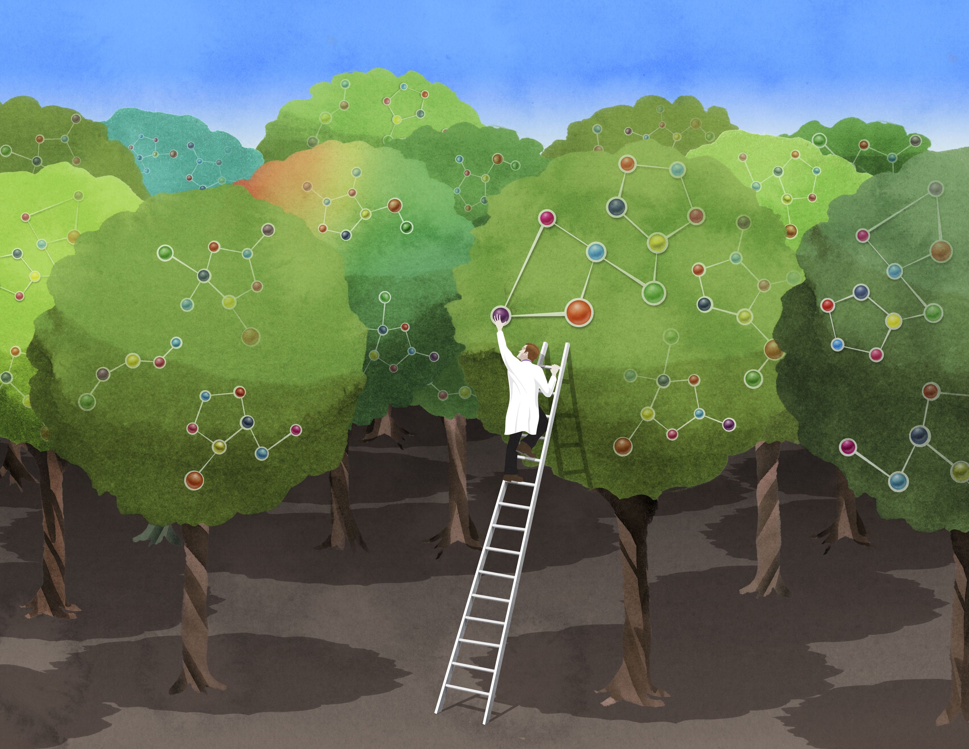
Research Summary
We aim to understand the code for RNA splicing: how the precise locations of introns and splice sites are identified in primary transcripts and how its specificity changes in different cell types. Toward this end, we are mapping the RNA-binding affinity spectra of dozens of human RNA-binding proteins and integrating this information with in vivo binding and activity data. We are also studying the functions of 3’ untranslated regions, including their roles in mRNA localization and microRNA regulation. The lab uses a combination of computational and experimental approaches to address these questions.
Awards
Schering-Plough Research Institute Award (ASBMB), 2007
Overton Prize for Computational Biology (ISCB), 2001
Education
PhD, 1990, University of California, Berkeley
BS, 1985, Integrated Science Program and Biochemistry, Molecular Biology and Cell Biology, Northwestern University
Research Summary
We focus on the events that occur at the starting points of chromosome duplication. These DNA sequences — called “origins of replication” — are found at multiple sites on each eukaryotic chromosome and direct the assembly of replisomes, which replicate the DNA on both sides of the origin. We study this assembly process to understand how chromosomes are replicated, and how these events are regulated during the cell cycle to ensure genome maintenance.
Awards
National Academy of Sciences, Member, 2017
National Academy of Sciences Award in Molecular Biology, 2009
Howard Hughes Medical Institute, HHMI Investigator, 2000
Education
PhD, 1993, Harvard University
BA, 1982, Biology, Goshen College
Research Summary
We study microRNAs and other small RNAs that specify the destruction and/or translational repression of mRNAs. We also study mRNAs, focusing on their untranslated regions and poly(A) tails, and how these regions recruit and mediate regulatory phenomena.
Awards
National Academy of Sciences, Member, 2011
Howard Hughes Medical Institute, HHMI Investigator, 2005
National Academy of Sciences Award in Molecular Biology, 2005
AAAS Newcomb Cleveland Prize, 2002
Education
PhD, 1965, Yale University
BA, 1962, Biology, Amherst College
Research Summary
We study the molecules that allow fungi to penetrate tissues and grow in a hostile environment. Using genetics, biochemistry and genomics, we answer questions such as: What makes Candida albicans such a successful pathogen? How do fungal pathogens evolve antibiotic resistance? How do they manage to change their genetic composition so rapidly?
The Fink lab is no longer accepting students.
Awards
Thomas Hunt Morgan Medal, Genetics Society of America, 2020
James R. Killian Jr. Faculty Achievement Award, 2018
American Association for the Advancement of Science, Fellow, 2015
Gruber International Prize in Genetics, 2010
American Philosophical Society, 2003
Yeast Genetics and Molecular Biology – Lifetime Achievement Award, 2002
George W. Beadle Award, Genetics Society of America, 2001
Ellison Medical Foundation, Senior Scholar Award, 2001
National Academy of Medicine, 1996
Wilbur Lucius Cross Medal, Yale University, 1992
Emil Christian Hansen Foundation Award for Microbiology, Denmark, 1986
American Academy of Arts and Sciences, Fellow, 1984
Yale Science and Engineering Award, 1984
National Academy of Sciences, Member, 1981
National Academy of Sciences Award in Molecular Biology, 1981
John Simon Guggenheim Memorial Foundation, Guggenheim Fellowship, 1974
Education
PhD,1995, University of Michigan
BS, 1985, Chemistry, Vassar College
Research Summary
We use X-ray crystallography to investigate the structure and function of enzymes that are medically important in environmental remediation. We are particularly interested in metalloprotein biochemistry, and in the role of conformational change in catalysis.
Awards
National Academy of Sciences, 2023
American Society for Biochemistry and Molecular Biology, Fellow, 2021
American Academy of Arts and Sciences, Member, 2020
Dorothy Crowfoot Hodgkin Award, Protein Society, 2020
Margaret MacVicar Faculty Fellow, 2015-2025
Howard Hughes Medical Institute, HHMI Investigator, 2008
Howard Hughes Medical Institute, HHMI Professor, 2006
Whitehead Institute researchers detect the chemical mistakes of a common herbicide-resistance enzyme, then successfully re-engineer it for enhanced precision.
Nicole Davis | Whitehead Institute
November 29, 2017
A research team led by MIT’s Whitehead Institute for Biomedical Research has harnessed metabolomic technologies to unravel the molecular activities of a key protein that enables plants to withstand a common herbicide.
Their findings reveal how the protein — a kind of catalyst or enzyme first isolated in bacteria and introduced into plants such as corn and soybeans in the 1990s — can sometimes act imprecisely, and how it can be successfully re-engineered to be more precise. The new study, which appears online in the journal Nature Plants, raises the standards for bioengineering in the 21st century.
“Our work underscores a critical aspect of bioengineering that we are now becoming technically able to address,” says senior author Jing-Ke Weng, a member of the Whitehead Institute and an assistant professor of biology at MIT. “We know that enzymes can behave indiscriminately. Now, we have the scientific capabilities to detect their molecular side effects, and we can leverage those insights to design smarter enzymes with enhanced specificity.”
Plants provide an extraordinary model for scientists to study how metabolism changes over time. Because they cannot escape from predators or search for new food sources when supplies run low, plants must often grapple with an array of environmental insults using what is readily available — their own internal biochemistry.
“Although they appear to be stationary, plants have rapidly evolving metabolic systems,” Weng explains. “Now, we can gain an unprecedented view of these changes because of cutting-edge techniques like metabolomics, allowing us to analyze metabolites and other biochemicals on a broad scale.”
Key players in this evolutionary process, and a major focus of research in Weng’s laboratory, are enzymes. Traditionally, these naturally occurring catalysts have been viewed as mini-machines, taking the proper starting material (or substrate) and flawlessly converting it to the correct product. But Weng and other scientists now recognize that they make mistakes, often by latching on to an unintended substrate.
“This concept, known as enzyme promiscuity, has a variety of implications, both in enzyme evolution and more broadly, in human disease,” Weng says.
It also has implications for bioengineering, as Bastien Christ, a postdoctoral fellow in Weng’s laboratory, and his colleagues recently discovered.
Christ, then a graduate student in Stefan Hörtensteiner’s lab at the University of Zurich in Switzerland, was studying a particular strain of the flowering plant Arabidopsis thaliana as part of a separate project when he made a puzzling observation. He found that two biochemical compounds were present at unusually high levels in the plant’s leaves.
Strangely, these compounds (called acetyl-aminoadipate and acetyl-tryptophan) weren’t present in any of the normal, so-called wild type plants. As he and his colleagues searched for an explanation, they narrowed in on the source: an enzyme, called BAR, that was engineered into the plants as a kind of chemical beacon, enabling scientists to more readily study them.
But BAR is more than just a tool for scientists. It is also one of the most commonly deployed traits in genetically modified crops such as soybeans, corn, and cotton, enabling them to withstand a widely-used herbicide (known as phosphinothricin or glufosinate).
For decades, scientists have known that BAR, originally isolated from bacteria, can render the herbicide inactive by tacking on a short string of chemicals, made of two carbons and one oxygen (also called an acetyl group). As the researchers describe in their Nature Plants paper, BAR has a promiscuous side, and can work on other substrates, too, such as the amino acids tryptophan and aminoadipate (a lysine derivative).
That explains why they can detect the unintended products (acetyl-tryptophan and acetyl-aminoadipate) in crops genetically engineered to carry BAR, such as soybeans and canola.
Their research included detailed studies of the BAR protein, including crystal structures of the protein bound to its substrates. This provided them with a blueprint for how to strategically modify BAR to make it less promiscuous, and favor only the herbicide as a substrate and not the amino acids. Christ and his colleagues created several versions that lack the non-specific activity of the original BAR protein.
“These are natural catalysts, so when we borrow them from an organism and put them into another, they may not necessarily be perfect for our purposes,” Christ says. “Gathering this kind of fundamental knowledge about how enzymes work and how their structure influences function can teach us how to select the best tools for bioengineering.”
There are other important lessons, too. When the BAR trait was first evaluated by the U.S. Food and Drug Administration (FDA) in 1995 for use in canola, and in subsequent years for other crops, metabolomics was largely non-existent as a technology for biomedical research. Therefore, it could not be applied toward the characterization of genetically engineered plants and foods, as part of their regulatory review. Nevertheless, acetyl-aminoadipate and acetyl-tryptophan, which are normally present in humans, have been reviewed by the FDA and are safe for human and animal consumption.
Weng and his colleagues believe their study makes a strong case for considering metabolomics analyses as part of the review process for future genetically engineered crops.
“This is a cautionary tale,” Weng says.
The work was supported by the Swiss National Science Foundation, the EU-funded Plant Fellows program, the Pew Scholar Program in the Biomedical Sciences, and the Searle Scholars Program.
Education
PhD, 1969, University of Illinois, Urbana-Champaign
BA, 1966, Chemistry and Math, Union College
Research Summary
We investigate small, non-coding RNAs called microRNAs (miRNAs), which regulate over half of the genes in mammalian cells at the stages of translation and mRNA stability. We are also interested in the processes underlying transcription from the anti-sense strand (so-called “divergent” transcription), as well as the relationship between elongation of transcription, RNA splicing, and chromatin modifications.
Awards
AACR Award for Lifetime Achievement in Cancer Research, 2020
AACR Distinguished Award for Extraordinary Scientific Innovation and Exceptional Leadership in Cancer Research and Biomedical Science, 2018
Royal Society of London, Foreign Fellow, 2011
National Science Foundation, National Medal of Science, 2004
The Nobel Foundation, Nobel Prize in Physiology or Medicine, 1993
National Academy of Medicine, Member, 1991
American Association for the Advancement of Science, Fellow, 1987
American Academy of Arts and Sciences, Fellow, 1987
National Academy of Sciences, Member, 1983
Education
PhD, 2002, University of California, Berkeley
BS, 1997, Biology, Duke University
Research Summary
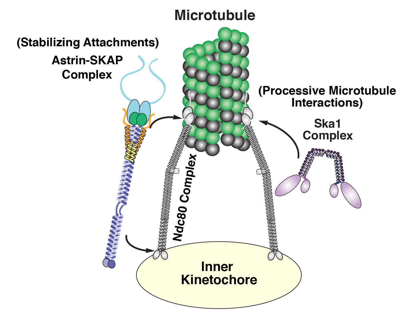
Our lab is fascinated by the molecular machinery that directs core cellular processes, and in particular how these processes are modulated and rewired across different physiological contexts. Our work has focused on the proteins that direct chromosome segregation and cell division, including the macromolecular kinetochore structure that mediates chromosome-microtubule interactions. Although cell division is an essential cellular process, this machinery is remarkably flexible in its composition and properties, which can vary dramatically between species and are even modulated within the same organism — over the cell cycle, during development, and across diverse physiological situations. To define the basis by which the kinetochore and other core cellular structures are rewired to adapt to diverse situations and functional requirements, we are currently investigating diverse transcriptional, translational, and post-translational mechanisms that act to generate proteomic variability both within individual cells and across tissues, cell state, development, and disease.
Awards
Global Consortium for Reproductive Longevity and Equality (GCRLE) Scholar Award, 2020
MIT Undergraduate Research Opportunities Program (UROP) Outstanding Mentor – Faculty, 2019
American Society for Cell Biology (ASCB) Early Career Life Scientist Award, 2012
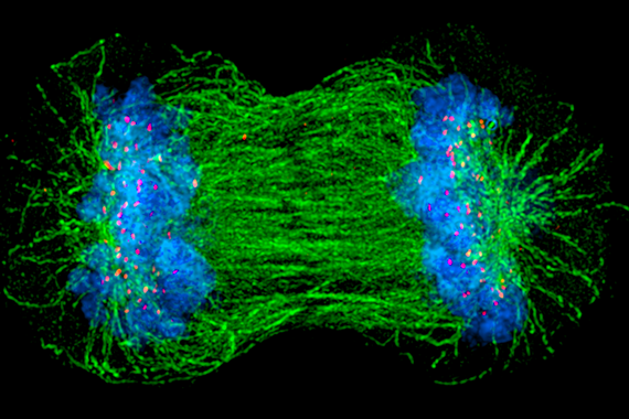
Whitehead researchers unravel fundamental molecular machinery that propels chromosome movement
November 16, 2017
Each day, billions of cells in the human body undergo a vital ritual, wherein one cell divides to form two. This process, known as cell division, is as beautiful as it is essential, undergirding the body’s growth in times of both health and disease. Despite the fact that cell division (or “mitosis”) has been a basic topic in high school biology classes for the past 70 years, the mechanisms by which cells conduct this critical event remain poorly understood. In particular, there are lingering uncertainties about how chromosomes — large units of DNA that include our genes — get properly allocated so that both daughter cells receive intact, complete copies of their genetic blueprint.
“People have been watching chromosomes move, align, and segregate for more than a century — it’s such a fundamental aspect of biology,” says Iain Cheeseman, a member of the Whitehead Institute for Biomedical Research and an associate professor of biology at Massachusetts Institute of Technology. “It’s also much more elegant and complicated than we ever anticipated.”
Cheeseman and members of his Whitehead laboratory have discovered many of the molecular movers and shakers that ensure chromosomes get to the right place at the right time. These components assemble together — like the parts of an engine — to establish robust connections with chromosomes and ultimately power their movement within cells. “The most important unanswered question in the field is how do these components work together? That is, how do you build a machine that is more than the sum of its parts?” he says.
Now, in two recent papers in the journals eLife and Current Biology, Cheeseman and his colleagues help shed light on this central question.
Back to the drawing board
As recently as 15 years ago, scientists assumed that in dividing cells, chromosomes move the way many other cellular objects move — transported by tiny molecular motors. These mini-motors are specifically designed to travel along roads made from rod-like structures called microtubules. Like a car cruising on a highway, they can carry cargo over long distances. Although such microtubule-based vehicles seemed a logical suspect, when scientists inactivate them in human cells, chromosomes can still move and segregate just fine. So something else must contribute the necessary molecular muscle.
“We basically had to throw out the major hypothesis that was out there and go back to the drawing board,” says Cheeseman.
Over the last several years, he and other scientists in the field have helped develop a clearer view of how this process works and who the key players are that enable a very different type of movement. Consider a car sitting motionless on a highway. Rather than revving its own engine to generate motion, the highway itself moves, shrinking or growing while the car hangs on. “It is a radically different way of imagining this movement process,” says Cheeseman. “An important part of my lab’s mission has been to figure out how do you build a motor like that? What are the factors required and how do they act?”
Of course, the highway — or more precisely, the microtubule — must grow and shrink as needed. But even more important, there must also be an apparatus that can enable chromosomes to hold on to such a dynamic structure. As Cheeseman and his colleagues have uncovered, this coupling requires a suite of highly sophisticated molecular players.
Building an unusual machine
Cheeseman and his laboratory have focused on three key groups or complexes of proteins that play essential roles in chromosome segregation in human cells. These components assemble together to form a kind of molecular tether point on chromosomes (called the kinetochore) where microtubules attach.
Diagram of the kinetochore/microtube interface Courtesy: David Kern/Whitehead Institute
Among this trio of parts, the most critical is the Ndc80 complex. “It is the major connection between the kinetochore and the microtubule,” says Cheeseman. As a postdoc, he discovered the biochemical properties that enable this Ndc80 complex to grab on to microtubules, research that sparked his lab’s quest to study the various pieces of the kinetochore machinery and how they work.
While Ndc80 forms a critical linkage, it lacks some key capabilities, like processivity — the ability to keep ahold of something while it moves. In a series of papers, one published in 2009, another in 2012, and a new one in Current Biology, Cheeseman’s team revealed that Ska1 can perform this crucial function. That is, it has the biochemical capacity to enable chromosomes to hang onto microtubules while they grow and as they shrink, an activity that it can impart to the Ndc80 complex. “These are pretty powerful properties,” says Cheeseman.
Diving even deeper into Ska1’s bag of tricks, Julie Monda and Ian Whitney, lead authors of the Current Biology paper, went on to decipher the precise molecular features that enable the complex’s dynamic capabilities, uncovering multiple surfaces that associate with microtubules and enable Ska1 to undergo something akin to molecular somersaults. These somersaults are what allow it to maintain its association with microtubules.
The third complex, Astrin-SKAP, also plays a unique role. As Cheeseman’s team described in their recent eLife paper, led by first author David Kern, it serves as a master stabilizer — like a final drop of superglue to secure everything in place. “It’s the last thing that comes in and helps lock down these interactions, so you can stabilize and maintain them,” says Cheeseman.
Uncovering its role was no easy feat. Astrin-SKAP proved to be rather temperamental biochemically, complicating Kern’s efforts to purify and manipulate it in the laboratory. Also, as he and his colleagues discovered, a tiny piece of the structure had previously gone undetected; it works alongside the rest of the complex and is required for its normal function. Perhaps the most important revelation was that Astrin-SKAP doesn’t just work alone — it also coordinates with Ndc80. “This is an important finding for how we think about these components as a whole,” saysCheeseman.
Although questions remain about how all of these parts work together and how other pieces may come into play, Cheeseman believes these studies provide an exciting start. “The first human kinetochore component wasn’t identified until 1987, when many of the other key processes in the cell had already been intensively studied,” he says. “There are so many exciting questions that are accessible now that we have these tools and knowledge.”
Now, he and his colleagues will continue to meld approaches in cell biology and biochemistry to decode the inner workings of the kinetochore. That includes understanding how the various components operate not only in individual cells, but also in multicellular organisms.
“We are currently thinking a lot about the physiological context— that is, what matters to cells and to an organism,” says Cheeseman. “The work that our lab and others have conducted over the past two decades has given us a molecular handle on this problem. I’m excited to be able to apply these finding to understanding the ways that cell division is altered in development and in disease states.”
Written by Nicole Davis
* * *
Iain Cheeseman’s primary affiliation is with Whitehead Institute for Biomedical Research, where his laboratory is located and all his research is conducted. He is also an associate professor of biology at Massachusetts Institute of Technology.
* * *
Full citations:
“Astrin-SKAP complex reconstitution reveals its kinetochore interaction with microtubule-bound Ndc80”
eLife 2017;6:e26866 August 25, 2017. DOI: 10.7554/eLife.26866
David M Kern (1,2), Julie K Monda (1,2), Kuan-Chung Su (1), Elizabeth M Wilson-Kubalek (3), and Iain M Cheeseman (1,2).
1. Whitehead Institute for Biomedical Research, 455 Main Street, Cambridge, MA 02142, USA
2. Department of Biology, Massachusetts Institute of Technology, Cambridge, MA 02142, USA
3. Department of Cell Biology, The Scripps Research Institute, La Jolla, CA 92037, USA
“Microtubule tip tracking by the spindle and kinetochore protein Ska1 requires diverse tubulin-interacting surfaces”
Current Biology, online November 16, 2017. DOI: 10.1016/j.cub.2017.10.018
Julie K. Monda (1,2,6), Ian P. Whitney (1,6), Ekaterina V. Tarasovetc (3,4), Elizabeth Wilson-Kubalek (5), Ronald A. Milligan (5), Ekaterina L. Grishchuk (3), and Iain M. Cheeseman (1,2).
1. Whitehead Institute for Biomedical Research, 455 Main Street, Cambridge, MA 02142, USA
2. Department of Biology, Massachusetts Institute of Technology, Cambridge, MA 02142, USA
3. Department of Physiology, Perelman School of Medicine, University of Pennsylvania, Philadelphia, PA 19104, USA
4. Center for Theoretical Problems of Physicochemical Pharmacology, Russian Academy of Sciences, Moscow, Russia
5. Department of Cell Biology, The Scripps Research Institute, La Jolla, CA 92037, USA
6. These authors contributed equally
Researchers have identified a key nutrient sensor in the mTOR pathway that links nutrient availability to cell growth.
Nicole Giese Rura | Whitehead Institute
November 9, 2017
To survive and grow, a cell must properly assess the resources available and couple that with its growth and metabolism — a misstep in that calculus can potentially cause cell death or dysfunction. At the crux of these decisions is the mTOR pathway, a cellular pathway connecting nutrition, metabolism, and disease.
The mTOR pathway incorporates input from multiple factors, such as oxygen levels, nutrient availability, growth factors, and insulin levels to promote or restrict cellular growth and metabolism. But when the pathway runs amok, it can be associated with numerous diseases, including cancer, diabetes, and Alzheimer’s disease. Understanding the various sensors that feed into the mTOR pathway could lead to novel therapies for these diseases and even aging, as dialing down the mTOR pathway is linked to longer lifespans in mice and other organisms.
Although the essential amino acid methionine is one of the key nutrients whose levels cells must carefully sense, researchers did not know how it fed into the mTOR pathway — or if it did at all. Now, Whitehead Institute Member David Sabatini and members of his laboratory have identified a protein, SAMTOR, as a sensor in the mTOR pathway for the methionine derivative SAM (S-adenosyl methionine). Their findings are described in the current issue of the journal Science.
Methionine is essential for protein synthesis, and a metabolite produced from it, SAM, is involved in several critical cellular functions to sustain growth, including DNA methylation, ribosome biogenesis, and phospholipid metabolism. Interestingly, methionine restriction at the organismal level has been linked to increased insulin tolerance and lifespan, similar to the antiaging effects associated with inhibition of mTOR pathway activity. But the connection between mTOR, methionine, and aging remains elusive.
“There are a lot of similarities between the phenotypes of methionine restriction and mTOR inhibition,” says Sabatini, who is also a Howard Hughes Medical Institute investigator and a professor of biology at MIT. “The existence of this protein SAMTOR provides some tantalizing data suggesting that those phenotypes may be mechanistically connected.”
Sabatini identified mTOR as a graduate student and has since elucidated numerous aspects of its namesake pathway. He and his lab recently pinpointed the molecular sensors in the mTOR pathway for two key amino acids: leucine and arginine. In the current line of research, co-first authors of the Science paper Xin Gu and Jose Orozco, both graduate students Sabatini’s lab, identified a previously uncharacterized protein that seemed to interact with components of the mTOR pathway. After further investigation, they determined that the protein binds to SAM and indirectly gauges the pool of available methionine, making this protein — SAMTOR — a specific and unique nutrient sensor that informs the mTOR pathway.
“People have been trying to figure out how methionine was sensed in cells for a really long time,” Orozco says. “I think that this is the first time in mammalian cells a mechanism has been found to describe the way methionine can regulate a major signaling pathway like mTOR.”
The current research indicates that SAMTOR plays a crucial role in methionine sensing. Methionine metabolism is vital for many cellular functions, and the Sabatini lab will further investigate the potential links between SAMTOR and the extended lifespan and increased insulin sensitivity effects that are associated with low methionine levels.
“It is very interesting to consider mechanistically how methionine restriction might be associated in multiple organisms with beneficial effects, and identification of this protein provides us a potential molecular handle to further investigate this question,” Gu says. “The nutrient-sensing pathway upstream of mTOR is a very elegant system in terms of responding to the availability of certain nutrients with specific mechanisms to regulate cell growth. The currently known sensors raise some interesting questions about why cells evolved sensing mechanisms to these specific nutrients and how cells treat these nutrients differently.”
This work was supported by the National Institutes of Health, the Department of Defense, the National Science Foundation, and the Paul Gray UROP Fund.