Grantees will spend the 2019-2020 academic year pursuing research and teaching opportunities abroad.
Julia Mongo | Office of Distinguished Fellowships
May 17, 2019
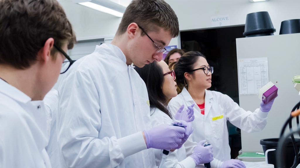
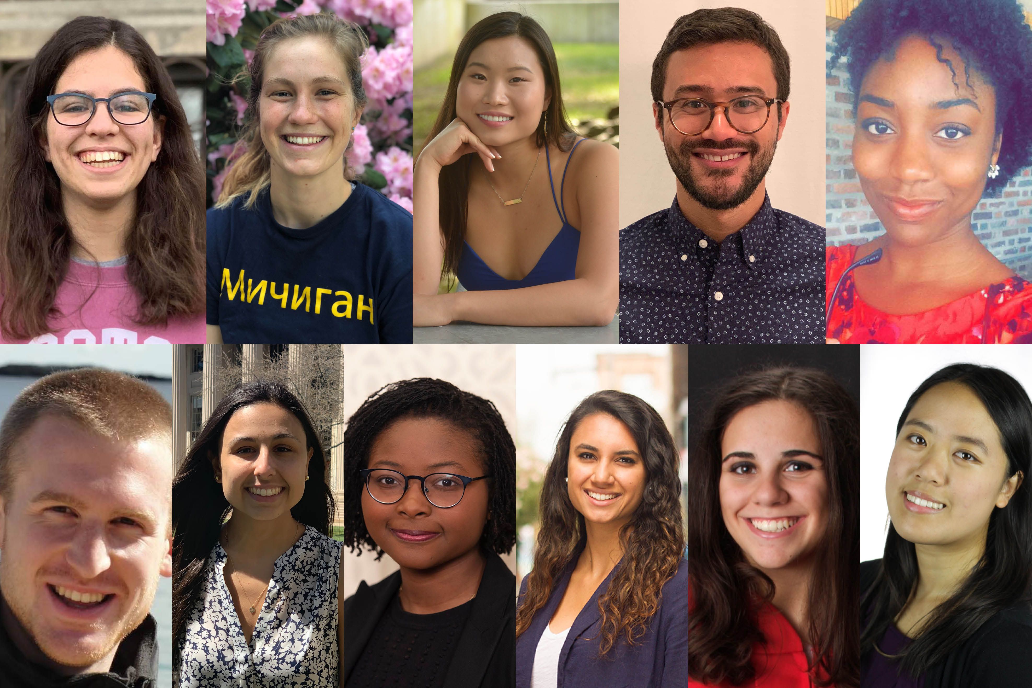
Eleven MIT graduating seniors and current graduate students have been named winners in the 2019-2020 Fulbright U.S. Student Fellowship Program. In addition to the 11 students accepting their awards, three applicants from MIT were selected as finalists but decided to decline their grants.
MIT’s newest Fulbright Students will engage in independent research and English teaching assignments in Brazil, the Netherlands, Spain, Russia, Taiwan, and Senegal.
Sponsored by the U.S. Department of State’s Bureau of Educational and Cultural Affairs, the mission of Fulbright is to promote cultural exchange, increase mutual understanding, and build lasting relationships among people of the world. The Fulbright U.S. Student Program offers grants in over 140 countries.
The MIT students were supported in the application process by the Presidential Committee on Distinguished Fellowships, chaired by professors Rebecca Saxe and Will Broadhead, and by MIT’s Distinguished Fellowships Office within Career Advising and Professional Development. The MIT winners are:
Annamarie “Anna” Bair ’18 earned a bachelor of science in computer science and engineering in June 2018 and will receive her master of engineering degree in computer science later this year. In Barcelona, Spain, Bair will engage in complex systems research.
Abigail “Abby” Bertics will graduate in June with a bachelor of science in electrical engineering and computer science. Her research in Yekaterinburg, Russia, will focus on natural language processing methods for understanding English second language acquisition by Russian speakers.
Hope Chen is a senior graduating with a bachelor of science in mechanical engineering. She will be going to Taiwan as an English Teaching Assistant in primary school classrooms. After completing her Fulbright program and returning to the U.S., Chen will matriculate in medical school.
Alexis D’Alessandro will graduate this spring with a bachelor of science in mechanical engineering. For her research in Aracaju, Brazil, she will develop an educational program and chemical sensing tool to promote water safety awareness among children.
Sarah DiIorio will earn her bachelor of science in biological engineering in June. She is headed to Eindhoven, the Netherlands, to conduct medical research related to cartilage regeneration for osteoarthritis.
Katie Fisher is a senior in MIT’s Scheller Teaching Education Program graduating with a bachelor of science in urban studies and planning with a concentration in education. As an English teaching assistant in the Netherlands, Fisher will work with students at a vocational college in Amsterdam.
Miranda McClellan ’18 received a bachelor of science in computer science and engineering in June 2018 and will earn her master of engineering degree in computer science this spring. McClellan will research automated scaling of 5G computer network resources in Barcelona, Spain.
Samira Okudo will graduate in June with a joint bachelor of science in computer science and comparative media studies. As an English teaching assistant in Brazil, she will work with university students training to be English-language instructors.
James Pelletier is a PhD candidate in physics. For his Fulbright research in Madrid, Spain, he will develop biophysical models to investigate how plants process information for cellular resource allocation and agricultural efficiency.
Jonars Spielberg is a third-year doctoral student in the Department of Urban Studies and Planning’s international development program. In Senegal, he will examine how the personal interactions of bureaucrats and farmers shape agricultural policy implementation in the country’s main irrigated regions.
Catherine Wu will graduate in June with a bachelor of science in biology. She will be working with university students in Brazil as a Fulbright English Teaching Assistant.
MIT students interested in applying to the Fulbright U.S. Student Program should contact Julia Mongo in Distinguished Fellowships.