Placement: Promote to Homepage
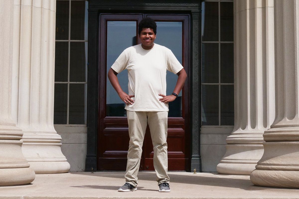
Undergraduate Meucci Ilunga spent 10 weeks investigating protein interactions, exploring career options, and making new friends.
Saima Sidik
September 4, 2019
Meucci Ilunga seems to know something about everything. He’s a videographer who’s branching out into podcasting. He’s researched cancer therapies and volunteered in a hospital. He grew up on a Navajo reservation, and he’s a year away from completing a biochemistry degree at the University of Arizona. “I’m excited about life in general,” he says. At the moment, though, he’s especially excited about a cellular conundrum that he investigated during the 10-week internship in the MIT Department of Biology that he completed as part of the MIT Summer Research Program in Biology (MSRP-Bio).
“Your cells are really, really complicated,” he says. “They’re packed with lots of different kinds of proteins. Yet when you look at how proteins interact, they’re specific.” How do proteins find the appropriate binding partners amongst all the noise? Ilunga and his MSRP-Bio supervisor, biology and biological engineering Professor Amy Keating, think that short sequences of amino acids — the units that comprise proteins — can mediate binding interactions more intricate than researchers had previously appreciated.
Just as proteins home in on their binding partners, Ilunga has always been drawn to science. As a kid, he told everyone he wanted to be an astrophysicist. “I had no idea what that meant,” he says, “but I loved the idea of exploring the unknown and being able to generate knowledge.”
Ilunga grew up on the Navajo reservation in Kinlichee, Arizona, however, and he didn’t have the same opportunities to engage in science as kids in urban centers. “Only about 60 percent of people on the reservation have running water and electricity,” he says, “so most people are pressed with more urgent matters than following their curiosities.”
Ilunga notes the myriad of difficulties his reservation faces, from prevalent diabetes to corrupt politicians and poor school systems, but says that the hardest part about being Navajo is feeling like his people’s problems are invisible to those outside the tribe. “A lot of us feel very forgotten about,” he says.
Ilunga quickly exhausted the opportunities that his high school in Fort Defiance, Arizona, had to offer, leading him to graduate early and leave for the University of Arizona at age 16. But he was determined to remember his roots. Balancing his love of science with his connection to the reservation — and finding a career that will let him return — has proven challenging.
“You can become an engineer, but there are no engineering jobs on the reservation. You can become a computer scientist, but there are no computer science jobs,” he says. So he decided to pursue biochemistry, as it would lay the foundation for medical school, and the reservation is always in need of doctors.
At his university, Ilunga started shadowing physicians and volunteering in a hospital. His path to medical school seemed clear. There was only one problem: He found medicine unfulfilling. “There’s so much more I could be doing. So I started looking at what else I could do to get back home,” he says.
This desire for balance is what made Ilunga choose to join the MSRP-Bio program, for which he received sponsorship from the Gould Fund. Ilunga met the MSRP-Bio coordinator, Mandana Sassanfar, at a conference for minority students, and she told him that MSRP-Bio promotes a balance between lab work and life. “What sold me on this program is that it understands that I’m more than just a scientist,” he says.
Over the summer, Ilunga has spoken with many MIT professors about the diverse professional paths scientists can take, and these conversations have inspired him to consider a career in policy.
“I could be someone who goes to Congress to fight — not only for Native American affairs, but also for scientific affairs,” he says.
Ilunga plans to pursue a PhD in life sciences in preparation for this career, possibly studying protein interactions like the ones he’s been working on all summer. He finds research most interesting when it has a clear clinical application, and understanding protein interactions lets researchers design drugs that disrupt them.
The protein interactions that Ilunga researched are mediated by sequences called short linear motifs, or SLiMs, which consist of contiguous stretches of only three to 10 amino acids — a small subset of the hundreds of amino acids that make up the typical protein. While larger domains are able to form tighter and more sustained interactions, SLiMs mediate weaker, transient interactions.
SLiMs make up in speed what they lack in strength. Allowing proteins to quickly bind and release each other is beneficial for some biological processes, and SLiMs can also evolve rapidly and let organisms adapt to change quickly. Researchers think this is why SLiMs have persisted in many different organisms over the course of evolution, despite being relatively unintuitive tools for forming protein complexes. The Keating lab noticed that sometimes proteins that contain SLiMs recognize their binding partners with a specificity that’s unexpected, given that so many proteins contain these short sequences.
Ilunga spent his summer looking into how small domains and short sequences can play a large role in protein pairing. His weeks began with culturing large quantities of bacteria that were used to produce SLiM-containing peptides; then he isolated these peptides and used a technique called biolayer interferometry to determine how tweaking their amino acid sequences affected how strongly they bound their target protein.
When he altered the amino acid sequence directly adjacent to the SLiMs, Ilunga found that the strength of their binding interactions could vary quite wildly. The Keating lab doesn’t understand how this occurs, and Ilunga’s findings pave the way for testing different biochemical mechanisms to explain this phenomenon.
When he wasn’t isolating proteins or chatting with the MIT faculty, Ilunga got to know the MIT community. “At a lot of top schools there’s a sense of prestige that fills the air, but it wasn’t like that at MIT. Everyone here is so humble,” he says.
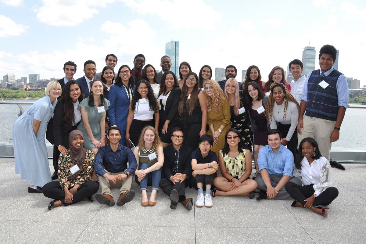
He especially enjoyed getting to know his fellow MSRP-Bio students. Whether they were going on a boat cruise along the Charles River or helping each other troubleshoot lab work, he says it was an amazing group of people to spend the summer with.
As he heads back to the University of Arizona, Ilunga is taking many technical skills back with him, as well as a new outlook on life. He has always been hopeful that life will get easier for Navajos and other minorities. Now he’s confident that the medical and technological advances that institutions like MIT are creating can improve living conditions for people like his family back on the reservation.
“I used to think my optimism was blind,” he says. “Now I think my optimism is informed.”
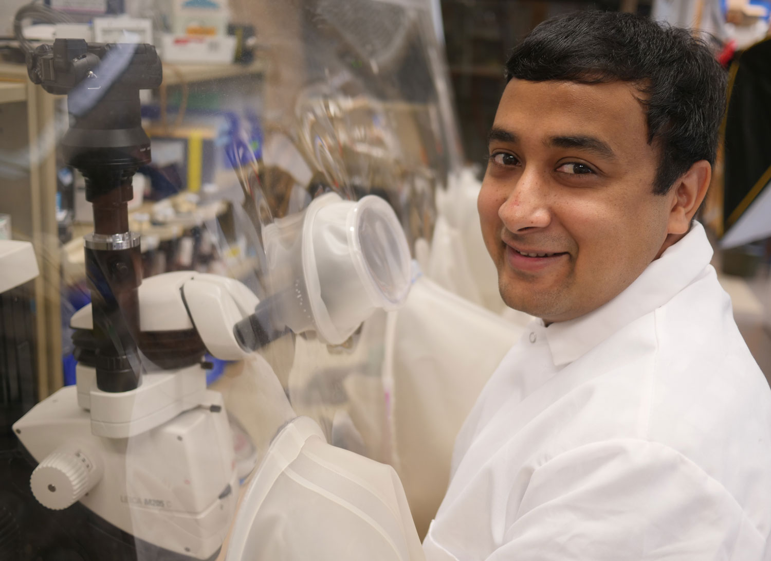
Graduate student Rohan Jonnalagadda analyzes the 3D shapes of iron-containing enzymes to parse their role in cellular processes.
Raleigh McElvery
August 27, 2019
Raised in a computer-savvy family well-versed in software and information technology, Rohan Jonnalagadda had a strong desire to “decode” the world around him. But his kind of code, the genetic one, consists of four repeating letters: A, T, C, and G. “Just like a computer runs on software, I wanted to investigate the code behind the molecular hardware that gives rise to life,” he says. Now a sixth-year graduate student in the Drennan lab, he works to decrypt the structure of metal-containing proteins, in order to determine the roles they play in vital cellular reactions.
When Jonnalagadda was an undergraduate biochemistry major at the University of California, Berkeley, it became clear to him that the genetic code was more than just a string of letters; it also serves as the blueprint for all the proteins in the entire organism. These proteins fold into complex 3D structures, which ultimately beget function.
At UC Berkeley, he joined a lab studying the iron-containing protein Heme-Nitric Oxide/Oxygen (H-NOX) that senses nitric oxide gas in bacterial and eukaryotic cells. When H-NOX binds to nitric oxide, it must change its 3D shape in the process. Jonnalagadda used a technique known as X-ray crystallography to freeze H-NOX in various stages of this conformational change to determine how it binds the gas molecules.
“I think we sometimes ignore the fact that we need trace metals in order to survive,” he says. “I was interested in continuing to think about what different metals could do in the cell. And using metals opens up a whole new world of chemical reactions that you generally don’t learn about in class.”By the time he graduated and began his PhD at MIT Biology, Jonnalagadda had been using X-ray crystallography for over two years. Today, as a member of Catherine Drennan’s lab, he continues to leverage this same method to parse the structure of additional metal-containing proteins.
In fact, the two projects that he’s devoted most of his time to over the past five years involve reactions that he’d never even heard of before he arrived at MIT. The focus of his first undertaking was the iron-containing enzyme ribonucleotide reductase (RNR), which helps generate deoxyribonucleotides, the building blocks of DNA.
Jonnalagadda aims to understand how this enzyme is regulated to ensure the cell maintains the proper amount of each type of deoxyribonucleotide, in order to properly replicate and repair its genome. If those ratios are incorrect, the cell could experience detrimental stress.
Because the enzyme is regulated differently in humans than it is in bacteria, scientists hope to one day create antibiotics that target the bacterial RNR while leaving the human RNR unscathed. Jonnalagadda works with the human version, devising an assay that will allow him to better assess the differences between the two enzymes. RNR is notoriously difficult to work with, and so Jonnalagadda has spent much of his time developing ways to purify it so it remains stable.
His second project is a collaboration with researchers at his alma mater, UC Berkeley, investigating isonitriles — compounds containing a carbon atom tripled bonded to a nitrogen atom. Because isonitriles are used to make drugs like antibiotics, scientists have a keen interest in exploring new ways to produce them. The team discovered that one bacterium, Streptomyces coeruleorubidus, had a novel and mysterious way of synthesizing these compounds. Jonnalagadda wants to know exactly how these particular bacteria do it.
He is using X-ray crystallography to determine the structure of the iron-containing enzyme ScoE in S. coeruleorubidus, which is responsible for forming the carbon-nitrogen triple bond characteristic of isonitriles.
“It’s exciting to be working on a protein that’s only just been discovered,” he says. “There’s just so much more to learn about its fundamental biological function. I think that’s why basic research is so appealing to me; you never know where the work will take you, or the impacts it could have on human health later on.”
Extending the frontiers of any discipline requires some guesswork and metaphorical bushwhacking, and Jonnalagadda has learned almost as much from his failed experiments as he has from his successful ones. “I’m proud that I’ve been able to use what I’ve learned about experimental design to help others in my lab when they have questions,” he says.
As he considers life post-graduation, he hopes to use the biochemical and structural techniques he’s mastered over the years to secure a job in industry.
“Being part of a department with such broad and wide-ranging research interests has made it easy to see that my work doesn’t exist in a vacuum,” he says. “It connects to many different aspects of biology.”
Photo credit: Raleigh McElvery
Posted 8.23.19
August 28, 2019
Picower Institute
August 27, 2019
Education
- PhD, 2013, University of California, Berkeley
- BS, 2008, Genetics, University of Georgia
Research Summary
We study the interplay of gene expression and genome organization. Our work focuses on understanding how large molecular machineries involved in genome organization and gene transcription regulate each others’ function to ultimately determine cell fate and identity. We employ a broad range of approaches including single-particle cryo-electron microscopy (cryo-EM), X-ray crystallography, biochemistry, and genetics to mechanistically understand how these molecular assemblies regulate each other across molecular scales.
Awards
- New Innovator Award, National Institutes of Health Common Fund’s High-Risk, High-Reward Research Program, 2021
Molecules called ketone bodies may improve stem cells’ ability to regenerate new intestinal tissue.
Anne Trafton | MIT News Office
August 22, 2019
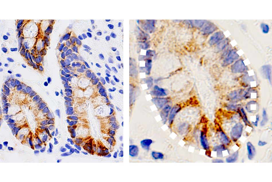
MIT biologists have discovered an unexpected effect of a ketogenic, or fat-rich, diet: They showed that high levels of ketone bodies, molecules produced by the breakdown of fat, help the intestine to maintain a large pool of adult stem cells, which are crucial for keeping the intestinal lining healthy.
The researchers also found that intestinal stem cells produce unusually high levels of ketone bodies even in the absence of a high-fat diet. These ketone bodies activate a well-known signaling pathway called Notch, which has previously been shown to help regulate stem cell differentiation.
“Ketone bodies are one of the first examples of how a metabolite instructs stem cell fate in the intestine,” says Omer Yilmaz, the Eisen and Chang Career Development Associate Professor of Biology and a member of MIT’s Koch Institute for Integrative Cancer Research. “These ketone bodies, which are normally thought to play a critical role in energy maintenance during times of nutritional stress, engage the Notch pathway to enhance stem cell function. Changes in ketone body levels in different nutritional states or diets enable stem cells to adapt to different physiologies.”
In a study of mice, the researchers found that a ketogenic diet gave intestinal stem cells a regenerative boost that made them better able to recover from damage to the intestinal lining, compared to the stem cells of mice on a regular diet.
Yilmaz is the senior author of the study, which appears in the Aug. 22 issue of Cell. MIT postdoc Chia-Wei Cheng is the paper’s lead author.
An unexpected role
Adult stem cells, which can differentiate into many different cell types, are found in tissues throughout the body. These stem cells are particularly important in the intestine because the intestinal lining is replaced every few days. Yilmaz’ lab has previously shown that fasting enhances stem cell function in aged mice, and that a high-fat diet can stimulate rapid growth of stem cell populations in the intestine.
In this study, the research team wanted to study the possible role of metabolism in the function of intestinal stem cells. By analyzing gene expression data, Cheng discovered that several enzymes involved in the production of ketone bodies are more abundant in intestinal stem cells than in other types of cells.
When a very high-fat diet is consumed, cells use these enzymes to break down fat into ketone bodies, which the body can use for fuel in the absence of carbohydrates. However, because these enzymes are so active in intestinal stem cells, these cells have unusually high ketone body levels even when a normal diet is consumed.
To their surprise, the researchers found that the ketones stimulate the Notch signaling pathway, which is known to be critical for regulating stem cell functions such as regenerating damaged tissue.
“Intestinal stem cells can generate ketone bodies by themselves, and use them to sustain their own stemness through fine-tuning a hardwired developmental pathway that controls cell lineage and fate,” Cheng says.
In mice, the researchers showed that a ketogenic diet enhanced this effect, and mice on such a diet were better able to regenerate new intestinal tissue. When the researchers fed the mice a high-sugar diet, they saw the opposite effect: Ketone production and stem cell function both declined.
Stem cell function
The study helps to answer some questions raised by Yilmaz’ previous work showing that both fasting and high-fat diets enhance intestinal stem cell function. The new findings suggest that stimulating ketogenesis through any kind of diet that limits carbohydrate intake helps promote stem cell proliferation.
“Ketone bodies become highly induced in the intestine during periods of food deprivation and play an important role in the process of preserving and enhancing stem cell activity,” Yilmaz says. “When food isn’t readily available, it might be that the intestine needs to preserve stem cell function so that when nutrients become replete, you have a pool of very active stem cells that can go on to repopulate the cells of the intestine.”
The findings suggest that a ketogenic diet, which would drive ketone body production in the intestine, might be helpful for repairing damage to the intestinal lining, which can occur in cancer patients receiving radiation or chemotherapy treatments, Yilmaz says.
The researchers now plan to study whether adult stem cells in other types of tissue use ketone bodies to regulate their function. Another key question is whether ketone-induced stem cell activity could be linked to cancer development, because there is evidence that some tumors in the intestines and other tissues arise from stem cells.
“If an intervention drives stem cell proliferation, a population of cells that serve as the origin of some tumors, could such an intervention possibly elevate cancer risk? That’s something we want to understand,” Yilmaz says. “What role do these ketone bodies play in the early steps of tumor formation, and can driving this pathway too much, either through diet or small molecule mimetics, impact cancer formation? We just don’t know the answer to those questions.”
The research was funded by the National Institutes of Health, a V Foundation V Scholar Award, a Sidney Kimmel Scholar Award, a Pew-Stewart Trust Scholar Award, the MIT Stem Cell Initiative, the Koch Institute Frontier Research Program through the Kathy and Curt Marble Cancer Research Fund, the Koch Institute Dana Farber/Harvard Cancer Center Bridge Project, and the American Federation of Aging Research.
Support from Squire Booker PhD ’94 and the Bernard S. and Sophie G. Gould Fund helps MSRP-bio students excel.
Laura Carter | School of Science
August 20, 2019
When you get a call offering you the chance to get involved in research at MIT, says Squire Booker PhD ’94, as he did when he was a student back home in Beaumont, Texas, with no summer plans, you don’t say no. This is how he joined seven other students from around the United States as the first class in the MIT Student Research Program (MSRP), even though the start date was only days away. “I was given the opportunity to get out of Texas, the opportunity to go to a big cosmopolitan city, the opportunity to go to MIT. So, I got a plane ticket and flew up a few days later,” says Booker.
Thirty-three summers later, back on campus to deliver the doctoral graduation ceremony speech, where he had lunch with several current members and fellow alumni of the program, Booker insists that he has no regrets with his decision.
Booker was one of three from that inaugural class who remained at MIT to pursue a PhD to continue the research he started during the program. He was incredibly fortunate, he notes, to get a “perfect match” placement, working with former professors of biology Bill Johnson and Chris Walsh on a project that aligned with his interests of combining chemistry and biology. He didn’t have much more of an idea of his preferred area of study than that.
Prior to arriving at MIT, given the lack of exposure to science, he didn’t know what research entailed, or what scientists did every day. But he says he quickly fell in love with the subject and his research group, even joining their summer lab softball team.
Although Walsh left MIT the year Booker was accepted as a PhD student, he easily shifted into the lab of Novartis Professor of Chemistry Emeritus JoAnne Stubbe, a new faculty member at the time, who was also working on the interface of chemistry and biology and provided the amount of hands-on support he needed as a new graduate student. “Ever since leaving the lab, she’s been my number one supporter,” he says of Stubbe.
Stubbe and her research inspired the direction Booker’s education took. He continues to conduct research revolving around proteins and catalysis reactions as a professor at Penn State University and a principal investigator with the Howard Hughes Medical Institute. Now, he heads a large lab group himself.
From mentee to mentor
Booker oversees an average of 10 group members at any given time, not including undergraduate students. Like his mentor, he tries to be very hands-on, resorting to email when he’s traveling — which is often. He admitted with a chuckle that his students keep track of where he is at any given time by following his Twitter account. Always trying to find ways to include motivated students who approach him about contributing to his research, the only time Booker turns them away is for their benefit — if they have a full course load and additional time on research will overload their schedules. He even considers high school students.
The first high school student to join his lab was Martin McLaughlin ’15, who Booker describes fondly as “aggressively motivated” and “trembling with excitement to do research.” Within the first week, McLaughlin was taking the initiative to use his lunch breaks from school to bike to Booker’s lab. Martin’s results, which were published in Science in collaboration with Professor Cathy Drennan in the MIT Department of Biology, introduced Booker into a new niche: crystallography.
When McLaughlin asked to continue working on the discovery with Drennan as an undergraduate at MIT, he didn’t hesitate to agree. McLaughlin had moved into Drennan’s lab a week into his first semester.
Research for all
Not all students share this drive to delve into research. Like Booker himself, many aren’t even aware of possibilities to get involved in science and consider a career in research. It’s still hard, he says, even though “people are more serious about this diversity thing,” as he calls it, than when he was first starting his education.
Booker tries to reach out, especially to other minority students, through several programs, much like the MSRP, an invaluable program. While on campus this past spring, Booker met with current and past MSRP students.
One of those students was Jeandele Elliot, a chemical engineering student at Howard University from Saint Lucia in the Caribbean, who is working in the Jing-Ke Weng Lab in the Department of Biology this summer on a molecule that can protect pollen grains. For her, meeting Booker was another connection the program affords her. “The MSRP program has been beneficial to me in a special way since it has connected me with people I can really relate to,” she said.
The advice he gave to Elliot, and the others in the same position he was in once, was to prepare for exciting careers. The program is not just a steppingstone into research, he proclaimed, but it places participants with the best mentors and being privy to the best frontiers. Booker was delighted that some of the 25 current and past participants then attended MIT for graduate school as he did.
Tsehai A.J. Grell PhD ’18, a current chemistry graduate student in Drennan’s research group and an alumnus of MSRP, calls Squire Booker a “labhold” name — a household name in the lab. “As an African-American professor of biochemistry, an alumnus of my department, and a leader in my field, he instantly became one of my role models,” Grell said. “This was further solidified when I found out that he was a part of the first cohort of MSRP students, the summer research program which is responsible for me enrolling in MIT’s graduate program.”
Grell reminisced on his experience and the spring luncheon with Booker. “Because MSRP was such a foundational experience in my career, I am always enthused to interact with the current MSRP cohort and to encourage them to make the most of this opportunity, as it can be a pivotal summer in their careers,” says Grell. In addition, he said, “the excitement of the students is palpable and contagious. It reenergizes me and gives me purpose.”
Elliott, Grell, and Booker are three of more than 800 students from institutions with limited research opportunities who have participated in the MSRP, which was divided into two subcategories in 2003: general and biology, the latter of which has hosted 450 students. Since 2003, the MRSP-Bio program has been administered by Mandana Sassanfar, a biology lecturer in charge of the Department of Biology’s diversity and outreach programs. Since then, nearly 70 MSRP alumni have, like Booker, continued their research as graduate students at MIT.
Going for Gould
Bernard “Bernie” Gould ’32, who received his BS from MIT, was a longstanding and beloved biochemistry professor in the Department of Biology, well known for being an incredibly dedicated mentor to biology and pre-med students at MIT for nearly 40 years. His wife, Sophia Gould CMP ’48, shared his passion for counseling students. To honor this investment in encouraging student learning, the Goulds’ son, Michael, and his wife, Sara Moss, founded the Bernard S. and Sophia G. Gould Fund in 2016. Gould is a philanthropist and the retired chairman and CEO of Bloomingdales. Moss is the vice chairman of Estée Lauder Companies. The Gould Fellow Fund sponsors students, such as Elliott, in MSRP-Bio. Each year, Gould and Moss return to the MIT campus to meet with students benefitting from their support.
Recently, the couple has designated a second fund, which will aid in extending the academic careers of students interested in the life sciences by providing support for MSRP-Bio alumni entering into the MIT biology graduate program.
Six of the 16 Gould Fellowship alumni who have graduated from college have already been admitted to MIT as graduate students. “This is an exceptionally high rate by any standards, which demonstrates the amazing success of this initiative,” says Sassanfar. “Gould Fellows are truly grateful for the generosity of Mike and Sara and are very eager to succeed and give back to their communities,” a goal that is always stressed by the founders.
With successful role models from previous MSRP cohorts, like Booker, combined with philanthropy from those like Gould and Moss, who believe strongly in supporting the education of our next generation of scientists, students are given the opportunity to thrive.
Nicole Davis, Whitehead Institute
August 15, 2019

Whitehead Institute scientists find chemical modification contributes to trafficking between non-membrane-bound compartments that control gene expression.
Nicole Davis | Whitehead Institute
August 9, 2019
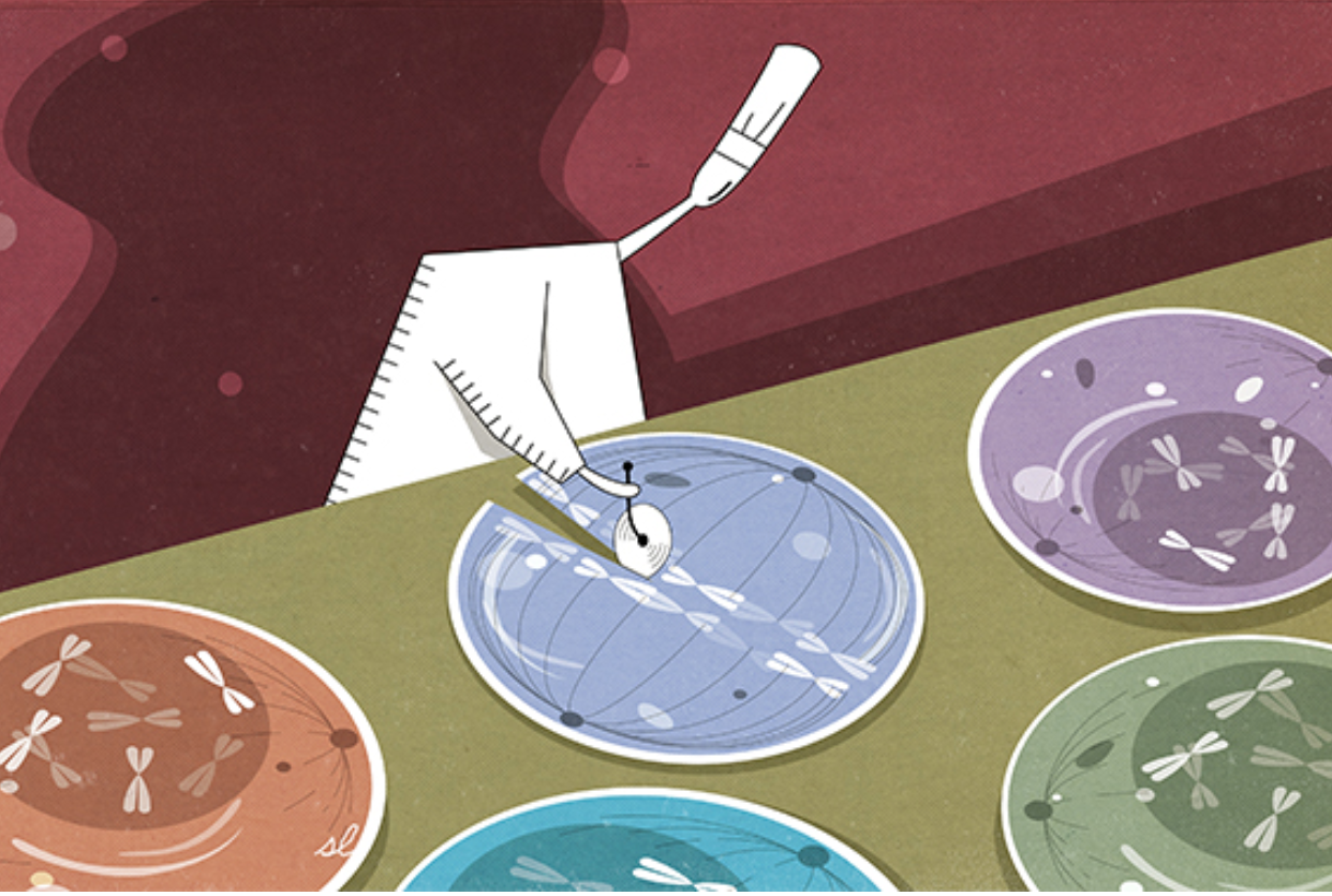
Cells often create compartments to control important biological functions. The nucleus is a prime example; surrounded by a membrane, it houses the genome. Yet cells also harbor enclosures that are not membrane-bound and more transient, like oil droplets in water. Over the past two years, these droplets (called “condensates”) have become increasingly recognized as major players in controlling genes. Now, a team led by Whitehead Institute scientists helps expand this emerging picture with the discovery that condensates play a role in splicing, an essential activity that ensures the genetic code is prepared to be translated into protein. The researchers also reveal how a critical piece of cellular machinery moves between different condensates. The team’s findings appear in the Aug. 7 online issue of Nature.
“Condensates represent a real paradigm shift in the way molecular biologists think about gene control,” says senior author Richard Young, a member of the Whitehead Institute and professor of biology at MIT. “Now, we’ve added a critical new layer to this thinking that enhances our understanding of splicing as well as the major transcriptional apparatus RNA polymerase II.”
Young’s lab has been at the forefront of studying how and when condensates form as well as their functions in gene regulation. In the current study, Young and his colleagues, including first authors Eric Guo and John Manteiga, focused their efforts on a key transition that happens when genes undergo transcription — an early step in gene activation whereby an RNA copy is created from the genes’ DNA template. First, all of the molecular machinery needed to make RNA, including a large protein complex known as RNA polymerase II, assembles at a given gene. Then, specific chemical modifications to RNA polymerase II allow it to begin transcribing DNA into RNA. This shift from so-called transcription initiation to active transcription also involves another important molecular transition: As RNA molecules begin to grow, the splicing apparatus must also move in and carry out its job.
“We wanted to step back and ask, ‘Do condensates play an important role in this switch, and if so, what mechanism might be responsible?’” explains Young.
For roughly three decades, it has been recognized that the factors required for splicing are stored in compartments called speckles. Yet whether these speckles play an active role in splicing, or are simply storage vessels, has remained unclear.
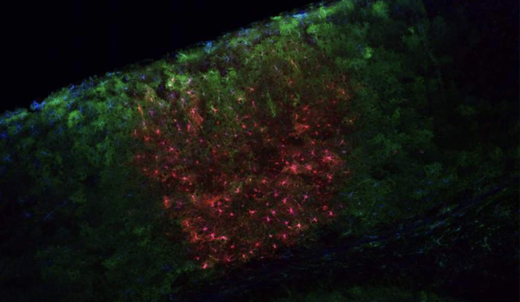
Using confocal microscopy, the Whitehead team discovered condensates filled with components of the splicing machinery in the vicinity of highly active genes. Notably, these structures exhibited similar liquid-like characteristics to those condensates described in prior studies from Young’s lab that are involved in transcription initiation.
“These findings signaled to us that there are two types of condensates at work here: one involved in transcription initiation and the other in splicing and transcriptional elongation,” said Manteiga, a graduate student in Young’s lab.
With two different condensates at play, the researchers wondered: How does the critical transcriptional machinery, specifically RNA polymerase II, move from one condensate to the other?
Guo, Manteiga, and their colleagues found that chemical modification, specifically the addition of phosphate groups, serves as a kind of molecular switch that alters the protein complex’s affinity for a particular condensate. With fewer phosphate groups, it associates with the condensates for transcription initiation; when more phosphates are added, it enters the splicing condensates. Such phosphorylation occurs on one end of the protein complex, which contains a specialized region known as the C-terminal domain (CTD). Importantly, the CTD lacks a specific three-dimensional structure, and previous work has shown that such intrinsically disordered regions can influence how and when certain proteins are incorporated into condensates.
“It is well-documented that phosphorylation acts as a signal to help regulate the activity of RNA polymerase II,” says Guo, a postdoc in Young’s lab. “Now, we’ve shown that it also acts as a switch to alter the protein’s preference for different condensates.”
In light of their discoveries, the researchers propose a new view of splicing compartments, where speckles serve primarily as warehouses, storing the thousands of molecules required to support the splicing apparatus when they are not needed. But when splicing is active, the phosphorylated CTD of RNA Pol II serves as an attractant, drawing the necessary splicing materials toward the gene where they are needed and into the splicing condensate.
According to Young, this new outlook on gene control has emerged in part through a multidisciplinary approach, bringing together perspectives from biology and physics to learn how properties of matter predict some of the molecular behaviors he and his team have observed experimentally. “Working at the interface of these two fields is incredibly exciting,” says Young. “It is giving us a whole new way of looking at the world of regulatory biology.”
Support for this work was provided by the U.S. National Institutes of Health, National Science Foundation, Cancer Research Institute, Damon Runyon Cancer Research Foundation, Hope Funds for Cancer Research, Swedish Research Council, and German Research Foundation DFG.