Research underscores infection is not a common hospital transmission.
Maria Iacobo | Department of Civil and Environmental Engineering
February 20, 2020
New research from MIT suggests the risk of becoming colonized by Clostridium difficile (C. difficile) increases immediately following gastrointestinal (GI) disturbances that result in diarrhea.
Once widely considered an antibiotic- and hospital-associated pathogen, recent research into C. difficile has shown the infection is more frequently acquired outside of hospitals. Now, a team of researchers has shown that GI disturbances, such as those caused by food poisoning and laxative abuse, trigger susceptibility to colonization by C. difficile, and carriers remain C. difficile-positive for a year or longer.
“Our work helps show why the hospital and antibiotic association of C. difficile infections is an oversimplification of the risks and transmission patterns, and helps reconcile a lot of the observations that have followed the more recent revelation that transmission within hospitals is uncommon,” says David VanInsberghe PhD ’19, a recent graduate of the MIT Department of Biology and lead author of the study. “Diarrheal events can trigger long-term Clostridium difficile colonization with recurrent blooms” in Nature Microbiology, published on Feb. 10.
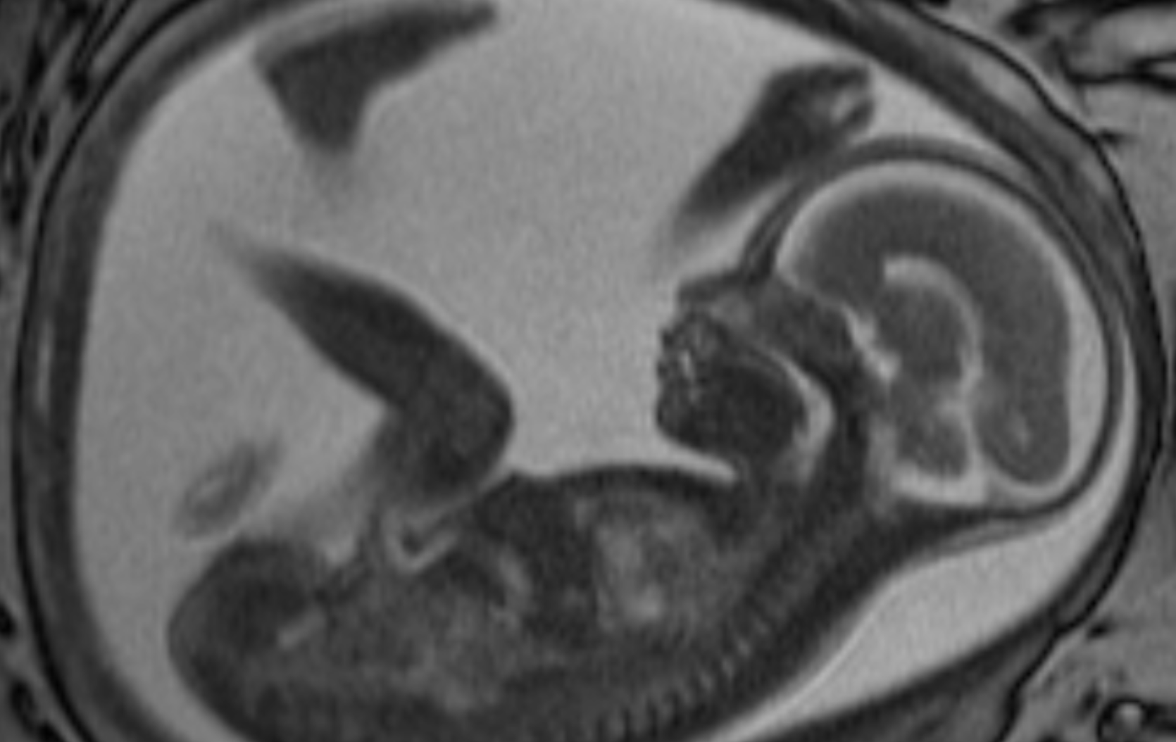
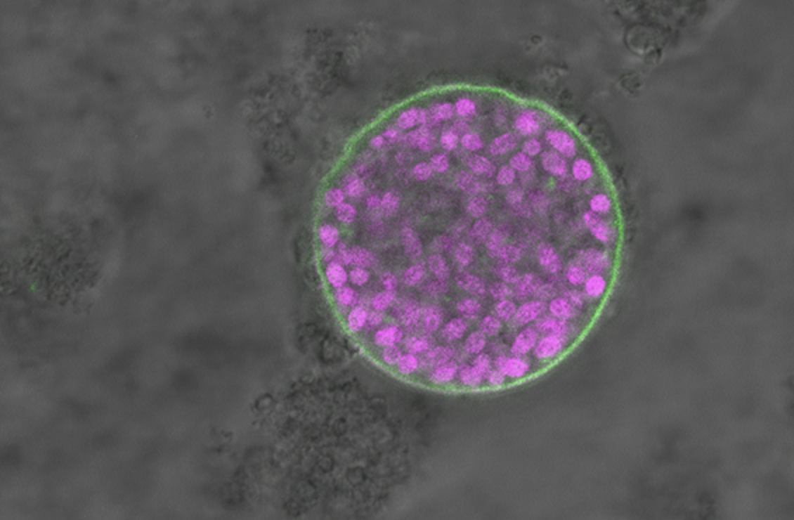
The researchers analyzed human gut microbiome time series studies conducted on individuals who had diarrhea illnesses and were not treated with antibiotics. Observing the colonization of C. difficile soon after the illnesses were acquired, they tested this association directly by feeding mice increasing quantities of laxatives while exposing them to non-pathogenic C. difficile spores. Their results suggest that GI disturbances create a window of susceptibility to C. difficile colonization during recovery.
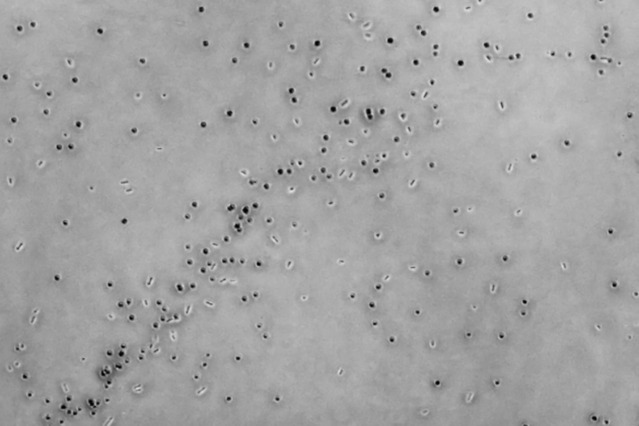
Further, the researchers found that carriers shed C. difficile in highly variable amounts day-to-day; the number of C. difficile cells shed in a carrier’s stool can increase by over 1,000 times in one day. These recurrent blooms likely influence the transmissibility of C. difficile outside of hospitals, and their unpredictability questions the reliability of single time-point diagnostics for detecting carriers.
“In our study, two of the people we followed with high temporal resolution became carriers outside of the hospital,” says VanInsberghe, who is now a postdoc in the Department of Pathology at Emory University. “The observations we made from their data helped us understand how people become susceptible to colonization and what the short- and long-term patterns in C. difficile abundance in carriers look like. Those patterns told us a lot about how C. difficile can spread between people outside of hospitals.”
“I believe that there is a lot of rethinking of C. diff infections at the moment and I hope our study will help contribute to ultimately better manage the risks associated with it,” says Martin Polz, senior author of the study and a visiting professor in MIT’s Parsons Laboratory for Environmental Science and Engineering within the MIT Department of Civil and Environmental Engineering.
The research team also included Joseph A. Elsherbini, a graduate student in the MIT Department of Biology; Bernard Varian, a researcher in MIT’s Division of Comparative Medicine; Theofilos Poutahidis, a professor in the Department of Pathology within the College of Veterinary Medicine at Aristotle University in Greece; and Susan Erdman, a principal research scientist in MIT’s Division of Comparative Medicine.