Placement: Promote to Homepage
Those selected for these positions receive additional support to pursue their research and develop their careers.
School of Science
November 2, 2021
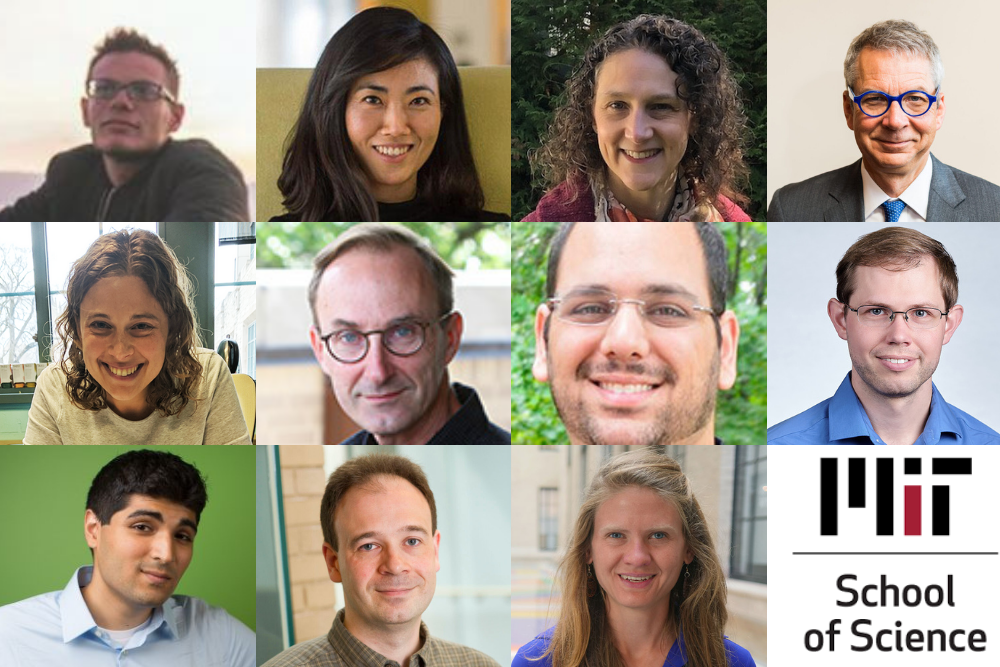
The School of Science has announced that 11 faculty members have been appointed to named professorships. These positions offer additional support to professors to advance their research and develop their careers.
Andrew Babbin was named a Cecil and Ida Green Career Development Professor. A marine biogeochemist, Babbin studies the processes that return fixed nitrogen in the ocean back to nitrogen gas, exploring marine nitrogen’s control on life in the ocean and its effects on climate. His research sheds light on the ocean’s potential to sustain life and store carbon. Babbin earned a BS degree from Columbia University in 2008 and a PhD from Princeton University in 2014. He came to MIT in 2014 as a postdoc in the Department of Civil and Environmental Engineering before joining the Department of Earth, Atmospheric and Planetary Sciences in January 2017.
Gloria Choi was selected as the Mark Hyman Jr. Career Development Professor. Choi, an associate professor in the Department of Brain and Cognitive Sciences and an investigator with the Picower Institute, examines the interaction of the immune system with the brain and the effects of that interaction on neurodevelopment, behavior, and mood. She also studies how social behaviors are regulated according to sensory stimuli, context, internal state, and physiological status, and how these factors modulate neural circuit function via a combinatorial code of classic neuromodulators and immune-derived cytokines. She received her bachelor’s degree from the University of California at Berkeley, and her PhD from Caltech, where she studied with David Anderson. She was a postdoc in the laboratory of Richard Axel at Columbia University. Choi joined the MIT faculty as an assistant professor in 2013.
Arlene Fiore joined MIT as the inaugural Peter H. Stone and Paola Malanotte Stone Professor in Earth, Atmospheric and Planetary Sciences in July 2021. Her research encompasses air pollution, chemistry-climate connections, trends and variability in atmospheric constituents, and biosphere-atmosphere interactions. Fiore’s group investigates regional meteorology and climate feedbacks due to aerosols versus greenhouse gases, future air pollution responses to climate change, as well as the factors controlling the oxidizing capacity of the atmosphere. After earning a bachelor’s degree and PhD from Harvard University, Fiore held a research scientist position at the Geophysical Fluid Dynamics Laboratory and was appointed as an associate professor with tenure at Columbia University in 2011.
Peter H. Fisher is now the Thomas A. Frank (1977) Professor of Physics. His interests include the detection of dark matter, development of new particle detectors, compact energy supplies, and wireless energy transmission. Currently serving as the head of the Department of Physics, Fisher also holds appointments in the Institute for Soldier Nanotechnologies, the Laboratory for Nuclear Science, and the Kavli Institute. He is involved in CERN’s Alpha Magnetic Spectrometer experiment to make high-precision measurements of cosmic rays and the development of new ideas for dark matter. After receiving a BS in engineering physics from the University of California at Berkeley in 1983 and a PhD in nuclear physics from Caltech in 1988, Fisher was at the Johns Hopkins University from 1989 to 1994 and joined MIT in 1994.
Danna Freedman has been named the Frederick George Keyes Professor of Chemistry. Freedman leverages inorganic chemistry to solve problems in physics. Her research focuses on creating spin-based quantum bits and synthesizing new emergent materials. Freedman received her bachelor’s degree from Harvard University and her PhD from the University of California at Berkeley, then conducted postdoctoral research at MIT before joining the faculty at Northwestern University as an assistant professor in 2012, where she was promoted to associate professor in 2018 and full professor in 2020. Freedman returned to MIT’s Department of Chemistry in 2021.
Michel Goemans has been named the RSA Professor of Mathematics. Goemans has been head of the Department of Mathematics since July 1, 2018, following a year as interim head. He received his undergraduate degree in applied mathematics from Université Catholique de Louvain in 1987 and completed his PhD at MIT in 1990. He has been on the faculty since 1992, receiving tenure in 1999, and held the Leighton Family Professorship from 2007 to 2017. The RSA cryptosystem is the brainchild of Ron Rivest, Adi Shamir, and Len Adleman, whose fruitful collaboration spanned the Laboratory for Computer Science — today the Computer Science and Artificial Intelligence Laboratory (CSAIL) — and the Department of Mathematics. Goemans is also a member of the Theory of Computation Group of CSAIL, and recently joined the Computing Council of the MIT Schwarzman College of Computing. Goemans’ research interests include combinatorics, optimization and algorithms. In particular, his pioneering use of semidefinite optimization and other techniques for designing approximation algorithms for hard combinatorial optimization problems has been rewarded with several awards, such as the Fulkerson, Farkas and Dantzig prizes.
Or Hen was named the Class of 1956 Career Development Associate Professor of Physics. He investigates quantum chromodynamic effects in the nuclear medium and the interplay between partonic and nucleonic degrees of freedom in nuclei. Specifically, Hen utilizes high-energy scattering of electron, neutrino, photon, proton, and ion off atomic nuclei to study short-range correlations: temporal fluctuations of high-density, high-momentum, nucleon clusters in nuclei with important implications for nuclear, particle, atomic, and astrophysics. He received his undergraduate degree in physics and computer engineering from the Hebrew University and earned his PhD in experimental physics at Tel-Aviv University. Hen was an MIT Pappalardo Fellow in Physics from 2015 to 2017 before joining the physics faculty in July 2017.
Brett McGuire is now the Class of 1943 Career Development Assistant Professor of Chemistry. He uses the tools of physical chemistry, molecular spectroscopy, and observational astrophysics to understand how the chemical ingredients for life evolve with and help shape the formation of stars and planets. His group aims to detect more new molecules in space and to better understand their significance, advancing the field of astrochemistry. McGuire obtained a bachelor’s degree from the University of Illinois at Urbana-Champaign in 2009, a master’s degree from Emory University in 2011, and a PhD from Caltech in 2015. McGuire joined the Department of Chemistry in 2020.
Iain W. Stewart has been selected for the Otto (1939) and Jane Morningstar Professorship in Science. Stewart is a professor of physics and the director of the Center for Theoretical Physics. His research interests involve theoretical nuclear and particle physics. In particular, he focuses upon the development and application of effective field theories to answer fundamental questions about interactions between elementary particles. Stewart earned a bachelor’s degree in physics and mathematics and a master’s degree in physics from the University of Manitoba in Canada. He then received his PhD from Caltech in 1999. Stewart joined the physics faculty at MIT in 2003, was promoted to associate professor with tenure in 2009, and became a full professor in 2013.
Ankur Moitra, a theoretical computer scientist, is now the Norbert Wiener Professor of Mathematics. The aim of his work is to bridge the gap between theoretical computer science and machine learning by developing algorithms with provable guarantees and foundations for reasoning about their behavior. Moitra received his bachelor’s degree in electrical and computer engineering from Cornell University in 2007 and his master’s degree and PhD from MIT in computer science in 2009 and 2011, respectively, then spent two years as a fellow at the Institute for Advanced Study and Princeton University. Moitra returned to MIT in 2013 as a professor in applied mathematics and a principal investigator in CSAIL.
Seychelle M. Vos has been named a Robert A. Swanson (1969) Career Development Professor of Life Sciences. Vos examines the interplay of genome organization and gene expression to gain insight into how the organization of a cell affects what it becomes. Vos’ lab examines these pieces at a molecular scale using varied approaches from single-particle cryo-electron microscopy to X-ray crystallography, biochemistry to genetics. This work can help to build a biological understanding of diseases such as developmental disorders or cancers. She received her BS in genetics in 2008 from the University of Georgia and her PhD in molecular and cell biology in 2013 from the University of California at Berkeley. Vos joined the Department of Biology in 2019.
October 30, 2021
Researchers decipher when and why immune cells fail to respond to immunotherapy, suggesting that T cells need a different kind of prodding to re-engage the immune response.
Grace van Deelen
October 29, 2021
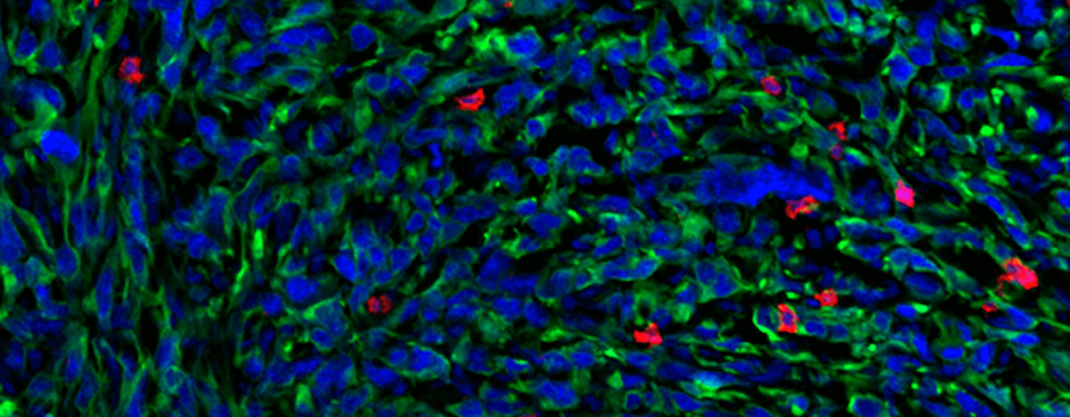
Non-small cell lung cancer (NSCLC) is the most common type of lung cancer in humans. Some patients with NSCLC receive a therapy called immune checkpoint blockade (ICB) that helps kill cancer cells by reinvigorating a subset of immune cells called T cells, which are “exhausted” and have stopped working. However, only about 35% of NSCLC patients respond to ICB therapy. Stefani Spranger’s lab at the MIT Department of Biology explores the mechanisms behind this resistance, with the goal of inspiring new therapies to better treat NSCLC patients. In a new study published on Oct. 29 in Science Immunology, a team led by Spranger lab postdoc Brendan Horton revealed what causes T cells to be non-responsive to ICB — and suggested a possible solution.
Scientists have long thought that the conditions within a tumor were responsible for determining when T cells stop working and become exhausted after being overstimulated or working for too long to fight a tumor. That’s why physicians prescribe ICB to treat cancer — ICB can invigorate the exhausted T cells within a tumor. However, Horton’s new experiments show that some ICB-resistant T cells stop working before they even enter the tumor. These T cells are not actually exhausted, but rather they become dysfunctional due to changes in gene expression that arise early during the activation of a T cell, which occurs in lymph nodes. Once activated, T cells differentiate into certain functional states, which are distinguishable by their unique gene expression patterns.
In order to determine why some tumors are resistant to ICB, Horton and the research team studied T cells in murine models of NSCLC. The researchers sequenced messenger RNA from the responsive and non-responsive T cells in order to identify any differences between the T cells. Supported in part by the Koch Institute Frontier Research Program, they used a technique called Seq-Well, developed in the lab of fellow Koch Institute member J. Christopher Love, the Raymond A. (1921) and Helen E. St. Laurent Professor of Chemical Engineering and a co-author of the study. The technique allows for the rapid gene expression profiling of single cells, which permitted Spranger and Horton to get a very granular look at the gene expression patterns of the T cells they were studying.
Seq-Well revealed distinct patterns of gene expression between the responsive and non-responsive T cells. These differences, which are determined when the T cells assume their specialized functional states, may be the underlying cause of ICB resistance.
Now that Horton and his colleagues had a possible explanation for why some T cells did not respond to ICB, they decided to see if they could help the ICB-resistant T cells kill the tumor cells. When analyzing the gene expression patterns of the non-responsive T cells, the researchers had noticed that these T cells had a lower expression of receptors for certain cytokines, small proteins that control immune system activity. To counteract this, the researchers treated lung tumors in murine models with extra cytokines. As a result, the previously non-responsive T cells were then able to fight the tumors — meaning that the cytokine therapy prevented, and potentially even reversed, the dysfunctionality.
Administering cytokine therapy to human patients is not currently safe, because cytokines can cause serious side effects as well as a reaction called a “cytokine storm,” which can produce severe fevers, inflammation, fatigue, and nausea. However, there are ongoing efforts to figure out how to safely administer cytokines to specific tumors. In the future, Spranger and Horton suspect that cytokine therapy could be used in combination with ICB.
“This is potentially something that could be translated into a therapeutic that could increase the therapy response rate in non-small cell lung cancer,” Horton says.
Spranger agrees that this work will help researchers develop more innovative cancer therapies, especially because researchers have historically focused on T cell exhaustion rather than the earlier role that T cell functional states might play in cancer.
“If T cells are rendered dysfunctional early on, ICB is not going to be effective, and we need to think outside the box,” she says. “There’s more evidence, and other labs are now showing this as well, that the functional state of the T cell actually matters quite substantially in cancer therapies.” To Spranger, this means that cytokine therapy “might be a therapeutic avenue” for NSCLC patients beyond ICB.
Jeffrey Bluestone, the A.W. and Mary Margaret Clausen Distinguished Professor of Metabolism and Endocrinology at the University of California-San Francisco, who was not involved with the paper, agrees. “The study provides a potential opportunity to ‘rescue’ immunity in the NSCLC non-responder patients with appropriate combination therapies,” he says.
This research was funded by the Pew-Stewart Scholars for Cancer Research, the Ludwig Center for Molecular Oncology, the Koch Institute Frontier Research Program through the Kathy and Curt Mable Cancer Research Fund, and the National Cancer Institute.
Eva Frederick | Whitehead Institute
October 25, 2021
Many people — around half of the adult population — are infected with a type of herpesvirus called human cytomegalovirus, or HCMV. Though mostly asymptomatic, the virus can be dangerous for immunocompromised people and unborn babies. Because HCMV is so widespread, the chance of a baby becoming infected in utero is around one in 200, and that infection can lead to problems with the baby’s brain, lungs and growth.
In a new paper from Whitehead Institute Member Jonathan Weissman published on October 25 in Nature Biotechnology, Weissman and colleagues turn cutting-edge CRISPR and single cell sequencing technologies on this virus, providing the most detailed picture yet on how viral and human genes interact to create an HCMV infection — and revealing new ways to potentially derail the virus’ progression through manipulating viral and host genes.
The research could provide an important road map for future studies of host-pathogen interactions, as well as inform antiviral drug design. Over the course of the project, the researchers generated a list of both viral and host genes that were either essential for the virus to replicate, or could potentially be manipulated to confer some immunity to the host cell. “Now that we have this list, we have a list of potential targets that one might now go ahead and develop drugs against,” said Marco Hein, the first author and a former postdoctoral researcher in the Weissman Lab.
Seeing both sides
Millions of years of evolution have created a complex web of interactions between virus and host. For example, viruses have their own set of genes, but they also depend on some human genes, called host factors. Hijacking these host factors allows the viruses to invade cells in the body and replicate their own genetic material.
Hein, who is now a researcher at the Chan Zuckerberg Biohub in San Francisco, and Whitehead Institute Member Jonathan Weissman, who is also a professor of biology at the Massachusetts Institute of Technology and Koch Institute and an investigator of the Howard Hughes Medical Institute, sought to gain a more thorough understanding of the web of host-viral interactions that arises throughout the course of an infection. “[We wanted to know] what actually happens when we [knock out or weaken] those critical factors,” Hein said. “Can we prevent infection? If so, what ‘goes wrong’ from the perspective of the virus?”
They chose HCMV as a test subject because, for one thing, the virus has a double-stranded DNA genome like humans. That means that CRISPR technologies that work by snipping DNA could theoretically work for both the virus and the host. “And because CMV is an important human pathogen and it’s such a complex and intricate virus, we thought we would have a chance to really discover something new,” said Weissman.
A series of screens
The researchers first set out, using a molecular technique called CRISPR screening, to determine whether any regions of the viral or host genomes in particular had an impact on the fate of infected host cells. By systematically knocking out individual genes in a large population of viruses and host cells, the researchers could then assess how essential each gene was to the infection.
The project took on a new dimension in 2016 with the development of accessible, large-scale single cell sequencing. “We had this idea to put together the CRISPR screening and the single cell sequencing, and [a screening method called PerturbSeq],” Hein said. “Basically, you perturb genes in a cell population and then you read out what happens to the cells, not just by measuring survival, but by actually looking at the pattern of gene expression in those cells over time.”
Combining these methods generated a huge set of data, which provided the researchers with a clearer view of which genes were important and when. “The single cell sequencing lets us watch the steps in the viral life cycle with much higher precision, and then the perturbation lets us understand how host and viral factors allow the virus to manipulate the host and complete its life cycle,” Weissman said.
The resultant data showed how the virus’ typical trajectory — from the initial waves of viral gene expression, to replication of the full viral genome, to the final step of budding off into newly-formed virions — could be derailed by altering specific viral genes. It also clued the researchers in to which host genes the virus depended on at what stages for a ‘successful’ infection.
“The course of infection is pre-programmed into the viral genome,” Hein said. “If you want to interfere with the course of infection you can do that by targeting a viral factor, or you can do it indirectly by targeting the host factor. And the outcomes are conceptually different. If you target a virus factor you derail the program that the virus would normally follow. If you target a host factor, the program itself is unchanged, but you change how far the virus gets in executing the program.”
These findings will be useful tools for the development of drugs that can be used as part of an antiviral “cocktail.” Because viruses and other pathogens are living creatures that can mutate and adapt to changing conditions, a common thread among antiviral treatments involves combining several drugs with different viral targets. This ensures the most complete eradication of viruses possible, reducing the chance that some will survive and create a new resistant population.
While the researchers’ list of essential viral genes provide parts of HCMV to target with drug cocktails, the list of contributing human genes could open the door for a more indirect therapy. “If you target a host factor to affect the virus, it’s much more difficult for the virus to escape because it can’t just mutate so the drug doesn’t bind anymore — it would have to mutate away from dependency on a host protein, which is much more complicated,” Hein said.
Of course, there are drawbacks to potentially targeting a human gene or protein to treat an infection, and much more work would need to be done for a viable treatment to emerge via this avenue of research. “If you target a host factor, you’re by definition targeting a protein that’s in our body, doing its normal job, so the risk of side effects is much higher,” Hein said.
Few drugs like this have made it past clinical trials; one famous example is hydroxychloroquine, which has been used successfully to treat malaria, and unsuccessfully to treat COVID-19.
In the future, Hein and Weissman hope to turn their multi-level approach for studying infection toward other viruses such as SARS-CoV-2. Although the novel coronavirus does not have double-stranded DNA that can be altered via CRISPR, the researchers can still investigate which host genes are essential at what stage of infection, and use their methods-driven approach to hopefully glean unexpected findings from a well-studied virus.
“I’m always driven by what technology can do,” Hein said. “I like to run a study in a systems-wide manner and then come up with some findings that you would have not found if you had only looked at one gene or protein at a time or looked at things more in the conventional way. This kind of high-level conclusion is what I personally always find the most exciting.”
Eva Frederick | Whitehead Institute
October 24, 2021
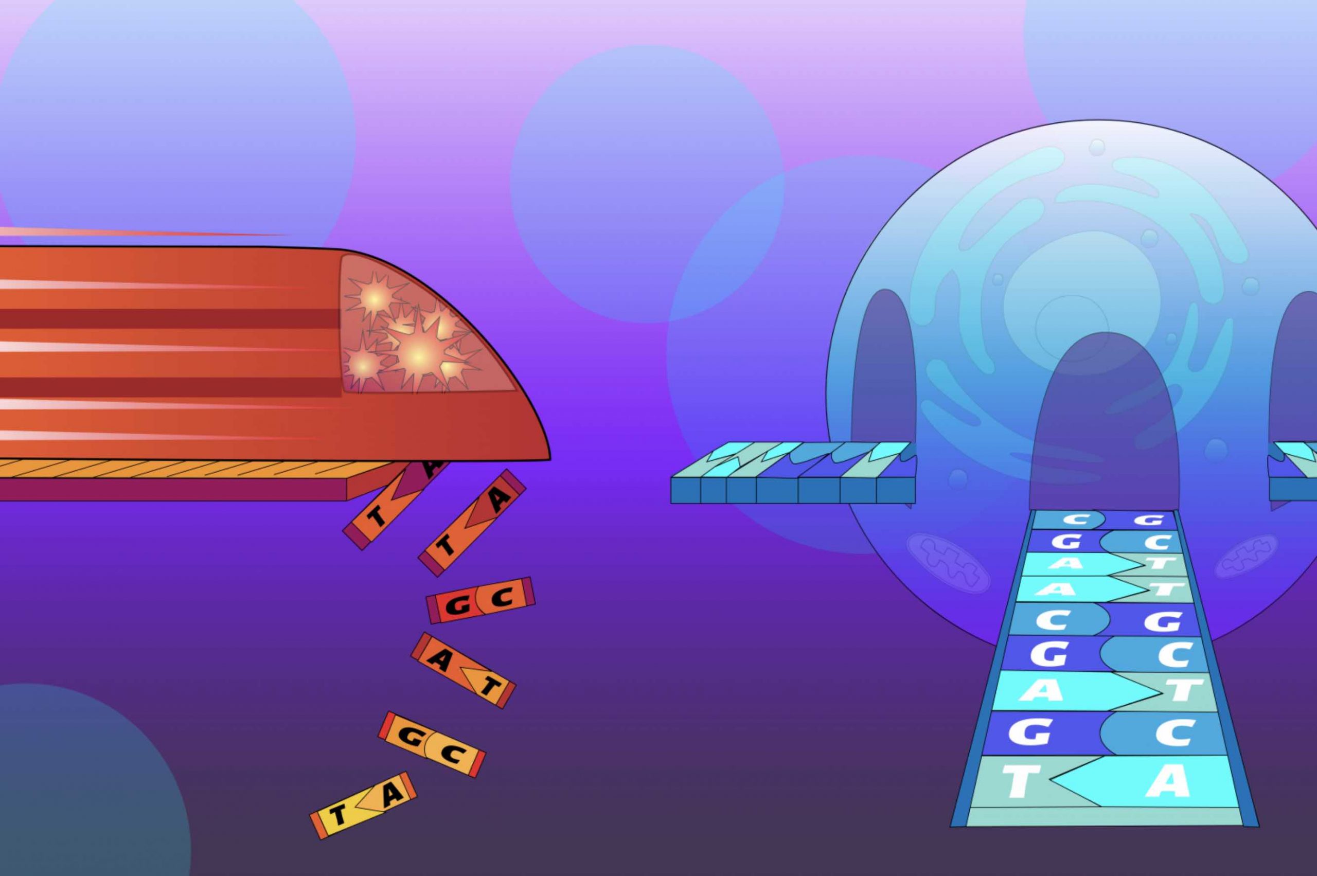
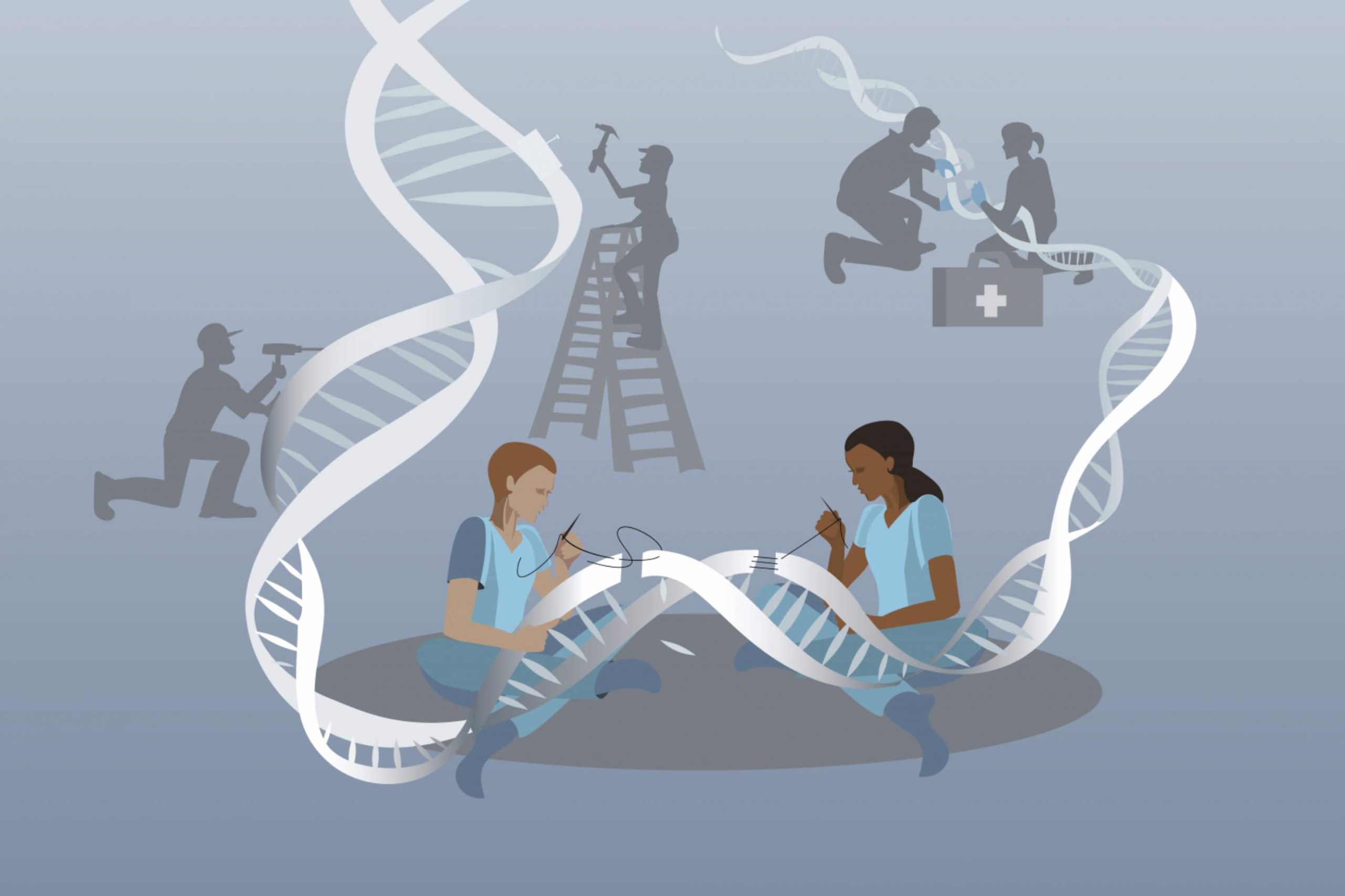
Gene editing methods often involve breaking a strand of DNA in order to make specific changes to the sequence. They then rely on the cell’s DNA repair pathways to mend the break. These cellular repair pathways, however, are not completely understood, and introduce an element of chance to gene editing; for example, a repair mechanism may patch up the edited strand, but also leave behind an unwanted mutation.
In a paper published online in Cell on October 20, 2021, a collaborative team of researchers in the lab of Whitehead Institute Member, Massachusetts Institute of Technology (MIT) biology professor and Howard Hughes Medical Institute Investigator Jonathan Weissman, the lab of Britt Adamson at Princeton University, and Cecilia Cotta-Ramusino, then a research scientist at Editas Medicine (now at Tessera Therapeutics), present a new experimental method that could help bridge this gap in our understanding.
The method, called Repair-seq, allows researchers to find out which genes and genetic pathways are involved in DNA repair mechanisms. It provides a useful tool for fundamental research on gene repair, as well as a way to test the action of new genome editing methods as they are developed, says Weissman Lab postdoc and first author Jeffrey Hussmann. A companion paper published concurrently in Cell in collaboration with researchers at the Broad Institute of MIT and Harvard provides a glimpse into the utility of Repair-seq when applied to new gene editing technologies.
“The field of gene editing has moved so quickly and people have been so creative developing new methods that our ability to apply them has dramatically outpaced our understanding of exactly how they work,” Hussmann said. “We think that Repair-seq will be a valuable tool going forward so that as new editing methods are developed, we can quickly do a better job of characterizing how they interact with different repair mechanisms.”
Studying repair mechanisms in one fell swoop
Cells have several different methods they use to repair breaks in DNA strands, and the path to any one method depends on a tree of decisions based on the circumstances. “Over decades, a huge number of people have worked out parts of these pathways through focused experiments,” said Adamson, a senior author on both papers and an assistant professor at Princeton University.
Together, the team of researchers saw an opportunity to harness existing CRISPR-based methods to take a broad look at repair pathways in the cell. The method they created combines several CRISPR-based technologies. First, the researchers used a method they previously developed called CRISPRi to inactivate hundreds of genes known to be involved in DNA repair across a cell population. Then, they induced double strand breaks in the cells’ DNA at specific places that the cell would need to heal.
As the cells mended the breaks, the researchers used targeted sequencing to examine the ‘repair outcomes’ — mutations or the lack thereof — in the DNA strand resulting from different methods of repair. Finally, they were able to extrapolate which genes were essential to various repair mechanisms and how they were involved in producing or preventing each type of resulting mutation. They also posted their data online in an interactive format so others can use it to investigate DNA repair genes and pathways.
“This combination of different CRISPR-based technologies has made it possible to, in one fell swoop, recapitulate a lot of the work that was done painstakingly over the past decades to study each repair pathway one at a time,” Hussmann said. “The high-level view of repair that our method produces shows us many of the things that people saw before, and at the same time reveals unexpected connections that we only get by having the comprehensive picture.”
These unforeseen relationships between repair genes may help fundamental researchers refine the decision tree of double strand break repair in the future, said Weissman. “One of the big themes that’s come out of this is that outcomes that superficially look similar can actually have very different mechanisms,” Weissman said.
A ‘prime’ example of Repair-seq’s utility
As the team was developing their Repair-seq methodology, Broad Institute of MIT and Harvard Core Member David Liu’s lab was working on prime editing, a gene editing method that promises more precise control over genetic outcomes than traditional CRISPR methods. Instead of snipping both strands of DNA’s double helix, prime editing makes a ‘nick’ in only one of the strands and introduces a short sequence template containing the desired genetic change.
“When Liu’s group came out with prime editing, our Repair-seq team realized that we had the perfect tool for quickly trying to understand exactly how it was working,” Hussmann said.
The three labs collaborated to use Repair-seq to identify which pathways were at play during the installation of mutations by prime editing, and identified one in particular, called the DNA mismatch repair pathway, that seemed to be interfering with the efficiency and accuracy of the method. When the researchers inhibited this pathway, the performance of Liu’s prime editing technique greatly improved.
“Working with Britt, Jonathan, and their labs has been a beautiful integration of basic science, tool application, and technology development—a real testament to the power of multidisciplinary collaboration” said Liu, also an Investigator of the Howard Hughes Medical Institute.
The researchers also applied Repair-seq to a base editor — a tool to swap specific bases in a DNA sequence — and were able to illuminate the DNA repair genes involved in swapping in particular base.
In the future, the researchers plan to continue adapting the method to new sequencing methods and applying it to new editors as they are developed. “We think Repair-seq is a really practical way of making better genome editors,” Weissman said.
“It has been rewarding to see the efforts of our collaboration come together,” said Adamson. “We hope the insights from our study and tools that those insights have led to will be widely useful to the research community.”
October 22, 2021
A new study finds cutting off cells’ supplies of lipids can slow the growth of tumors in mice.
Anne Trafton | MIT News Office
October 20, 2021
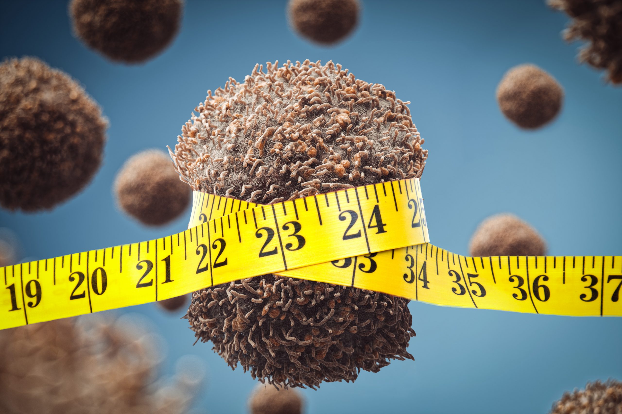
In recent years, there has been some evidence that dietary interventions can help to slow the growth of tumors. A new study from MIT, which analyzed two different diets in mice, reveals how those diets affect cancer cells, and offers an explanation for why restricting calories may slow tumor growth.
The study examined the effects of a calorically restricted diet and a ketogenic diet in mice with pancreatic tumors. While both of these diets reduce the amount of sugar available to tumors, the researchers found that only the calorically restricted diet reduced the availability of fatty acids, and this was linked to a slowdown in tumor growth.
The findings do not suggest that cancer patients should try to follow either of these diets, the researchers say. Instead, they believe the findings warrant further study to determine how dietary interventions might be combined with existing or emerging drugs to help patients with cancer.
“There’s a lot of evidence that diet can affect how fast your cancer progresses, but this is not a cure,” says Matthew Vander Heiden, director of MIT’s Koch Institute for Integrative Cancer Research and the senior author of the study. “While the findings are provocative, further study is needed, and individual patients should talk to their doctor about the right dietary interventions for their cancer.”
MIT postdoc Evan Lien is the lead author of the paper, which appears today in Nature.
Metabolic mechanism
Vander Heiden, who is also a medical oncologist at Dana-Farber Cancer Institute, says his patients often ask him about the potential benefits of various diets, but there is not enough scientific evidence available to offer any definitive advice. Many of the dietary questions that patients have focus on either a calorie-restricted diet, which reduces calorie consumption by 25 to 50 percent, or a ketogenic diet, which is low in carbohydrates and high in fat and protein.
Previous studies have suggested that a calorically restricted diet might slow tumor growth in some contexts, and such a diet has been shown to extend lifespan in mice and many other animal species. A smaller number of studies exploring the effects of a ketogenic diet on cancer have produced inconclusive results.
“A lot of the advice or cultural fads that are out there aren’t necessarily always based on very good science,” Lien says. “It seemed like there was an opportunity, especially with our understanding of cancer metabolism having evolved so much over the past 10 years or so, that we could take some of the biochemical principles that we’ve learned and apply those concepts to understanding this complex question.”
Cancer cells consume a great deal of glucose, so some scientists had hypothesized that either the ketogenic diet or calorie restriction might slow tumor growth by reducing the amount of glucose available. However, the MIT team’s initial experiments in mice with pancreatic tumors showed that calorie restriction has a much greater effect on tumor growth than the ketogenic diet, so the researchers suspected that glucose levels were not playing a major role in the slowdown.
To dig deeper into the mechanism, the researchers analyzed tumor growth and nutrient concentration in mice with pancreatic tumors, which were fed either a normal, ketogenic, or calorie-restricted diet. In both the ketogenic and calorie-restricted mice, glucose levels went down. In the calorie-restricted mice, lipid levels also went down, but in mice on the ketogenic diet, they went up.
Lipid shortages impair tumor growth because cancer cells need lipids to construct their cell membranes. Normally, when lipids aren’t available in a tissue, cells can make their own. As part of this process, they need to maintain the right balance of saturated and unsaturated fatty acids, which requires an enzyme called stearoyl-CoA desaturase (SCD). This enzyme is responsible for converting saturated fatty acids into unsaturated fatty acids.
Both calorie-restricted and ketogenic diets reduce SCD activity, but mice on the ketogenic diet had lipids available to them from their diet, so they didn’t need to use SCD. Mice on the calorie-restricted diet, however, couldn’t get fatty acids from their diet or produce their own. In these mice, tumor growth slowed significantly, compared to mice on the ketogenic diet.
“Not only does caloric restriction starve tumors of lipids, it also impairs the process that allows them to adapt to it. That combination is really contributing to the inhibition of tumor growth,” Lien says.
Dietary effects
In addition to their mouse research, the researchers also looked at some human data. Working with Brian Wolpin, an oncologist at Dana-Farber Cancer Institute and an author of the paper, the team obtained data from a large cohort study that allowed them to analyze the relationship between dietary patterns and survival times in pancreatic cancer patients. From that study, the researchers found that the type of fat consumed appears to influence how patients on a low-sugar diet fare after a pancreatic cancer diagnosis, although the data are not complete enough to draw any conclusions about the effect of diet, the researchers say.
Although this study showed that calorie restriction has beneficial effects in mice, the researchers say they do not recommend that cancer patients follow a calorie-restricted diet, which is difficult to maintain and can have harmful side effects. However, they believe that cancer cells’ dependence on the availability of unsaturated fatty acids could be exploited to develop drugs that might help slow tumor growth.
One possible therapeutic strategy could be inhibition of the SCD enzyme, which would cut off tumor cells’ ability to produce unsaturated fatty acids.
“The purpose of these studies isn’t necessarily to recommend a diet, but it’s to really understand the underlying biology,” Lien says. “They provide some sense of the mechanisms of how these diets work, and that can lead to rational ideas on how we might mimic those situations for cancer therapy.”
The researchers now plan to study how diets with a variety of fat sources — including plant or animal-based fats with defined differences in saturated, monounsaturated, and polyunsaturated fatty acid content — alter tumor fatty acid metabolism and the ratio of unsaturated to saturated fatty acids.
The research was funded by the Damon Runyon Cancer Research Foundation, the National Institutes of Health, the Lustgarten Foundation, the Dana-Farber Cancer Institute Hale Family Center for Pancreatic Cancer Research, Stand Up to Cancer, the Pancreatic Cancer Action Network, the Noble Effort Fund, the Wexler Family Fund, Promises for Purple, the Bob Parsons Fund, the Emerald Foundation, the Howard Hughes Medical Institute, the MIT Center for Precision Cancer Medicine, and the Ludwig Center at MIT.
By combining chemotherapy, tumor injury, and immunotherapy, researchers show that the immune system can be re-engaged to destroy tumors in mice.
Anne Trafton | MIT News Office
October 20, 2021
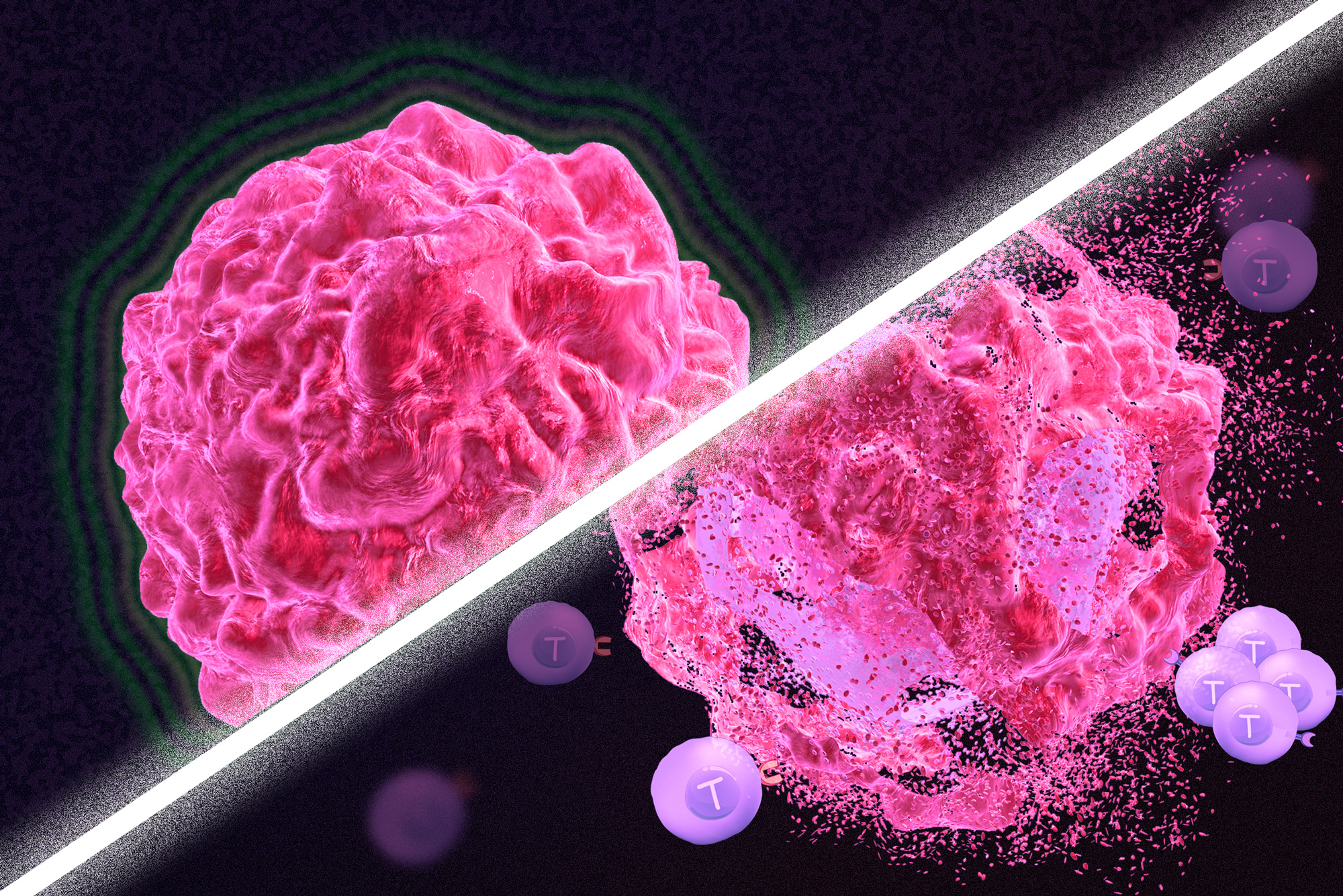
Immunotherapy is a promising strategy to treat cancer by stimulating the body’s own immune system to destroy tumor cells, but it only works for a handful of cancers. MIT researchers have now discovered a new way to jump-start the immune system to attack tumors, which they hope could allow immunotherapy to be used against more types of cancer.
Their novel approach involves removing tumor cells from the body, treating them with chemotherapy drugs, and then placing them back in the tumor. When delivered along with drugs that activate T cells, these injured cancer cells appear to act as a distress signal that spurs the T cells into action.
“When you create cells that have DNA damage but are not killed, under certain conditions those live, injured cells can send a signal that awakens the immune system,” says Michael Yaffe, who is a David H. Koch Professor of Science, the director of the MIT Center for Precision Cancer Medicine, and a member of MIT’s Koch Institute for Integrative Cancer Research.
In mouse studies, the researchers found that this treatment could completely eliminate tumors in nearly half of the mice.
Yaffe and Darrell Irvine, who is the Underwood-Prescott Professor with appointments in MIT’s departments of Biological Engineering and Materials Science and Engineering, and an associate director of the Koch Institute, are the senior authors of the study, which appears today in Science Signaling. MIT postdoc Ganapathy Sriram and Lauren Milling PhD ’21 are the lead authors of the paper.
T cell activation
One class of drugs currently used for cancer immunotherapy is checkpoint blockade inhibitors, which take the brakes off of T cells that have become “exhausted” and unable to attack tumors. These drugs have shown success in treating a few types of cancer but do not work against many others.
Yaffe and his colleagues set out to try to improve the performance of these drugs by combining them with cytotoxic chemotherapy drugs, in hopes that the chemotherapy could help stimulate the immune system to kill tumor cells. This approach is based on a phenomenon known as immunogenic cell death, in which dead or dying tumor cells send signals that attract the immune system’s attention.
Several clinical trials combining chemotherapy and immunotherapy drugs are underway, but little is known so far about the best way to combine these two types of treatment.
The MIT team began by treating cancer cells with several different chemotherapy drugs, at different doses. Twenty-four hours after the treatment, the researchers added dendritic cells to each dish, followed 24 hours later by T cells. Then, they measured how well the T cells were able to kill the cancer cells. To their surprise, they found that most of the chemotherapy drugs didn’t help very much. And those that did help appeared to work best at low doses that didn’t kill many cells.
The researchers later realized why this was so: It wasn’t dead tumor cells that were stimulating the immune system; instead, the critical factor was cells that were injured by chemotherapy but still alive.
“This describes a new concept of immunogenic cell injury rather than immunogenic cell death for cancer treatment,” Yaffe says. “We showed that if you treated tumor cells in a dish, when you injected them back directly into the tumor and gave checkpoint blockade inhibitors, the live, injured cells were the ones that reawaken the immune system.”
The drugs that appear to work best with this approach are drugs that cause DNA damage. The researchers found that when DNA damage occurs in tumor cells, it activates cellular pathways that respond to stress. These pathways send out distress signals that provoke T cells to leap into action and destroy not only those injured cells but any tumor cells nearby.
“Our findings fit perfectly with the concept that ‘danger signals’ within cells can talk to the immune system, a theory pioneered by Polly Matzinger at NIH in the 1990s, though still not universally accepted,” Yaffe says.
Tumor elimination
In studies of mice with melanoma and breast tumors, the researchers showed that this treatment eliminated tumors completely in 40 percent of the mice. Furthermore, when the researchers injected cancer cells into these same mice several months later, their T cells recognized them and destroyed them before they could form new tumors.
The researchers also tried injecting DNA-damaging drugs directly into the tumors, instead of treating cells outside the body, but they found this was not effective because the chemotherapy drugs also harmed T cells and other immune cells near the tumor. Also, injecting the injured cells without checkpoint blockade inhibitors had little effect.
“You have to present something that can act as an immunostimulant, but then you also have to release the preexisting block on the immune cells,” Yaffe says.
Yaffe hopes to test this approach in patients whose tumors have not responded to immunotherapy, but more study is needed first to determine which drugs, and at which doses, would be most beneficial for different types of tumors. The researchers are also further investigating the details of exactly how the injured tumor cells stimulate such a strong T cell response.
The research was funded, in part, by the National Institutes of Health, the Mazumdar-Shaw International Oncology Fellowship, the MIT Center for Precision Cancer Medicine, and the Charles and Marjorie Holloway Foundation.

Studying cancer in the Sharp lab helped Courtney JnBaptiste learn strategic thinking skills that he uses as a patent agent, transforming technology into successful biotech businesses.
Raleigh McElvery | Department of Biology
October 17, 2021
Courtney JnBaptiste PhD ’16 spent the first 19 years of his life on the idyllic Caribbean island of Saint Lucia, surrounded by clear waters, sandy beaches, and a robust agricultural community. His family owned their own farm, where they grew bananas and other crops for export. JnBaptiste and his six siblings spent hours each day after school and on the weekends helping to harvest the fruits of their labor. “We were better off than most, but it was a hard existence,” he says. “I had to fight to make something out of life. Where I am today is a big leap.”
He has since moved to the U.S. and completed his PhD at the MIT Department of Biology. Today, he uses the strategic thinking skills he learned during graduate school in his job as a patent agent, helping researchers protect their inventions and start biotech companies.
Despite his exceptional grades, JnBaptiste didn’t enjoy school growing up, and he’d often try to convince his parents that he didn’t need to go to class. His mother, a middle school teacher, was unfazed by his excuses and sent him off to school most days. Despite his protests, JnBaptiste understood his mother’s motto that “education is the key to success.” He knew he’d need good academic standing and self-motivation to attain the financially-stable life he envisioned.
During high school, cable TV became available to his community for the first time, and he was overwhelmed by the deluge of information. He became hooked on Animal Planet and Discovery Channel, and decided that he wanted to be like Jeff Corwin, a biologist, wildlife conservationist, and TV personality. JnBaptiste knew he’d need a college education to reach his career aspirations, and — due to its proximity and promise of opportunity — the U.S. seemed like an ideal destination.
He was accepted to Bethune-Cookman University in Florida and declared a major in biology. He was awarded an environmental research grant, which allowed him to spend several semesters studying the snail populations in Blue Spring State Park. But a summer internship at the University of Kansas Medical Center was what ultimately convinced him to pursue molecular biology rather than environmental sciences. During his junior year, a new biochemistry professor arrived: Christopher Ainsley Davis, a former postdoc in the lab of Cathy Drennan at the MIT Department of Biology.
“When the two of us spoke, he said something that shocked me,” JnBaptiste recalls. “He told me, ‘You’re good enough for MIT and you should apply to their summer research program.’ That just blew my mind. I never thought I was of the MIT caliber — no one had ever challenged me like that before.”
At Davis’ urging, JnBaptiste applied to the MIT Summer Research Program in Biology (MSRP-Bio), and was placed in the lab of Nobel laureate Phil Sharp. In the 1970s, Sharp had co-discovered splicing, a molecular process that happens after DNA has been transcribed into RNA. Segments of the RNA strand are removed, and the remaining parts are stitched back together and translated into proteins that perform vital functions inside the cell.
Under the supervision of graduate student and postdoc mentors, JnBaptiste spent the summer of 2009 investigating the role that RNA splicing plays in cancer. “It was the best time of my life,” he says. “I just I loved it. I loved the environment. I loved the lab. I loved MIT. And that experience had a profound impact on me.”
He enjoyed campus so much that he returned just one year later to begin his PhD. He was looking forward to returning to the lab bench, but what he didn’t anticipate was that his first semester of classes would be the most rigorous education he’d ever received. He excelled in biochemistry, but found 7.52 (Graduate Genetics) especially difficult. “It was the first time I’d ever failed an exam,” he recalls.
With the help of mentors, classmates, and tutors, JnBaptiste passed genetics and moved on to the stage of his PhD that he was most excited about: lab rotations. After testing out a few different research groups, he ultimately decided to return to the Sharp lab. In his own words: “It was home.”
JnBaptiste’s thesis project focused on a type of RNA known as microRNA (miRNA), which is never translated into a protein. Instead, it remains in its single-stranded RNA form and helps regulate gene expression. The Sharp lab found that removing all the miRNAs from adult cells prompted dramatic activation of embryonic genes. These genes are typically turned off in adult cells, and only expressed during early development when rapid cell division is required. But they can also be hijacked during cancer to create tumors.
JnBaptiste was surprised to find that adding the miRNAs back into the cells didn’t shut down these embryonic genes. In fact, restoring the miRNAs made the cells divide even more rapidly and increased their ability to form tumors — suggesting “global miRNA restoration” would not be a viable approach to treat cancer.
“This model that we developed showed miRNAs control a very important network in the context of both development and cancer,” JnBaptiste explains. “Cancer occurs when normal cellular processes go awry, so understanding those fundamental molecular interactions is critical to fighting the disease.”
By the time he graduated from MIT in 2016, JnBaptiste knew he enjoyed science, but didn’t have ambitions to run his own lab. Instead, he was curious about how lab experiments and research questions engender companies.
When he was still an MSRP-Bio student, JnBaptiste had met an intellectual property lawyer who’d come to speak on campus. He’d been saving her business card ever since, and reached out to her as his time at MIT was drawing to a close. With her assistance, JnBaptiste was offered a job as a scientific advisor at Goodwin Procter, the international law firm where she worked.
JnBaptiste has since transitioned to a similar role as a patent agent at Pabst Patent Group. There, he collaborates with lawyers to write patents protecting new research technology. While scientists are focused on the minutia of their day-to-day lab experiments, JnBaptiste is tasked with understanding the bigger picture, and how those experiments might lay the foundation for successful businesses that could revolutionize therapeutic approaches.
“As a grad student at MIT, I learned a lot about what it takes to be a strategic thinker in science,” JnBaptiste says. “People like my mentor, Phil Sharp, not only recognize a discovery, they look beyond it to envision its future potential as the next biotech company. That’s a skill I’m still honing as I work to develop my business acumen.”
Looking back at his career trajectory thus far, JnBaptiste is struck by the “beauty and diversity” that comes with earning a degree in biology. “Follow your passions,” he advises, “and surround yourself with people who can see the potential and value in you — even when you cannot yet see it yourself.”