Certain genes are “selfish," cheating the rules of inheritance to increase their chances of being transmitted. Researchers in the Yamashita Lab have uncovered a unique "self-limiting" mechanism keeping the selfish gene Stellate in check
Shafaq Zia | Whitehead Institute
May 7, 2025
When a species reproduces, typically, each parent passes on one of their two versions, or alleles, of a given gene to their offspring. But not all alleles play fair in their quest to be passed onto future generations.
Certain alleles, called meiotic drivers, are “selfish”—they cheat the rules of inheritance to increase their chances of being transmitted, often at the expense of the organism’s fitness.
The lab of Whitehead Institute Member Yukiko Yamashita investigates how genetic information is transmitted across generations through the germline—cells that give rise to egg and sperm. Now, Yamashita and first author Xuefeng Meng, a graduate student in the Yamashita Lab, have discovered a meiotic driver that operates differently from previously known drivers.
The researchers’ findings, published online in Science Advances on May 7, reveal that the Stellate (Ste) gene—which has multiple copies located close to one another—on the X chromosome in Drosophila melanogaster, a fruit fly species, is a meiotic driver that biases the transmission of the X chromosome. However, it also has a unique “self-limiting” mechanism that helps preserve the organism’s ability to have male offspring.
“This mechanism is an inherent remedy to the gene’s selfish drive,” says Yamashita, who is also a professor of biology at Massachusetts Institute of Technology and an investigator of the Howard Hughes Medical Institute. “Without it, the gene could severely skew the sex ratio in a population and drive the species to extinction—a paradox that has been recognized for a long time.”
Fatal success
Meiosis is a key process underlying sexual reproduction. This is when cells from the germline undergo two rounds of specialized cell division—meiosis I and meiosis II—to form gametes (egg and sperm cells). In males, this typically results in an equal number of X-bearing and Y-bearing sperm, which ensures an equal chance of having a male or female offspring.
Meiotic drivers located on sex chromosomes can skew this sex ratio by selectively destroying gametes that do not carry the driver allele. Among them is the meiotic driver Ste.
In male germline cells of fruit flies, Ste is kept in check by small RNA molecules, called piRNAs, produced by Suppressor of Stellate (Su(Ste)) located on the Y chromosome. These RNA molecules recruit special proteins to silence Ste RNA. This prevents the production of Ste protein that would otherwise disrupt the development of Y-bearing sperm, which helps maintain the organism’s ability to have male offspring.
“But the suppressing mechanism isn’t foolproof,” Meng explains. “When the meiotic driver and its suppressor are located on different chromosomes, they can get separated during reproduction, leaving the driver unchecked in the next generation.”
A skewed sex ratio toward females offers a short-term advantage: having more females than males could increase a population’s reproductive potential. But in the long run, the meiotic driver risks fatal success—driving the species toward extinction through depletion of males.
Interestingly, prior research suggests that un-silencing Ste only modestly skews a population’s sex ratio, even in the absence of the suppressor, unlike other meiotic drivers that almost exclusively produce females in the progeny. Could another mechanism be at play, keeping Ste’s selfish drive in check?
Practicing self-restraint
To explore this intriguing possibility, researchers in the Yamashita Lab began by examining the process of sperm development. Under moderate Ste expression, pre-meiotic germ cell development and meiosis proceeded normally but defects in sperm development began to emerge soon after. Specifically, a subset of spermatids—immature sperm cells produced after meiosis—failed to incorporate essential DNA-packaging proteins called protamines, which are required to preserve the integrity of genetic information in sperm.
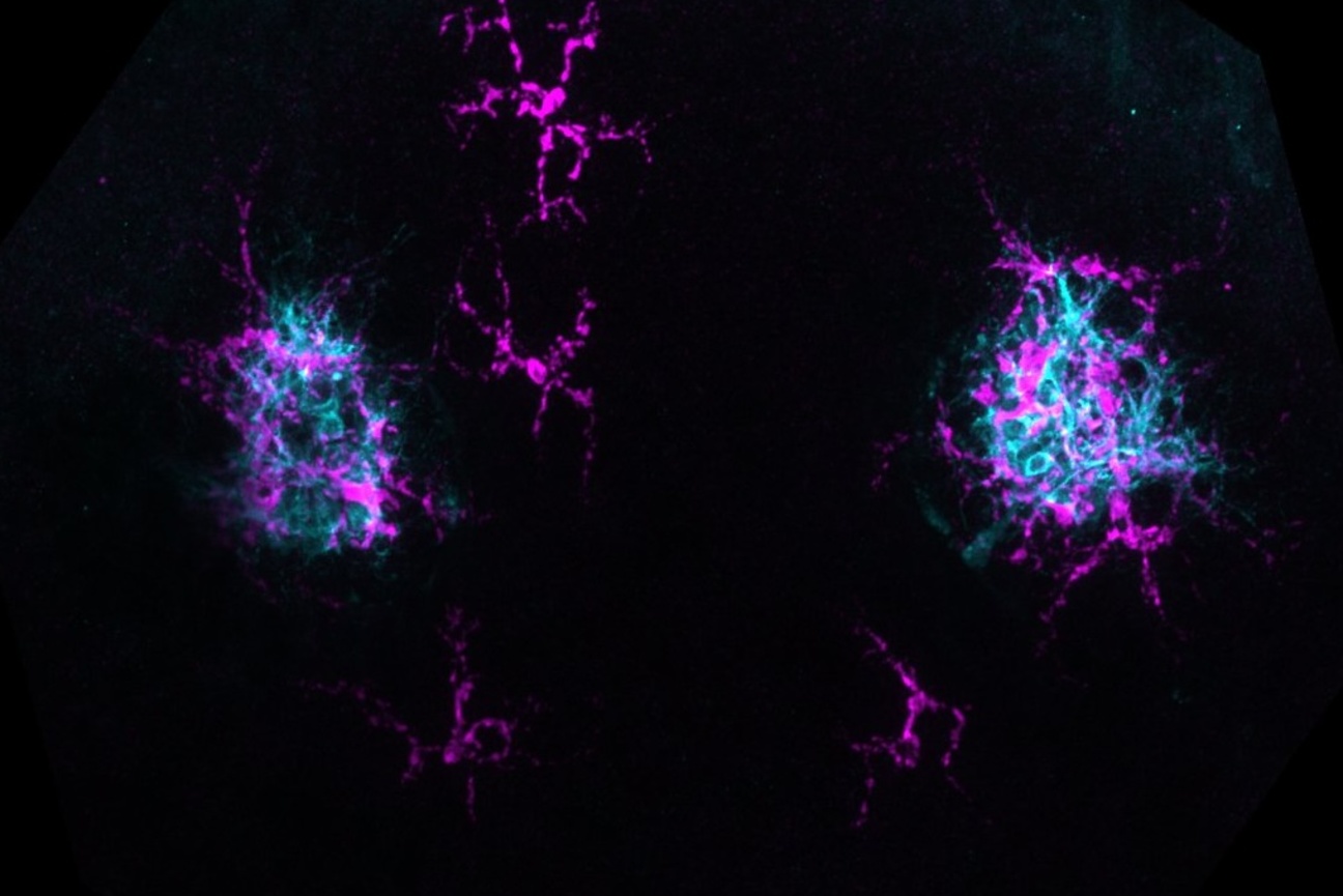
To confirm if the spermatids impacted were predominantly those that carried the Y chromosome, the researchers used an imaging technique called immunofluorescence staining, which uses antibodies to attach fluorescent molecules to a protein of interest, making it glow. They combined this with a technique called FISH (fluorescence in-situ hybridization), which tags the X and Y chromosomes with fluorescent markers, allowing researchers to distinguish between cells that will become X-bearing or Y-bearing following meiosis.
Indeed, the team found that while Ste protein is present in all spermatocytes before meiosis I, it unevenly divides between the two daughter cells—a phenomenon called asymmetric segregation—during meiosis I and gets concentrated in Y-bearing spermatids, eventually inducing DNA-packaging defects in these spermatids.
These findings clarified Ste’s role as a meiotic driver but the researchers still wondered why expression of Ste only led to a moderate sex ratio distortion. The answer soon became clear when they observed Ste undergo another round of asymmetric segregation during meiosis II. This meant that even if a secondary spermatocyte inherited Ste protein after meiosis I, only half of the spermatids produced in this round of cell division ended up retaining the protein. Hence, only half of the Y-bearing spermatids were going to be killed off.
“This self-limiting mechanism is the ultimate solution to the driver-suppressor separation problem,” says Yamashita. “But the idea is so unconventional that had it been proposed as just a theory, without the evidence we have now, it would’ve been completely dismissed.”
These findings have solved some questions and raised others: Unlike female meiosis, which is known to be asymmetrical, male meiosis has traditionally been considered symmetrical. Does the unequal segregation of Ste suggest there’s an unknown asymmetry in male meiosis? Do meiotic drivers like Ste trigger this asymmetry, or do they simply exploit it to limit their selfish drive?
Answering them is the next big step for Yamashita and her colleagues. “This could fundamentally change our understanding of male meiosis,” she says. “The best moments in science are when textbook knowledge is challenged and it turns out to have been tunnel vision.”