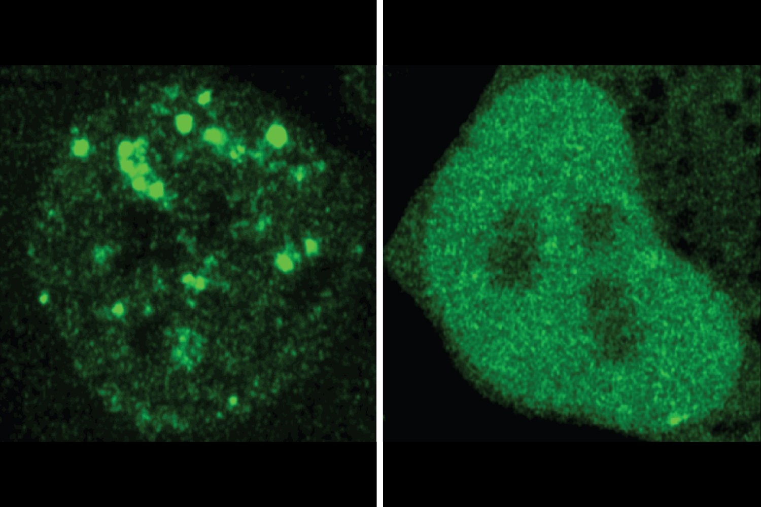
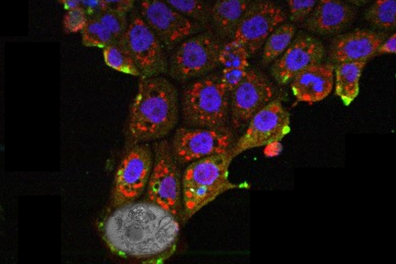
Bat cells possess a unique antiviral mechanism, preventing the SARS-CoV-2 virus from taking control

Bats have the amazing ability to coexist with viruses that are deadly to humans. New work from the Jaenisch Lab uncovers an antiviral mechanism that allows viruses to enter bat cells but prevents them from replicating.

Shafaq Zia | Whitehead Institute
October 14, 2024

Viruses are masters of stealth. From the moment a virus enters the host’s body, it begins hijacking its cells. First, the virus binds to a specific protein on the cell’s surface through a lock-and-key mechanism. This protein, known as a receptor, facilitates the entry of the virus’s genetic material into the cell. Once inside, this genetic code takes over the cell’s machinery, directing it to produce copies of the virus and assemble new viral particles, which can go on to infect other cells. Upon detecting the invasion, the host’s immune system responds by attacking infected cells in hopes of curbing the virus’s spread.

But in bats, this process unfolds differently. Despite carrying several viruses — Marburg, Ebola, Nipah, among others — bats rarely get sick from these infections. It seems their immune systems are highly specialized, allowing them to live with viruses that would typically be deadly in humans, without any clinical symptoms.

Since the onset of the COVID-19 pandemic, the lab of Whitehead Institute Founding Member Rudolf Jaenisch has been investigating the molecular basis of bats’ extraordinary resilience to viruses like SARS-CoV-2. In their latest study, published in the journal PNAS on Oct. 14 , Jaenisch lab postdoc Punam Bisht and colleagues have uncovered an antiviral mechanism in bat cells that allows viruses to enter the cells but prevents them from replicating their genome and completing the hijacking process.

“These cells have elevated expressions of antiviral genes that act immediately, neutralizing the virus before it can spread,” says Jaenisch, a professor of biology at the Massachusetts Institute of Technology. “What’s particularly interesting is that many of these antiviral genes have counterparts, or orthologs in humans.”

Striking a delicate balance

The innate immune system is the body’s first line of defense against foreign invaders like the SARS-CoV-2 virus. This built-in security system is always on alert, responding swiftly — within minutes to hours — to perceived threats.

Upon detecting danger, immune cells rush to the site of infection, where they target the virus with little precision in attempts to slow it down and buy time for the more specialized adaptive immune system to take over. During this process, these cells release small signaling proteins called cytokines, which coordinate the immune response by recruiting additional immune cells and directing them to the battleground.

If the innate immune response alone isn’t sufficient to defeat the virus, it signals the adaptive immune system for support. The adaptive immune system tailors its attacks to the exact pathogen it is fighting and can even keep records of past infections to launch a faster, more aggressive attack the next time it encounters the same pathogen.

But in some infections, the innate immune response can quickly spiral out of control before the adaptive immune response is activated. This phenomenon, called a cytokine storm, is a life-threatening condition characterized by the overproduction of cytokines. These proteins continue to signal the innate immune system for backup even when it’s not necessary, leading to a flood of immune cells at the site of infection, where they inadvertently begin damaging organs and healthy tissues.

Bats, on the other hand, are uniquely equipped to manage viral infections without triggering an overwhelming immune response or allowing the virus to take control. To understand how their innate immune system achieves this delicate balance, Bisht and her colleagues turned their attention to bat cells.

In this study, researchers compared how the SARS-CoV-2 virus replicates in human and bat stem cells and fibroblasts — a type of cell involved in the formation of connective tissue. While fibroblasts are not immune cells, they can secrete cytokines and guide immune response, particularly to help with tissue repair.

After exposing these cells to the SARS-CoV-2 virus for 48 hours, the researchers used a Green Fluorescent Protein (GFP) tag to track the virus’s activity. GFP is a fluorescent protein whose genetic code can be added as a tag to a gene of interest. This causes the products of that gene to glow, providing researchers with a visual marker of where and when the gene is expressed.

They observed that over 80% of control cells — derived from the kidneys of African green monkeys and known to be highly susceptible to SARS-CoV-2 — showed evidence of the virus replicating. In contrast, they did not detect any viral activity in human and bat stem cells or fibroblasts.

In fact, even after introducing the human ACE2 receptor — which SARS-CoV-2 uses to bind and enter cells — into bat cells, the infected bat fibroblasts were able to replicate viral RNA and produce viral proteins, but at much lower levels compared to infected human fibroblasts.

These bat fibroblasts, however, could not assemble these viral proteins into fully infectious virus particles, suggesting an abortive infection, where the virus is able to initiate replication but fails to complete the process and produce progeny viruses.

Using electron microscopy to look inside bat and human cells, they began to understand why: in human cells, SARS-CoV-2 had created special structures called double-membrane vesicles (DMV). These vesicles acted like a bubble, shielding the viral genome from detection and providing it safe space to replicate more effectively. However, these “viral replication factories” were absent in bat fibroblasts.

When the researchers examined the gene expression profiles of these bat fibroblasts and compared them those of infected human cells, they found that although both human and bat cells have genes regulating the release of a type of cytokine called interferons, these genes are already turned on in bat fibroblasts — unlike in human cells — even before virus infection occurs.

These findings suggest that bat cells are in a constant state of vigilance. This allows their innate immune system to stop the SARS-CoV-2 virus in its tracks early on in the replication process before it can entirely hijack cellular machinery.

Surprisingly, this antiviral mechanism does not protect bat cells against all viruses. When the researchers infected bat fibroblasts with Zika virus, the virus was able to replicate and produce new viral particles.

“This means there are still many questions unanswered about how bat cells resist infection,” says Bisht. “COVID-19 continues to circulate, and the virus is evolving quickly. Filling in these gaps in our knowledge will help us develop better vaccines and antiviral strategies.”
The researchers are now focused on identifying the specific genes involved in this antiviral mechanism, and exploring how they interact with the virus during infection.

Brain cell types are affected differently by Rett Syndrome mutation

New research from Jaenisch Lab postdoc Danielle Tomasello focuses on an understudied question: how Rett Syndrome affects cell types in the human brain other than neurons.

Greta Friar | Whitehead Institute
September 6, 2024

Rett Syndrome is a X-chromosome-linked neurodevelopmental disorder; it can lead to loss of coordination, mobility, ability to speak, and use of the hands, among other symptoms. The syndrome is typically caused by mutations within the gene MECP2. Researchers in Whitehead Institute Founding Member Rudolf Jaenisch’s lab have studied Rett Syndrome for many years in order to understand the biological mechanisms that cause disease symptoms, and to identify possible avenues for treatments or a cure. Jaenisch and colleagues have gained many insights into the biology of Rett syndrome and developed tools that can rescue neurons from Rett syndrome symptoms in lab models.

However, much about the biology of Rett Syndrome remains unknown. New research from Jaenisch and postdoc in his lab Danielle Tomasello focuses on an understudied question: how Rett Syndrome affects cell types in the human brain other than neurons. Specifically, Tomasello investigated the effects of Rett Syndrome on astrocytes, a type of brain cell that supports and provides energy for neurons. The work, shared in the journal Scientific Reports on September 6, details changes that occur in Rett syndrome astrocytes, in particular in relation to their mitochondria, and shows how these changes directly impact neurons. The findings provide a new framework for thinking about Rett Syndrome and possible new avenues for therapies.

“By considering Rett Syndrome from a different perspective, this project expands our understanding of a multifaceted and thus far incurable disease,” says Jaenisch, who is also a professor of biology at the Massachusetts Institute of Technology.

Energy metabolism in Rett Syndrome

Mitochondria are organelles that generate energy, which cells use to carry out their functions, and mitochondrial dysfunction was known to occur in Rett Syndrome. Jaenisch and Tomasello found that mitochondria in astrocytes are particularly affected, even more so than mitochondria in neurons. Tomasello grew human stem-cell-derived astrocytes in 2D cultures and also grew 3D organoids: mini brain-like tissues that contain multiple cell types growing in a structure that resembles actual brain anatomy. This approach allowed Tomasello to use human cells, rather than an animal model, and to study how cells behave within a brain-like environment.

When the researchers observed Rett astrocytes grown in these conditions, they found that the mitochondria were misshapen: short, small circles instead of large, long ovals. Additional studies showed evidence of the mitochondria experiencing stress and not being able to generate enough energy through their usual processes. The mitochondria did not have enough of the typical proteins they use to make energy, and so began to break down the cell’s supply of the building blocks of proteins, amino acids, for parts to make up for the missing material. Additionally, the researchers observed an increase in reactive oxygen species, byproducts of mitochondrial metabolism that are toxic to the cell.

Further experiments suggested that the cells try to compensate for this mitochondrial stress by increasing transcription of mitochondrial genes. For example, Tomasello found that regions of DNA called promoters that can increase expression of key mitochondrial genes were more open for the cell to use in Rett astrocytes. Altogether, these findings paint a picture of severe mitochondrial dysfunction in Rett astrocytes.

Although mitochondria in Rett neurons did not have such severe defects, astrocytes and neurons have a close relationship. Not only do neurons rely on astrocytes to supply them with energy, they even accept mitochondria from astrocytes to use for themselves. Jaenisch and Tomasello found that neurons take up dysfunctional mitochondria from Rett astrocytes at a higher rate than they take up mitochondria from unaffected astrocytes. This means that the effects of Rett syndrome on astrocytes have a direct effect on neurons: the dysfunctional mitochondria from the astrocytes end up in the neurons, where they cause damage. Tomasello took mitochondria from Rett astrocytes and placed them on both healthy and Rett neurons. In either case, the neurons took up the dysfunctional mitochondria in large numbers and then experienced significant problems. The neurons entered a hyperexcitable state that is ultimately toxic to the brain. The neurons also contained higher levels of reactive oxygen species, the toxic byproducts of mitochondrial metabolism, which can cause widespread damage. These effects occurred even in otherwise healthy neurons that did not themselves contain a Rett-causing MECP2 mutation.

“This shows that in order to understand Rett Syndrome, we need to look beyond what’s happening in neurons to other cell types,” Tomasello says.

Learning about the role that astrocytes play in Rett Syndrome could provide new avenues for therapies. The researchers found that supplying affected astrocytes with healthy mitochondria helped them to recover normal mitochondrial function. This suggests to Tomasello that one possibility for future Rett Syndrome therapies could be something that either targets mitochondria, or supplies additional mitochondria through the bloodstream.

Together, these insights and their possible medical implications demonstrate the importance of taking a broader look at the foundational biology underlying a disease.

Whitehead Institute researchers uncover a new clue toward understanding the molecular basis of Parkinson’s disease

In Parkinson's disease, a mutation that causes protein misfolding can also turn the brain’s immune cells from friends to foes, possibly accelerating the progression of the disease. New Research from the Jaenisch Lab aims to uncover mechanisms that go awry in the brain, which may inform the development of new therapies that can halt or even reverse the progression of neurological conditions such as Parkinson's.

Shafaq Zia | Whitehead Institute
August 29, 2024

Dopamine is more than the “rush molecule”. This chemical messenger, produced by neurons in the midbrain, acts as a traffic controller that regulates the flow of electrical signals between neurons, assisting with brain functions like cognition, attention, movement, and behavior. But, in instances of Parkinson’s disease (PD), a progressive brain disorder, dopamine-producing neurons begin to die at an unprecedented rate, leading to dwindling levels of this vital chemical and impaired neural communication.

The lab of Whitehead Institute’s Founding Member Rudolf Jaenisch studies genetic and epigenetic factors — changes in gene expression that control which genes are turned on and off, and to what extent, without altering the DNA sequence itself — underlying neurological disorders like PD, Alzheimer’s disease, and Rett Syndrome. Their work aims to uncover the mechanisms that go awry in the brain, which may inform the development of new therapies that can halt or even reverse the progression of these conditions.

In their latest work, Jaenisch and former postdoctoral associate Marine Krzisch examine how a mutation in the gene that encodes for alpha-synuclein, a protein regulating the release of dopamine, affects the resident immune cells of the brain called microglia. The researchers’ detailed findings, published in the journal Biological Psychiatry on August 29, reveal that the mutation renders microglia extremely sensitive, worsening the problem of inflammation in the brain and potentially exacerbating damage to neurons in Parkinson’s disease.

“In fact, even when these mutant microglia are transplanted into a healthy, young brain, they have heightened activation upon stimulation, and low levels of the protective antioxidant catalase,” Krzisch says. “This tells us that in Familial Parkinson’s disease, which is due to genetic mutations, these microglia may be playing an important role in neuron degeneration.”

When nature’s origami falters

The human body is home to tens of thousands of unique proteins, each essential for processes sustaining life. These proteins are composed of linear chains of smaller building blocks called amino acids that are linked together in a specific sequence. For the proteins to perform their functions, the amino acid chains must crumple, rotate, and twist into stable three-dimensional structures. The stakes are high — just as precise folds and creases are crucial to the art of origami, even minor errors in the protein folding process can result in dysfunctional proteins that contribute to disease.

To date, scientists have identified over 20 causative genes in which mutations can result in Familial Parkinson’s disease, a rare, genetically inherited form of PD affecting individuals under or around the age of 50. Among them is SNCA, which encodes for alpha-synuclein, a small protein abundant in dopamine-producing neurons.

The A53T mutation in SNCA promotes the formation of dysfunctional alpha-synuclein proteins that clump together — almost like a ball of yarn — within dopamine-producing neurons. The accumulation of these protein clumps, also known as Lewy bodies, triggers inflammatory signaling in the brain, eventually killing the affected neurons. However, prior research has also shown that the A53T mutation accelerates the progression of PD, or the rate at which neurons die, although the full molecular mechanisms underlying this process are not yet fully understood.

To uncover pathways involved in this progression, researchers in the Jaenisch Lab turned their attention to star-shaped patrollers called microglia that protect the brain from foreign invaders and respond to injuries, including protein aggregates within neurons. This immune response includes activated microglia trying to clear out Lewy bodies by digesting them, recruiting additional immune cells to the site of neurons with protein aggregates, and even killing off diseased neurons to limit damage to the brain.

But these friends can quickly turn to foes. Over-activated microglia can also degrade healthy neurons in the brain, prompting Jaenisch, Krzisch, and colleagues to investigate if excessive microglia activation is one pathway that contributes to progression in PD.

Microglia go rogue

To explore how the A53T mutation in the SNCA gene affects microglia function in PD, scientists at the Jaenisch Lab began by growing human myeloid precursors — the cells that eventually develop into microglia — in lab culture and transplanting them into the brains of immune-deprived mice.

Given the complexity of the brain, it’s common for researchers to study brain cells in the Petri dish. “But in cell cultures, microglia do not have the same morphology [form] as in the brain, show signs of chronic activation, and they don’t survive for a very long time,” says Krzisch. “When we transplant them in mice, the precursors differentiate into microglia that look and function like those in the human brain, and survive for the mouse’s lifespan.”

Using this method, the researchers compared the gene expression profiles of A53T-mutant microglia with those that did not carry the mutation, revealing differences in pathways linked to inflammation, microglia activation, and DNA repair. Additionally, when A53T-mutant microglia were exposed to an immune activator called lipopolysaccharide, they exhibited a heightened inflammatory response compared to non-mutant microglia.

In fact, even in non-inflammatory conditions, A53T-mutant microglia had decreased expression of catalase, an enzyme that helps break down harmful reactive oxygen species produced in response to protein aggregates in PD.

Understanding the molecular basis of progression in PD is challenging, which explains why there are currently no drugs to alter the disease’s course. With these findings in hand, researchers at the Jaenisch Lab are now eager to explore how factors like aging also influence microglia function and contribute to an increased rate of progression in PD.

“Overactivation of microglia isn’t the only cause of neuron death in Parkinson’s,” says Jaenisch. “But if we can decrease their activation, it will help us get to the point where we can slow down or actually stop the disease.”


Study reveals the benefits and downside of fasting

Fasting helps intestinal stem cells regenerate and heal injuries but also leads to a higher risk of cancer in mice, MIT researchers report.

Anne Trafton | MIT News
August 21, 2024

Low-calorie diets and intermittent fasting have been shown to have numerous health benefits: They can delay the onset of some age-related diseases and lengthen lifespan, not only in humans but many other organisms.

Many complex mechanisms underlie this phenomenon. Previous work from MIT has shown that one way fasting exerts its beneficial effects is by boosting the regenerative abilities of intestinal stem cells, which helps the intestine recover from injuries or inflammation.

In a study of mice, MIT researchers have now identified the pathway that enables this enhanced regeneration, which is activated once the mice begin “refeeding” after the fast. They also found a downside to this regeneration: When cancerous mutations occurred during the regenerative period, the mice were more likely to develop early-stage intestinal tumors.

“Having more stem cell activity is good for regeneration, but too much of a good thing over time can have less favorable consequences,” says Omer Yilmaz, an MIT associate professor of biology, a member of MIT’s Koch Institute for Integrative Cancer Research, and the senior author of the new study.

Yilmaz adds that further studies are needed before forming any conclusion as to whether fasting has a similar effect in humans.

“We still have a lot to learn, but it is interesting that being in either the state of fasting or refeeding when exposure to mutagen occurs can have a profound impact on the likelihood of developing a cancer in these well-defined mouse models,” he says.

MIT postdocs Shinya Imada and Saleh Khawaled are the lead authors of the paper, which appears today in Nature.

Driving regeneration

For several years, Yilmaz’s lab has been investigating how fasting and low-calorie diets affect intestinal health. In a 2018 study, his team reported that during a fast, intestinal stem cells begin to use lipids as an energy source, instead of carbohydrates. They also showed that fasting led to a significant boost in stem cells’ regenerative ability.

However, unanswered questions remained: How does fasting trigger this boost in regenerative ability, and when does the regeneration begin?

“Since that paper, we’ve really been focused on understanding what is it about fasting that drives regeneration,” Yilmaz says. “Is it fasting itself that’s driving regeneration, or eating after the fast?”

In their new study, the researchers found that stem cell regeneration is suppressed during fasting but then surges during the refeeding period. The researchers followed three groups of mice — one that fasted for 24 hours, another one that fasted for 24 hours and then was allowed to eat whatever they wanted during a 24-hour refeeding period, and a control group that ate whatever they wanted throughout the experiment.

The researchers analyzed intestinal stem cells’ ability to proliferate at different time points and found that the stem cells showed the highest levels of proliferation at the end of the 24-hour refeeding period. These cells were also more proliferative than intestinal stem cells from mice that had not fasted at all.

“We think that fasting and refeeding represent two distinct states,” Imada says. “In the fasted state, the ability of cells to use lipids and fatty acids as an energy source enables them to survive when nutrients are low. And then it’s the postfast refeeding state that really drives the regeneration. When nutrients become available, these stem cells and progenitor cells activate programs that enable them to build cellular mass and repopulate the intestinal lining.”

Further studies revealed that these cells activate a cellular signaling pathway known as mTOR, which is involved in cell growth and metabolism. One of mTOR’s roles is to regulate the translation of messenger RNA into protein, so when it’s activated, cells produce more protein. This protein synthesis is essential for stem cells to proliferate.

The researchers showed that mTOR activation in these stem cells also led to production of large quantities of polyamines — small molecules that help cells to grow and divide.

“In the refed state, you’ve got more proliferation, and you need to build cellular mass. That requires more protein, to build new cells, and those stem cells go on to build more differentiated cells or specialized intestinal cell types that line the intestine,” Khawaled says.

Too much of a good thing

The researchers also found that when stem cells are in this highly regenerative state, they are more prone to become cancerous. Intestinal stem cells are among the most actively dividing cells in the body, as they help the lining of the intestine completely turn over every five to 10 days. Because they divide so frequently, these stem cells are the most common source of precancerous cells in the intestine.

In this study, the researchers discovered that if they turned on a cancer-causing gene in the mice during the refeeding stage, they were much more likely to develop precancerous polyps than if the gene was turned on during the fasting state. Cancer-linked mutations that occurred during the refeeding state were also much more likely to produce polyps than mutations that occurred in mice that did not undergo the cycle of fasting and refeeding.

“I want to emphasize that this was all done in mice, using very well-defined cancer mutations. In humans it’s going to be a much more complex state,” Yilmaz says. “But it does lead us to the following notion: Fasting is very healthy, but if you’re unlucky and you’re refeeding after a fasting, and you get exposed to a mutagen, like a charred steak or something, you might actually be increasing your chances of developing a lesion that can go on to give rise to cancer.”

Yilmaz also noted that the regenerative benefits of fasting could be significant for people who undergo radiation treatment, which can damage the intestinal lining, or other types of intestinal injury. His lab is now studying whether polyamine supplements could help to stimulate this kind of regeneration, without the need to fast.

“This fascinating study provides insights into the complex interplay between food consumption, stem cell biology, and cancer risk,” says Ophir Klein, a professor of medicine at the University of California at San Francisco and Cedars-Sinai Medical Center, who was not involved in the study. “Their work lays a foundation for testing polyamines as compounds that may augment intestinal repair after injuries, and it suggests that careful consideration is needed when planning diet-based strategies for regeneration to avoid increasing cancer risk.”

The research was funded, in part, by a Pew-Stewart Trust Scholar award, the Marble Center for Cancer Nanomedicine, the Koch Institute-Dana Farber/Harvard Cancer Center Bridge Project, and the MIT Stem Cell Initiative.

New approach enables a closer look at brain cell organelle

Microglia are involved in brain development, as well as neurodegeneration and brain cancer. A new approach from the Jaenisch Lab allows researchers to isolate and analyze microglia phagosomes.

Greta Friar | Whitehead Institute
August 14, 2024

Microglia are the immune system’s front-line enforcers in the brain. They are cells that patrol the brain and destroy anything harmful that they encounter, from invading bacteria to cellular debris. They also remove plaques and prune dysfunctional synapses between neurons. Microglia eliminate their targets by eating them: they envelope material and seal it in bubble-like organelles called phagosomes. A phagosome can then fuse with other organelles that break down its contents.

Microglial phagosomes play important roles in brain development, brain function and a plethora of brain diseases, including neurodegeneration and brain cancer. Therefore, understanding microglial phagosome biology could help to develop new therapies for currently untreatable brain diseases. However, microglia and their organelles have been difficult to study because existing stem cell and animal models insufficiently resemble microglia in the human brain, and because microglia, as vigilant immune patrollers, react to even subtle stimuli and so experimental conditions can trigger changes in the cells that confound analyses.

To overcome those issues, Whitehead Institute Founding Member Rudolf Jaenisch, also a professor of biology at the Massachusetts Institute of Technology; University of Freiburg Professor of Neuropathology Marco Prinz; and University of Freiburg neuropathologist Emile Wogram, who began this project as a postdoctoral researcher in Jaenisch’s lab, have developed a method to isolate and analyze microglia phagosomes in a rapid, gentle, and unbiased fashion.

In research shared in the journal Immunity on August 15, the researchers describe how they can isolate and profile phagosomes from stem cell-derived microglia and fresh human brain tissue. They also share new insights into phagosome biology in the human brain, regarding synaptic pruning and generation of NAD+, a broadly used molecule in the brain, by microglia.

The method that the researchers developed to isolate phagosomes from cells uses immunoprecipitation, in which antibodies latch on to a specific target protein on an organelle’s surface. When the antibodies are collected, they pull the organelles with them. This technique avoids many chemical perturbations that might alter the microglial profile. Sometimes researchers genetically engineer a target for the antibodies, but in order to isolate phagosomes from human brain tissue, Wogram had to find a naturally expressed target. Eventually, he and colleagues found one: the protein CD68.

The researchers first isolated phagosomes from stem cell-derived microglia. They co-cultured the microglia with other brain cell types to create a more brain-like environment, which led to a better match between brain and stem cell-derived microglia gene expression. They triggered some of the microglia to enter an inflammatory or disease-like state to see how that affected the phagosomes. Additionally, Wogram collaborated with the neurosurgery department at the University of Freiburg to get access to brain tissues immediately after their removal during surgery. He isolated phagosomes from brain tissue within a half hour of its removal, allowing him to profile the organelles before their contents could change much.

The profiles that the researchers built included what proteins and metabolites the phagosomes contained, and the whole-cell gene expression profile. The profiles differed significantly between sets of phagosomes, but the researchers identified a core of consistent proteins, including many known and also some unknown phagosome proteins. The results showed that phagosomes contain sensitive signaling molecules that allow them to react quickly to even subtle environmental stimuli.

Additionally, the protein contents of the co-cultured microglia provided strong evidence that when microglia prune synapses, they predominantly prune the side that sends a signal and not the side that receives one. This insight could be useful for understanding how microglia interact with synapses in health and disease.

The researchers also gained insights into a key metabolic pathway that occurs inside of microglia. In excess, the molecule quinolinic acid can be toxic to neurons; it is implicated as involved in many neurodegenerative diseases. However, cells can use quinolinic acid to make NAD+, a molecule broadly used to carry out essential cellular functions. Microglia are the only brain cells that generate NAD+. Wogram and colleagues found that key steps in this process occur in phagosomes. Phagosomes are therefore necessary both for removing excess quinolinic acid to prevent toxicity and for helping to generate NAD+ in the brain.

Finally, Wogram used brain tissues to compare phagosomes from within a tumor to those in the surrounding healthy tissue. The phagosomes in the tumor contained excess quinolinic acid. Although follow-up studies would be needed to confirm the results, these findings are consistent with research that suggests cancer cells use quinolinic acid to fuel their growth.

Collectively, these findings illuminate aspects of phagosome biology and the roles that phagosomes may play in normal brain development and maintenance, as well as in cancer and neurodegeneration. The researchers also anticipate that their method could prove useful for profiling other organelles, especially when the organelles need to be rapidly isolated from human tissue.

In immune cells, X marks the spot(s)

By researching the effects of sex chromosomes on two types of immune cells, researchers in the Page Lab explore the biological underpinnings of sex biases in immunity and autoimmune disease

Greta Friar | Whitehead Institute
August 6, 2024

There are many known sex differences in health and disease: cases in which either men or women are more likely to get a disease, experience a symptom, or have a certain drug side effect. Some of these sex differences are caused by social and environmental factors: for example, when men smoked more than women, men were more likely to develop lung cancer. However, some have biological underpinnings. For example, men are more likely to be red-green colorblind because the relevant gene is on the X chromosome, of which men with XY chromosomes have no backup copy for a dysfunctional version.

Often, the specific factors contributing to a sex difference are hard to tease apart; there may not be a simple way to tell what is caused by sex chromosomes versus sex hormones versus environment. To address this question, researchers in Whitehead Institute Member David Page’s lab previously developed an approach to identify the contributions of the sex chromosomes to sex differences. Now, Page and former postdoc in his lab Laura Blanton have built on that work by measuring the effects of the sex chromosomes on two types of immune cells. The work, published in the journal Cell Genomics on August 6, shows that sex chromosome gene expression is consistent across cell types, but that its effects are cell type specific.

Sex differences are common in the function and dysfunction of our immune system. Examples include the typically weaker male immune response to pathogens and vaccines, and the female-biased frequency of autoimmune diseases. Page and Blanton’s work in immune cells examines several genes that have been implicated in such sex differences.

Developing a method to measure sex chromosome influence

The approach that the researchers used is based on several facts about sex chromosomes. Firstly, although females typically have two X chromosomes and males typically have one X and one Y, there are people with rare combinations of sex chromosomes, who have anywhere from 1-5 X chromosomes and 0-4 Y chromosomes. Secondly, there are two types of X chromosome: The active X chromosome (Xa) and the inactive X chromosome (Xi). They are genetically identical, but many of the genes on Xi are either switched off or have their expression level dialed way down.

Xa does not really function as a sex chromosome since everyone in the world has exactly one Xa regardless of their sex. In people with more than one X chromosome, any additional X chromosomes are always Xi. Furthermore, Page and Blanton’s research demonstrates that Xa responds to gene expression by Xi and Y—the sex chromosomes—in the same manner as do the other 22 pairs of non-sex chromosomes—the autosomes.

With these facts in mind, the researchers collected cells from donors with different combinations of sex chromosomes. Then they measured the expression of every gene in these cells, across the donor population, and observed how the expression of each gene changed with the addition of each Xi or Y chromosome.

This approach was first shared in a Cell Genomics paper by Page and former postdoc Adrianna San Roman in 2023. They had cultured two types of cells, fibroblasts and lymphoblastoid cell lines, from donor tissue samples. They found that the effects of Xi and Y were modular—each additional chromosome changed gene expression by about the same amount. This approach allowed the researchers to identify which genes are sensitive to regulation by the sex chromosomes, and to measure the strength of the effect for each responsive gene.

In that and a following paper, Page and San Roman looked at how Xi and Y affect gene expression from Xa and the autosomes. Blanton expanded the study of Xi and Y by using the same approach in two types of immune cells, monocytes and CD4+ T cells, taken directly from donors’ blood. Studying cells taken directly from the body, rather than cells cultured in the lab, enabled the researchers to confirm that their observations applied in both conditions.

In all three papers, the researchers found that the sex chromosomes have significant effects on the expression levels of many genes that are active throughout the body. They also identified a particular pair of genes as driving much of this effect in all four cell types. The genes, ZFX and ZFY, found on the X and Y chromosomes respectively, are transcription factors that can dial up the expression of other genes. The pair originates from the same ancestral gene, and although they have grown slightly apart since the X and Y chromosomes diverged, they still perform the same gene regulatory function. The researchers found that they tended to affect expression of the same gene targets by similar though not identical amounts.

In other words, the presence of either sex chromosome causes roughly the same effect on expression of autosomal and Xa genes. This similarity makes sense: carefully calibrated gene regulation is necessary in every body, and so each sex chromosome must maintain that function. It does, however, make it harder to spot the cases in which sex chromosomes contribute to sex differences in health and disease.

“Sex differences in health and disease could stem from the rare instances in which one gene responds very differently to Xi versus Y—we found cases where that occurs,” Blanton says. “They could also stem from subtle differences in the gene expression changes caused by Xi and Y that build up into larger effects downstream.”

Blanton then combined her and San Roman’s data in order to look at how the effects of sex chromosome dosage—how many Xs or Ys are in a cell—compared across all four cell types.

The effects of sex chromosomes on immune cells

 Blanton found that gene expression from the sex chromosomes was consistent across all four cell types. The exceptions to this rule were always X chromosome genes that are only expressed on Xa, and so could be regulated by Xi and Y in the way that autosomal genes are. This contrasts with speculation that different genes on Xi might be silenced in different cells.

However, each cell type had a distinct response to this identical sex chromosome gene expression. Different biological pathways were affected, or the same biological pathway could be affected in the opposite direction. Key immune cell processes affected by sex chromosome dosage in either monocytes or T cells included production of immune system proteins, signaling, and inflammatory response.

The cell type specific responses were due to different genes responding to the sex chromosomes in each cell type. The researchers do not yet know the mechanism causing the same gene to respond to sex chromosome dosage in one cell type but not another. One possibility is that access to the genes is blocked in some of the cell types. Regions of DNA can become tightly packed so that a gene, or a DNA region that regulates the gene, becomes inaccessible to transcription factors such as ZFX and ZFY, and so they cannot affect the gene’s expression. Another possibility is that the genes might require specific partner molecules in order for their expression level to increase, and that these partners may be present in one cell type but not the other.

Blanton also measured how X chromosome dosage affected T cells in their inactive state, when there is no perceived immune threat, versus their activated state, when they begin to produce an immune response and replicate themselves. Increases in X chromosome dosage led to heightened activation, with increased expression of genes related to proliferation. This finding highlights the importance of looking at how sex chromosomes affect not just different cell types, but cells in different states or scenarios.

“As we learn what pathways the sex chromosomes influence in each cell type, we can begin to make sense of the contributions of the sex chromosomes to each cell type’s functions and its roles in disease,” Blanton says.

Although Page and Blanton found that the presence of an Xi or Y chromosome had very similar effects on most genes, the researchers did identify one interesting case in which response to X and Y differed. FCG2RB is a gene involved in immunity that has been implicated in and thought to contribute to the female bias in developing systemic lupus erythematosus (SLE). Blanton found that unlike most genes, FCGR2B is sensitive to X and not Y chromosome dosage. This strengthens the case that higher expression of FCGR2B could be driving the SLE female bias.

FCGR2B provides a promising opportunity to study the contributions of the sex chromosomes to a sex bias in disease, and to learn more about the biology of a chronic disease that affects many people around the world,” Page says.

In other cases, the researchers found that genes which have been suspected to contribute to female bias in disease did not have a strong response to X chromosome dosage. For example, TLR7 is thought to contribute to female bias in developing autoimmunity, and CD40LG is thought to contribute to female bias in developing lupus. Neither of the genes showed increased expression as X chromosome dosage increased. This suggests that other mechanisms may be driving the sex bias in these cases.

Because of the limited pool of donors, the researchers were not able to identify every gene that responds to sex chromosome dosage, and future research may uncover more sex-chromosome-sensitive genes of interest. Meanwhile, the Page lab continues to investigate the sex chromosomes’ shared role as regulators of gene expression throughout the body.

“We’ve got to recalibrate our thinking from the view that X and Y are mainly involved in differentiating males and females, to understanding that they also have largely shared functions that are important throughout the body,” Page says. “At the same time, I think that uncovering the biology of Xi is going to be incredibly important for understanding women’s health and sex differences in health and disease.”

New findings activate a better understanding of Rett syndrome’s causes

Rett syndrome is caused by mutations to the gene MECP2, which is highly expressed in the brain and appears to play important roles in maintaining healthy neurons. Researchers led by Rudolf Jaenisch have used cutting-edge techniques to create an epigenome map of MECP2, which may help guide future research on the disease.

Greta Friar | Whitehead Institute
April 25, 2024
Ragon faculty finds intricate functions of Resident Tissue Macrophages (RTM’s) extend beyond immune defense

The lab of Ragon Institute faculty @hernandezmsilva published a review in Science Immunology regarding resident tissue macrophages (RTMs), shedding light on these cells’ multifaceted roles.

April 15, 2024
Uncovering answers to longstanding questions about sex differences in autoimmune and neurodegenerative diseases

Whitehead Institute researchers including those in the Page Lab and Corradin Lab are investigating the role of X and Y chromosomes beyond sex determination, paying close attention to conditions that mostly — or distinctly — affect females, and mentoring the next generation of researchers to challenge the status quo for a better world.

Shafaq Zia | Whitehead Institute
March 12, 2024
How phase separation is revolutionizing biology

Postdocs from Whitehead Institute Member Richard A. Young's lab found that imaging and molecular manipulation reveal how biomolecular condensates form and offer clues to the role of phase separation in health and disease.

February 27, 2024