Whether seeking a career change or rediscovering intellectual pursuits, learners worldwide turn to <i>MITx</i> courses.
Kate Stringer | MIT Open Learning
Despite the extraordinary pressures of adapting to the realities of the Covid-19 pandemic, learners have increasingly sought out MITx courses as a way to stay intellectually active, work toward longstanding goals, and affect change in themselves and in the world around them. MITx courses have seen over 500,000 enrollments since the start of the pandemic.
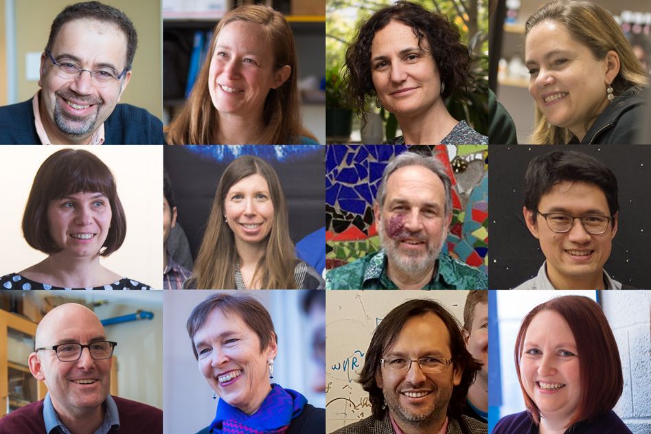
“It’s been humbling to witness the role our courses have played in learners’ lives these past few months,” says Dana Doyle, director of the MITx Program. “The number of people who are using their time at home to learn something new or make a change in their lives is inspiring.”
MITx instructors and staff have heard from learners from over a dozen countries across the globe, sharing their experiences during the pandemic. Some have used MITx courses to rediscover subjects they had once been passionate about; some are leveraging a career change; still others hope to pass on new knowledge to the next generation. The following represent just a few of their stories.
Between careers and countries
Paula Unger was just finishing up an internship in Peru when Covid-19 hit. “The first case was discovered in March, and the lockdown began eight days later,” she recalls. Unger, who recently received her degree in agricultural studies from the University of Bonn, had spent several months analyzing DNA sequencing data at the International Potato Center in Lima.
A Peruvian national, Unger had planned to return to her home in Aachen, Germany immediately following the internship to begin looking for jobs. Instead, she sheltered in place with her family in Lima, where lockdown was strictly enforced. “You could not even go outside for a walk, it’s totally prohibited,” she says.
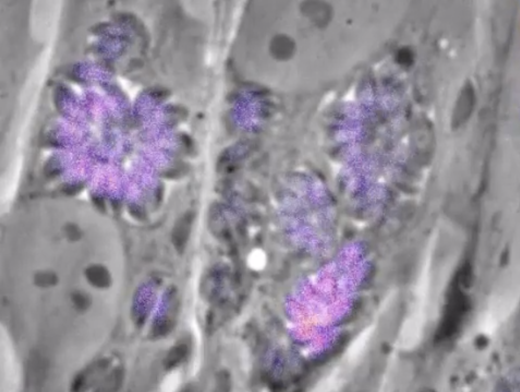
Unable to leave the house, Unger turned to a project she’d been putting off for some time: taking Professor Eric Lander’s Introduction to Biology MITx course. Though she earned her degree in a science-based field, Unger had spent a few years moving between majors and universities across Germany, and felt that a stronger background in biology would help her career. She didn’t count on how much she would enjoy the course for its own sake.
“I’m mind-blown by how well the course is made,” she says, citing Lander’s engaging lectures and the course’s challenging, interactive problems sets as particularly valuable. “A lot of universities should learn to create courses that are as well-conceived pedagogically as these are.” Thanks to her MITx learning journey, Unger felt she was able to keep moving forward even while stuck in one place: “I could keep growing as a person, even though my life had been put on hold.”
Happily, Unger’s life and career were able to resume sooner than expected. Not long into lockdown, the Max Planck Institute for Plant Breeding Research in Cologne contacted her about a position, conducted an e-interview, and hired her with the promise that they would wait for her until she could return to Germany.
Now back in Aachen, Unger has started her new job, but has no plans to abandon her learning journey. She enrolled in the MITx Quantitative Biology Workshop, and plans eventually to return to school to complete a master’s degree. “I wish more people would realize the potential of what’s possible through online learning,” she says.
Between flights, Australian pilot learns to engineer spacecraft
When he’s not flying U.S. and Australian citizens back to their home countries as part of pandemic-related repatriation efforts, Sydney-based pilot Andrew Wangler necessarily has a lot of time on his hands.
While Wangler’s company maintains the “minimum viable international network” of flights, he’s been on and off furlough throughout the pandemic. When called up, he commutes 10 hours to Melbourne International Airport before flying to San Francisco or Los Angeles to drop off American nationals and pick up returning Australians.
Wangler joined Qantas after a 15-year career in the Royal Australian Air Force. He graduated from the Australian Defence Force Academy with a double major in mathematics and political science, and minors in physics and computer science, before completing an MBA; it’s safe to say that he loves to learn. So when he found himself stuck in a pattern of self-isolation at home and in hotels before and after each flight, Wangler was thrilled to find MITx courses that helped him rediscover yet another academic passion: spaceflight.
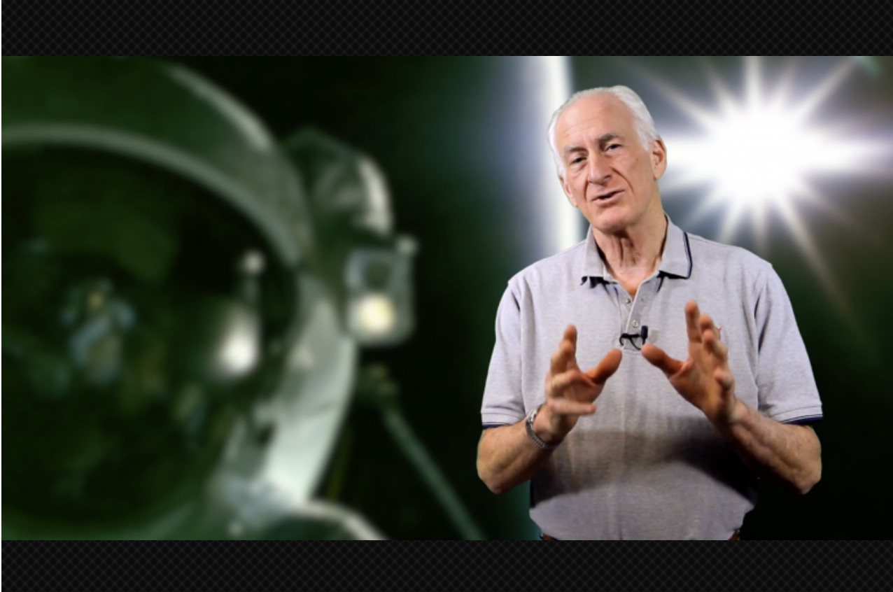
“Professor Hoffmann’s passion for the subject material and teaching style are very infectious and engaging,” says Wangler. Finding Hoffman’s Introduction to Aerospace Engineering course brought back fond memories of his interest in the subject as an undergraduate. These days, Wangler hopes to channel his own enthusiasm and what he’s learned from MITx to help his 12 year-old son, “hell-bent on being an engineer,” to find the right learning resources.
“As my son gets older, it will be helpful to have the engineering background, just to open his eyes and point him in the right direction,” says Wangler. Last year, father and son visited the Boeing facilities near Seattle as well as the Museum of Flight, including a session in the Space Shuttle Crew Trainer. They are planning more educational trips in the future, including Houston, Texas and Cape Canaveral, Florida.
In the meantime, Wangler’s enthusiasm for his online learning journey shows no signs of abating: while preparing for another flight to LAX, he emails, “I am actually enjoying Professor Hoffman’s archived course on Engineering the Space Shuttle as we speak!”
Under lockdown in Madrid, retiree rediscovers a love of physics
Miguel Doñate has witnessed the effects of Covid-19 more directly than many. Under strict lockdown since March 15 in Madrid, Spain, Doñate is surrounded by reminders of the pandemic’s worst outcomes.
“We have been in a very difficult situation here, with a lot of deaths, including people I know,” Doñate says. “Five hundred meters away from where I live, they created a morgue within a shopping mall.” Police keep tight control of the streets, regulating all forms of traffic. Doñate hasn’t been able to leave the house except to buy necessities.
Doñate feels fortunate to have found intellectual stimulation and a welcome distraction in MITx courses on quantum mechanics, taught by Professor Barton Zwiebach. After retiring last year from a long career in information technology, Doñate, who earned his undergraduate degree in physics in 1978, turned to online courses as a way to reconnect with the field. After exploring a variety of options, he gravitated toward MITx courses for their rigor, engaging problem sets, and the support of the professor and an online community of learners.
When the pandemic began, all these qualities became even more important to him. “I’m very grateful to be able to do what I enjoy,” Doñate says. “These courses prevent me from turning on the TV to watch the news, or from looking at my phone, seeing people post negative things,” noting that deep political divisions have sprung up in his country.
Physics coursework has become an integral part of Doñate’s daily routine, helping him stay focused on the things that make him happy. He studies every weekday morning for three to four hours before moving on to chores and other household activities. This “productive isolation” allows him to stay positive, instead of dwelling on circumstances outside his control, including the future of his wife’s optics business, which has suffered as a result of the crisis.
Still, unlike many in his situation, Doñate says he is determined to take life one day at a time: “I’m not just counting the days until this is over.” After 40 years away from the field, he’s fully occupied catching up on physics: “I’m very focused on the present; I have a lot of things to do.”