Placement: Promote to Homepage
November 26, 2020

Whitehead Institute
November 18, 2020
In a new study, published Nov. 18 in the journal Genome Research, scientists in the lab of Whitehead Institute Member David Page present the first ever full, high-resolution sequence of the Y chromosome of a Hereford bull. The research, more than a decade in the making, suggests that bulls’ Y chromosomes have evolved dozens of copies of the same genes in a selfish attempt to make more males — a move that is countered in the female-determining X chromosome.
“When you have an X and a Y chromosome, it’s a setup for conflict,” said Page, who is also a professor of biology at the Massachusetts Institute of Technology and investigator with the Howard Hughes Medical Institute. “Seeing this full blown competition between the cattle X and Y means we have to think more deeply about this conflict as a constant and general feature of sex chromosomes in mammals.”
This insight into the forces that govern sex chromosome behavior and evolution will help scientists in Page’s lab study genetic differences between males and females and how they play out in health and disease across every part of the body, Page added.
Of mice, men and cattle
Sex chromosomes — the X and the Y — evolved from a regular pair of symmetrical chromosomes some 200 million years ago. Those born biologically female have two X chromosomes. Those born biologically male have one X and one Y.
Page’s lab successfully sequenced the human Y chromosome in 2003, and afterwards the researchers wanted to be able to compare the sequence to its counterparts in other animals in order to help understand how they have evolved and diverged over time.
To make these comparisons, researchers in Page’s lab laid out a list of several mammals — including chimps, opossums, and mice — that occupied different branches of the mammalian family tree. One after another, the scientists began sequencing these creatures’ Ys, using a high-resolution sequencing method called SHIMS — short for Single-Haplotype Iterative Mapping and Sequencing — to obtain a level of detail that other techniques, like shotgun sequencing, can’t.
This powerful sequencing technology allowed the researchers to observe a strange peculiarity of Y chromosomes: in some species, nearly all of the genetic material on the Y is made up of sequences of DNA that have been amplified dozens or hundreds of times over — “like a hall of mirrors,” Page said.
In mice, for example, repeats of just a few testis-specific genes make up nearly 98 percent of the Y chromosome. In humans, however, repeats make up only about 45 percent. “We wanted to know if this was just a peculiarity of rodents, or if other Y chromosomes might come close,” Page said.
That’s where the bull came in. “Outside of primates and rodents, the next branch off the mammalian tree includes bull,” said Jennifer Hughes, a researcher in Page’s lab and the first author of the paper. “We didn’t know if the bull’s Y chromosome would look like a mouse Y or a human Y or something else entirely.”
The running of the bull’s (sequencing data)
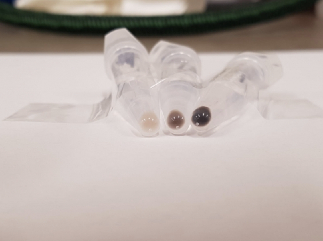
It took the Page Lab and collaborators at Baylor College of Medicine’s Human Genome Sequencing Center, the McDonnell Genome Institute at Washington University, Texas A&M University, and other institutions more than a decade to tease apart the complexities of the bull Y chromosome. In fact, it turned out to be the most gene-dense of any Y chromosome ever mapped — largely due to the fact that 96 percent of its genetic material was made up of repetitive sequences.
As in the mouse, most of the bull’s “hall of mirrors” repeats appeared to be expressed in the testis. But the question remained: Why? “What drives it can’t just be purely making more sperm, because that’s just overkill, right?” Hughes said. “You don’t really need hundreds of copies of a gene to accomplish that task.”
The researchers found a clue when they took a closer look at the bovine X chromosome: the female-determining sex chromosome also had a few copies of these testis-specific genes. “We don’t really know the mechanism in the bull, but the thought is that somehow the amplification of these genes in the Y has to do with helping the Y get passed on — and the X copies are amplified to compete against that tendency and help the X,” Hughes said.
A selfish pursuit
This X-Y arms race has been proven to happen in mice: somehow, repetitive genes on the Y chromosome give it an extra edge when it comes to ending up in the sperm during gamete formation. In a 2012 study, researchers knocked out the Y-chromosome repeats. Without the extra genes, more X chromosomes than Ys ended up in sperm cells, and the sex ratio of offspring skewed female. Over years of evolution, the X has developed repeats as well — its own way to get a leg up in the race.
Competition between X and Y chromosomes is selfish, Hughes said, because it’s not a good thing for the species to have a skewed sex ratio. Thus, these alterations benefit only the lucky chromosome that ends up in the fertilized egg. The fact that a selfish — and even detrimental — mechanism would continue for millions of years in disparate branches of the evolutionary tree suggests that these conflicts may be an inevitable side effect of having a pair of asymmetrical sex chromosomes. “These X-Y arms races have probably been around for as long as mammals have been around,” Page said.
Evolutionary theory aside, knowing the mechanisms controlling the sex ratios of cattle could be of practical use in the coming years. “It could be of great interest to breeders, because they would love to be able to manipulate the sex of cattle offspring,” Hughes said. “For example, dairy farmers would prefer more females and meat farmers would prefer more males.”
Right now, the lab is working on leafing out the branches of their Y chromosome evolutionary tree. The bull’s is the seventh sex chromosome to be completely sequenced using the SHIMS method. Hughes, Page and the lab are also eyeing members of other animal groups, including reptiles.
“Our lab is focused on sex differences across the human body, and all of that work really is inspired by lessons that we’ve learned by comparing the Y chromosomes of different animals with our own,” Page said. “It’s like when you go to an art gallery and just sit on a bench and look and feel inspired — these sequences are an infinite source of inspiration in the work we are doing. And we can now add the bull to our gallery.”
***
Hughes, J. et al. “Sequence analysis in Bos taurus reveals pervasiveness of X-Y arms races in mammalian lineages.” Genome Research, Nov. 18, 2020. DOI: 10.1101/gr.269902.120
Greta Friar | Whitehead Institute
November 18, 2020
Melanosomes are the organelles, or structures, inside our cells, that produce melanin, the molecule that gives our skin, hair and eyes their color. Melanosomes produce several different forms of melanin, including black/brown coloration and yellow/red coloration, and the many variations in levels at which each coloration can be produced in an individual generate the wide variety of skin, hair, and eye colors in the world.
Many genes that have been associated with skin color encode proteins that are active in melanosomes, but their specific functions are unknown, leaving gaps in researchers’ understanding of the underlying biology of skin color. In order to help researchers get a more detailed understanding of melanosome biology, Whitehead Institute Member David Sabatini’s lab has developed a tool, called MelanoIP, with which researchers can rapidly and specifically isolate melanosomes from the cell and analyze their contents. Using this tool, researchers can uncover the identity of the proteins at work there and explain mechanistically how genetic variation contributes to differences in skin color. In research published in Nature on November 18, Sabatini and graduate student Charles Hank Adelmann unveil MelanoIP and explain how they used it to crack the identity of melanosome protein MFSD12.
MelanoIP is the latest in a series of tools based on a method that Sabatini, who is also a professor of biology at Massachusetts Institute of Technology and an investigator with the Howard Hughes Medical Institute, and collaborators developed to rapidly extract specific organelles from the cell for investigation. Sabatini and former graduate student Walter Chen first developed the method to isolate mitochondria. The process starts with researchers creating a tag that localizes to the organelle type of interest. Then they expose the contents of the whole cell to beads covered in antibodies that latch onto the tags, which pull the organelles with them when they are collected. The lab has since adapted this process to use on lysosomes, the recycling centers of the cell, and peroxisomes, organelles important in several metabolic processes—and now, melanosomes.
The first melanosome protein that Sabatini and Adelmann turned their attention to, MFSD12, was known to be linked to the production of red coloration or pheomelanin. When MFSD12 is suppressed, this leads to darker skin color in humans and mice, because the melanosomes are generating brown/black melanin but not any of the lighter red melanin. However, MFSD12’s exact role was unknown. Using MelanoIP, Adelmann discovered that MFSD12 is required for the import of the amino acid cysteine into melanosomes, which is a necessary component in red melanin synthesis. Adelmann’s research suggests that MFSD12 is itself the transporter, but further work is needed to confirm whether it works alone or in conjunction with other molecules.
One reason that the Sabatini lab picked the melanosome as the next organelle to apply their IP toolkit to is because of its close relation to the lysosome, one of the organelles for which the lab had already built such a tool. This close relation proved relevant in Adelmann’s research on MFSD12, when he discovered that the protein is also required for the transport of cysteine into lysosomes. People with the rare genetic disorder cystinosis are affected by the buildup of cystine, another form of cysteine, in lysosomes. Adelmann found that by inhibiting MFSD12, and preventing cysteine from entering lysosomes, he could reverse the buildup of cystine in cells with the genetic mutation linked to cystinosis, suggesting a potential therapeutic use for MFSD12 inhibitors.
Adelmann is now turning his attention to cracking the identity of more of the proteins active in melanosomes and uncovering more of the biology underlying variation in skin color.
***
Written by Greta Friar
***
Adelmann, Charles H. et al. “MFSD12 mediates the import of cysteine into melanosomes and lysosomes.” Nature, Nov. 18, 2020. DOI: 10.1038/s41586-020-2937-x
Biologist Jianzhu Chen works to enhance immune response
Mark Wolverton | Spectrum
November 16, 2020
Jianzhu Chen, professor of biology and a member of the Koch Institute for Integrative Cancer Research at MIT, is pursuing a different strategy from most of his colleagues working on SARS-CoV-2, the virus that causes Covid-19. “We focus on the immune system and fundamental mechanisms as well as their application in cancer immunotherapy, vaccine development, and metabolic diseases,” he explains. Rather than trying to develop a specific vaccine, Chen is pursuing vaccine platform technologies that can be used to enhance any vaccine.
This effort is built on Chen’s previous work on dengue fever, a severe tropical disease transmitted by mosquitos. “We have been working to improve a vaccine against dengue virus infection,” he says, “which has this phenomenon called antibody-dependent enhancement,” in which “non-neutralizing” antibodies bind to the virus but do not destroy it. The immune system’s pathogen-eating macrophages then consume these virus-antibody complexes and become infected themselves, making a subsequent infection worse.
Chen’s team has identified vaccine adjuvants, or enhancing agents, that can increase neutralizing (that is, effective) antibodies while reducing non-neutralizing antibody response in mice and nonhuman primates. The team is confident that using a similar strategy against Covid-19 would improve any vaccine’s effectiveness.
Addressing cytokine storm
Chen is also focusing on the dangerous hyperinflammatory response seen in Covid-19: the cytokine storm that can result when the immune system overreacts to infection.
“We have been working on macrophage biology for quite some time,” Chen says. “SARS-CoV-2 infection is a hyperinflammatory response, and macrophages probably play a critical role in that response.”
“We have identified many compounds, including FDA-approved drugs, bioactive compounds, and natural products that can modulate macrophage activity to become anti-inflammatory,” he says. Such macrophage modulation would likely be used in conjunction with other treatments as a therapeutic strategy for already-infected patients.
A promising result from either research project could be used along with a Covid-19 vaccine to enhance immune response while preventing or reducing the severity of any possible reinfection. But it’s too early to tell what might happen. “We don’t have a vaccine yet,” Chen notes. “It’s not clear when we’ll have one. Even when we have one, it’s not clear how well it will work. It could be 95% protection; it could be 50%. Some of them may not confer much protection at all. But even 50% or 60% is a significant number of people.”
Another challenge, Chen acknowledges, is that medical research must move from theory to lab and ultimately into the real world. Vaccines can be designed and modeled on computers but eventually “we have to test them to see if they work as we expect,” he says. “You have to immunize mice or some other animals and then challenge them with SARS-CoV-2 to see whether the vaccine protects the animals from infection or dramatically minimize disease symptoms. These kinds of studies can’t be modeled computationally.”
Chen also hopes that his particular contributions will have benefits beyond the pandemic. “We’re aiming to develop a vaccine platform prototyped on SARS-CoV-2 that can be used for the development of many other vaccines as well, using the most appropriate technologies.” If that happens, science will have dug at least one substantial jewel out of the depths of an unprecedented public health crisis.
November 14, 2020
Whitehead Institute
November 12, 2020
MicroRNAs are short RNA sequences that maintain a tight control on which genes are expressed and when. They do this by regulating which messenger RNA (mRNA) transcripts — the single-stranded templates for proteins — are actually read by the cell. But what controls these cellular controllers?
In a new study published Nov. 12 in Science, researchers in David Bartel’s lab at Whitehead Institute show that mRNAs and other RNAs often turn the tables on their microRNA regulators — and show that the path to microRNA degradation is not what scientists expected it to be.
“A lot of people know that microRNAs repress mRNAs — that’s textbook,” said Charlie Shi, a graduate student in Bartel’s lab and first author on the paper. “But in certain cases, this logic is reversed. And I think that’s really interesting and weird, this idea that often the tables are turned.”
When transcripts attack
MicroRNAs typically control gene expression by binding to mRNA transcripts, and then working together with a protein called Argonaute to “silence” those transcripts by causing them to be more rapidly degraded. Because microRNAs are held cozily inside of the Argonaute protein, they are shielded from destructive enzymes in the cell, and are thus fairly long-lived by cellular standards. They can persist for up to a week, causing the destruction of many mRNA molecules over that time.
Sometimes, however, a microRNA binds to a special target site on an mRNA transcript that leads to premature destruction of the microRNA. This phenomenon — called target-directed microRNA degradation, or TDMD — happens naturally in cells, and is a way to control how much of certain microRNAs are allowed to persist at any given time.
Bartel’s lab began studying this form of degradation after researchers in the lab discovered that an RNA called CYRANO, which doesn’t code for any proteins, leads to the degradation of a specific microRNA called miR-7. This interaction was interesting to the researchers because the mechanism did not seem to line up with the current theories about TDMD.
Previous models of TDMD suggested that special target sites, like the one in CYRANO, cause one end of the microRNA to stick out of Argonaute and become vulnerable to the addition and subtraction of nucleotides by cytoplasmic enzymes. This process, called tailing and trimming, was thought to be a key step in the path to degradation of the microRNA.
“But when you knock out the enzyme that causes tailing of miR-7, it has no impact on the degradation,” Shi said. “So that’s curious, right? So how can we really perturb this supposedly responsible system and have no impact?”
A new model
In order to further probe the mechanism of TDMD, the researchers focused in on this interaction between the CYRANO noncoding RNA and miR-7. Shi designed a CRISPR screen to identify genes essential for the microRNA’s degradation when it encountered a CYRANO transcript.
The screen yielded one gene that was essential to the microRNA’s degradation, called ZSWIM8. When they looked up the gene’s function, the researchers found that it codes for a component of a ubiquitin ligase. Ubiquitin — so named because it is found in virtually all types of cells — serves as a flag to mark proteins for degradation in a cellular garbage disposal called the proteasome.
The finding of the ZSWIM8 ubiquitin ligase implied that CYRANO-mediated microRNA degradation involves destruction of the Argonaute protein. In this new molecular model for TDMD, the regulating RNA, CYRANO, binds to the microRNA, mir-7, encased in its protective Argonaute protein, and then recruits the ZSWIM8 ubiquitin ligase. This ligase then sticks a few ubiquitin molecules onto the microRNA’s Argonaute, leading Argonaute to be degraded, and thereby exposing its microRNA cargo to be destroyed by enzymes in the cell. Importantly, this process does not require any trimming and tailing of the microRNA.
“The discovery of this unanticipated pathway for TDMD illustrates the power of CRISPR screens, which can simultaneously query essentially every protein in the cell, including those that you never dreamed would be involved,” said Bartel, who is also an investigator of the Howard Hughes Medical Institute and a professor of biology at Massachusetts Institute of Technology.
A multitude of microRNAs
When the researchers looked at other known examples of TDMD, they found the ZSWIM8 was essential in all of them. Having identified this key part of the degradation pathway allowed them to seek out more microRNAs that are subject to this regulation.
“When we started this project, there were only around four examples in nature of endogenous RNAs that are encoded by the cell that can perform TDMD,” Shi said. “We had a feeling that there would be many more, and so by finding a factor that was required for TDMD in a general way — ZSWIM8 –we were then able to ask and answer the question, ‘how widespread this phenomenon?’”
As it turns out, TDMD is fairly common in multicellular organisms. The researchers looked for evidence of the microRNA degradation mechanism in different cell types — two from mice, and one from fruit flies — and found that in any given cell, up to 20 different microRNAs were regulated by TDMD out of a couple hundred total microRNAs in the cell.
The researchers also observed this mechanism in human cells and nematodes, suggesting that TDMD as a method for regulating microRNAs dates back to the common ancestor of these disparate species. That definitely creates a lot of questions for us,” Shi said. “Each one of these microRNAs is a story.”
Raleigh McElvery
November 10, 2020
In 2019, cancer researchers from MIT found a drug that targeted an elusive molecular interaction previously considered “undruggable.” In so doing, they opened up a new realm of chemotherapy. This drug, called JH-RE-06, sensitized tumors to treatment, but the scientists didn’t fully understand how it exerted its effects. Now, in a pair of studies published in PNAS, the same team is closer to determining which cellular processes this drug alters to enhance cancer therapy.
Many widely-used chemotherapies, like cisplatin, kill tumors by damaging their DNA and inducing programmed cell death. But cells are resilient, and many can continue to function with the help of repair enzymes that simply bypass the damage and continue to replicate the DNA. As a result, some tumors not only defy death, but gain mutations that render them more resistant to treatment or spur new malignancies elsewhere.
“If you’re not making a patient better, it’s very likely you’re making them worse,” says Michael Hemann, associate professor of biology and co-author on both studies. Rather than fully replacing conventional DNA-damaging treatments — which could take decades — Hemann suggests an effective “medium-term” solution: augmenting low doses of cisplatin with safer agents that strengthen the chemotherapy’s tumor-killing capacity.
The team had tested this approach back in 2019, when they first identified JH-RE-06 and saw it enhanced chemotherapy treatment. These experiments revealed that JH-RE-06 bound to an especially shallow (and infamously undruggable) pocket of one DNA repair enzyme called REV1. This barred REV1 from interacting with another key enzyme, and prevented the cancer cells from recovering after cisplatin treatment. But what happened next to cause the tumors to shrink was unclear.
As they began their next round of experiments, the researchers expected to find that the drug would simply enhance the way cisplatin kills tumors via programmed cell death.
Nimrat Chatterjee, Walker’s former postdoc and lead author of the first study, treated mice and individual cells with a combination of cisplatin and JH-RE-06. She expected to see signs of programmed cell death, but for months, she saw no such markers.“We thought that if we blocked the DNA repair process with the JH compound, we’d see more programmed cell death,” says co-author Graham Walker, American Cancer Society Professor and Howard Hughes Medical Institute Professor. “As it turns out, we did see more cell death — just not the kind we were expecting.”
One evening, just as she was about to head home for the day, she peeked through her microscope at the cancer cells treated with JH-RE-06 and cisplatin. She noticed they were fluorescing a strange green color.
“At first, I didn’t know what I was seeing,” she recalls. But after some follow-up, it became clear that the mysterious green color was coming from lipid-containing residues that usually appear as cells age and stop dividing. The cells appeared to be in a permanently dormant state known as senescence — not yet dead but unable to proliferate. JH-RE-06 was altering cisplatin function by triggering a second molecular pathway independent of programmed cell death.
“That was one of the best ‘aha’ moments of my scientific career so far,” Chatterjee says. “REV1, the DNA repair enzyme that JH-RE-06 binds, may have other novel biological functions and a larger role in cancer cell etiology than we originally thought. We’re now grappling with more questions about REV1 than ever before.”
Around the same time, Faye-Marie Vassel PhD ’20, Walker and Hemann’s former joint graduate student and lead author of the second study, witnessed a similar phenomenon in her own experiments. She was investigating a different way of inhibiting the two key DNA repair enzymes that enable cancer cells to survive chemotherapy. Instead of probing JH-RE-06, which latches onto REV1, she tried deleting REV1’s binding partner, called REV7. This protein is particularly influential because it serves an important role in fixing double-stranded breaks in addition to interacting with REV1.
When Vassel deleted REV7 from mice with non-small cell lung cancer, the tumors became more sensitive to cisplatin, as expected. But, like Chatterjee, she saw signs of senescence rather than programmed cell death. The two studies had converged on a common biology: adding JH-RE-06 or deleting REV7 strengthened the effects of cisplatin by inducing this dormant state.
Cancer detection and treatment methods have improved dramatically in the last two decades, but drug-resistant cancers like non-small cell lung cancer remain difficult to combat, Vassel says. “Our experiments are the first to show that senescence induction is likely a consequence of REV7 inhibition,” she adds. “Inhibiting REV7 in tandem with cisplatin therapy may prove to be an effective strategy for enhancing a chemotherapeutic response.”
Chemotherapies that trigger programmed cell death have been the mainstay of cancer treatment for decades. But studies like these show that triggering senescence may be a promising complementary strategy. Most senescent cells are eventually cleared by the immune system, and the researchers suspect this is how cancer cells treated with JH-RE-06 or REV7 inhibitors would be eliminated from the body.
Walker and Hemann agree that, at the moment, their sister studies raise more questions than answers. As Walker explained, “We’ve pried open a new discovery, and hopefully set the stage for many exciting experiments to come.”
Top image: Genetically-engineered mouse model for lung cancer. Credit: Credit: National Cancer Institute, National Institutes of Health. NIH Image Gallery/Flickr (CC BY-NC)
The REV7 image was originally published in: “Structural basis of Rev1-mediated assembly of a quaternary vertebrate translesion polymerase complex consisting of Rev1, heterodimeric polymerase (Pol) ζ, and Pol κ.”
Journal of Biological Chemistry, online August 2, 2012, DOI: 10.1074/jbc.M112.394841
Jessica Wojtaszek, Chul-Jin Lee, Sanjay D’Souza, Brenda Minesinger, Hyungjin Kim, Alan D. D’Andrea, Graham C. Walker, and Pei Zhou.
Citations:
“REV1 inhibitor JH-RE-06 enhances tumor cell response to chemotherapy by triggering senescence hallmarks”
Proceedings of the National Academy of Sciences, online November 9, 2020, DOI: 10.1073/pnas.2016064117
Nimrat Chatterjee , Matthew A Whitman, A Harris , Sophia M Min , Oliver Jonas , Evan C Lien , Alba Luengo , Matthew G Vander Heiden , Jiyong Hong , Pei Zhou , Michael T Hemann , and Graham C Walker
“Rev7 loss alters cisplatin response and increases drug efficacy in chemotherapy-resistant lung cancer”
Proceedings of the National Academy of Sciences, online November 3, 2020, DOI: 10.1073/pnas.2016067117
Faye-Marie Vassel, Ke Bian, Graham C. Walker, and Michael T. Hemann