Cells are known by the company they keep
Eva Frederick
March 2, 2021

In the paper, published online March 1 in the journal Cell Metabolism, researchers at Whitehead Institute and the Morgridge Institute for Research performed CRISPR-based genetic screens of cells cultured in either traditional media or a new physiologic medium previously designed in the Sabatini Lab at Whitehead Institute designed to more closely reflect the nutrient composition of human blood. The screen revealed that different genes became essential for survival and reproduction in the various conditions.

“This work underscores the importance of using more human-like, physiologically relevant media for culturing human cancer cell lines,” said Whitehead Institute Member and co-senior author David Sabatini, who is also a professor of biology at the Massachusetts Institute of Technology and an investigator of the Howard Hughes Medical Institute. “The information we can learn from screens in human plasma-like media — or media designed to mimic other fluids throughout the body — may inform new therapeutic methods down the line.”

The widespread use of a human plasma-like medium could open the door for many researchers to conduct experiments in the lab that could have more relevance to human disease, but without complicated methods or prohibitive costs.

“Medium composition is both relatively accessible and quite flexible,” said co-senior author Jason Cantor, an Investigator at the Morgridge Institute for Research and an assistant professor of biochemistry at the University of Wisconsin-Madison, and a former postdoc in Sabatini’s lab. “Not all researchers have access to specialized tissue culture incubators, nor can everyone easily pursue some of the more complex 3D and co-culture methods, but most can get their hands on a bottle of media.”

The big screen

The idea that different environmental conditions may lead to different genes being essential is not a new one. “People have done this in microorganisms, and shown that if you throw [bacteria] into different growth conditions — the contributions of different genes to cell fitness can change,” Cantor said.

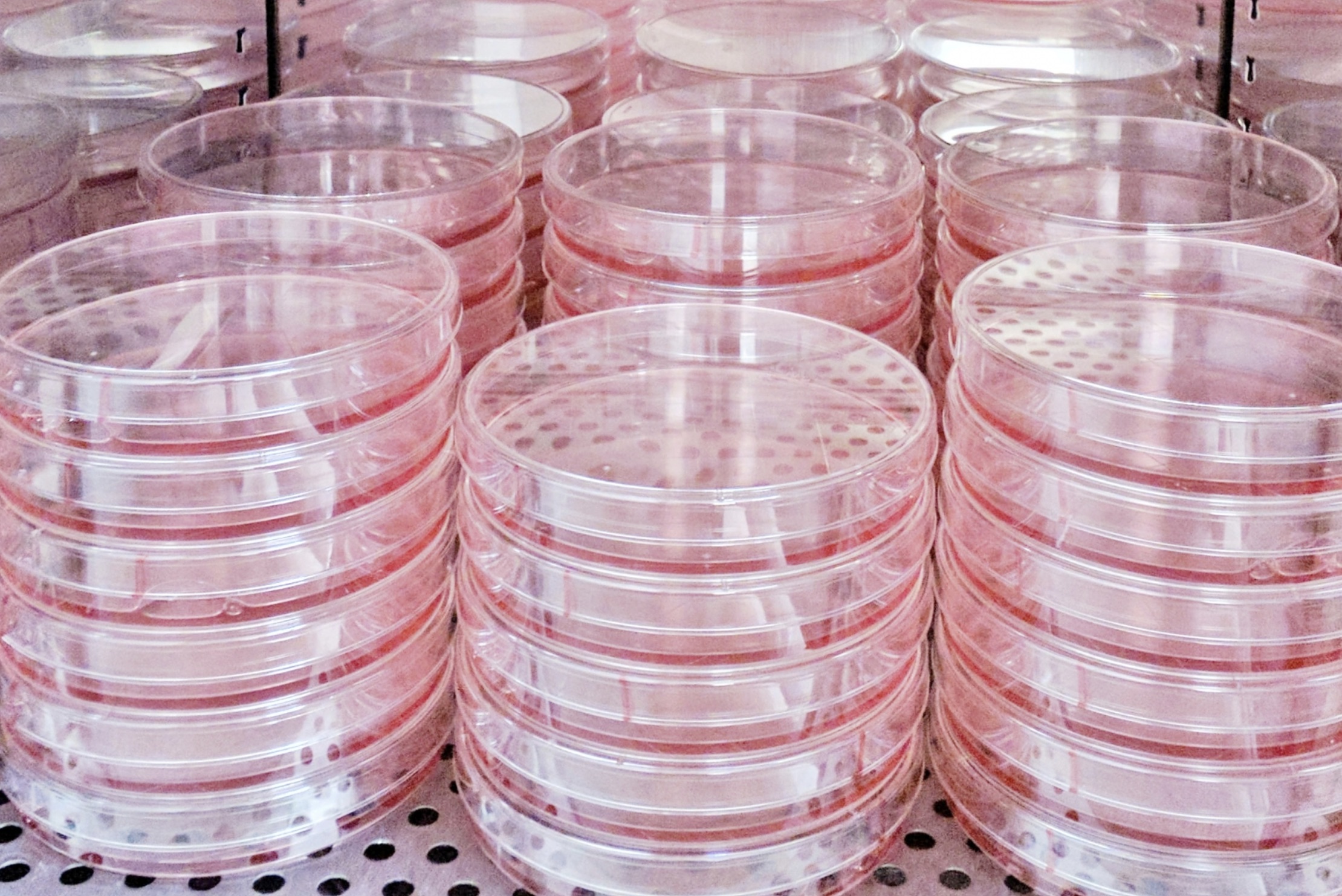
With this reasoning in mind — that medium composition could affect which genes become necessary for human cells to grow — the researchers set up screens to identify essential genes in a single leukemia cell line in different kinds of culture media. One batch was grown in a traditional medium, and another cultured in the lab’s new medium called Human Plasma-Like Medium, or HPLM, which has a metabolic composition more reflective of that in human blood.

The approach they used — called a CRISPR screen —  takes advantage of CRISPR-Cas9 gene editing technology to systematically snip and knock out genes across the genome, with the goal of creating a population of cells in which every possible gene knockout is represented. The expression of some genes is essential to survival, and cells grow substantially slower or die when those genes are deleted. Other cells may have trouble functioning, and some may grow even faster. Once the pooled cells have had a chance to grow and proliferate, researchers sequence the genetic material of the entire population to determine which genes were critical for survival within the given screen.

Once they completed the initial screens, the researchers identified hundreds of genes that were conditionally essential — that is, necessary for cell growth in one medium versus another. Interestingly, these medium-dependent essential genes collectively had roles in a number of different biological processes.

To determine how much these genes were dependent on the type of cells studied, the researchers then ran similar screens across a panel of human blood cancer cell lines, and then pursued follow-up work to understand why certain genes were identified as conditionally essential.

Ultimately, they uncovered the underlying gene-nutrient interactions, and specifically for these hit genes, traced the effects to availability of certain metabolites — the nutrients and small molecules necessary for metabolism — that are uniquely defined in HPLM versus the traditional media.

The next steps

CRISPR screens can help scientists identify potential drug targets and map out important cellular interactions to inform cancer therapies. “There are so many ways that people use CRISPR screens right now,” said Cantor. “What this study is showing is that the availability metabolites can have a major impact on which genes are important for cell growth, and so I think there are a lot of implications here in terms of how these types of screens could be performed in the future in order to potentially increase the fidelity of what we see in the lab and what might happen in the body.”

The research also establishes more nuanced relationships between cells’ genes and their environment. “What this allows us to do down the line, theoretically, is to tune how important a gene is — how important the encoded protein is — by manipulating metabolite levels in the blood,” said Cantor. “That’s one of our bigger-picture ideas.”

In the future, these relationships could inform cancer treatments. For example, if scientists could “tune” the importance of a specific gene for cancer cell growth, then the protein encoded by that gene could become a more promising drug target — in effect, tricking cancer cells into revealing possible context-dependent vulnerabilities. “The idea of targeting metabolites to treat cancer isn’t itself new — in fact, it [underlies] a well-established anti-cancer therapeutic enzyme still in use today — but I think our work maybe enables us to look for ways to couple this type of approach with other targeted therapies.”

“At our core, we are a basic cell biology lab,” added Nicholas Rossiter, a technician in Cantor’s lab and the first author of the study. “But whenever you’re studying basic cell biology, there’s the potential to translate it into therapeutic strategy. Our plan is just to keep on chugging along in our lab and studying how exactly cell biology can be influenced by these environmental factors. We do the basics, and then there will hopefully be some auspicious findings that can be carried on into therapeutics.”

Seychelle Vos investigates how the genome is organized so it can fit inside the cell — and how that careful organization affects gene expression.

February 24, 2021
How this biology lab class went virtual during the pandemic

Instructor Mandana Sassafar found creative ways to teach first-years experimental techniques and laboratory protocols remotely.

Department of Biology
February 23, 2021

Each January, MIT hosts a four-week term known as Independent Activities Period (IAP). This year, though, was different: All IAP activities were held online due to Covid-19 restrictions. Like many other IAP instructors, the Department of Biology’s director of outreach, Mandana Sassafar, was facing a dilemma. How could she transfer a fast-paced, hands-on lab class to the virtual realm?

Sassanfar has been teaching class 7.102 (Introduction to Molecular Biology Techniques) for over a decade. The class was originally designed to familiarize first-year undergraduates with lab equipment, troubleshooting, and basic methods in molecular biology in preparation for MIT’s Undergraduate Research Opportunities Program (UROP). She felt this goal was now more important than ever, given that too many students had already lost precious chances to work in labs due to the pandemic.

After weeks of consideration, she came up with a solution: create a remote version of the class called 7.S391 (Special Subject in Biology) using video clips. She filmed more than 15 graduate students and postdocs on her iPhone, maintaining at least 6 feet of distance as the trainees wore masks and demonstrated various lab techniques.

The 7.S391 students then watched the videos, described each experiment, compared techniques, and devised protocols based on their observations. Although they did not have lab equipment in their homes, observing researchers in action is the first step toward learning-by-doing, according to Sassanfar.

“Because so many first-years are eager to start UROPs, this seemed like the best way to prepare them,” Sassanfar said. “They were exposed to research and lab tools on a daily basis, and watching experiments helped them gain knowledge and confidence.”

The class was capped at 24 students, who met for two-and-a-half hours each day for 12 days. Thanks to Sassanfar’s videos, the students learned to grow bacteria; set up polymerase chain reaction (PCR) tests; design primers to construct recombinant plasmids; do tissue culture; and perform gel electrophoresis, western blots, and affinity chromatography. They also practiced interpreting the results.

During the final days of the class, MIT lab groups hoping to recruit UROPs gave short presentations about their research. In total, 10 labs from the departments of Biology, Brain and Cognitive Sciences, and Biological Engineering participated.

One graduate student presenter, Kristina Lopez ’18, had completed her undergraduate degree at MIT before beginning her PhD in the Knouse lab. She advised undergraduate students to find a lab they like and work there until they graduate. “I joined a lab my freshman year and stayed there all four years,” she said. “It allowed me to really delve into the project and make important contributions.”

First-year Alesandra (Alysse) Pusey says the class introduced her to an array of lab techniques for investigating biological questions. “I feel more prepared and eager to take on a UROP,” she adds. “Most virtual classes lack organic social interactions, but the implementation of breakout rooms and break time in this class allowed me to bond with my classmates, each of whom share an interest in biology with me.”

Her classmate Antonella Rebolledo-Ledesma also enjoyed the experience. “I was surprised by how much I could learn about lab work in a virtual setting,” she says. “I would highly recommend this course to anyone who is thinking about taking it next year — although hopefully it will be in-person!”

Eight from MIT named 2021 Sloan Research Fellows

Awards honor, support young professors in the Media Lab and departments of Biology, Brain and Cognitive Sciences, Chemical Engineering, EECS, and Mathematics.

MIT News Office
February 19, 2021

The Alfred P. Sloan Foundation announced Feb. 16 that it has awarded Sloan Research Fellowships to eight MIT professors in the MIT Media Lab and in the departments of Biology, Brain and Cognitive Sciences, Chemical Engineering, Electrical Engineering and Computer Science, and Mathematics. The fellowships, which honor pre-tenure faculty members, will support their research with two-year, $75,000 awards.

“The Sloan Research Fellowship Program recognizes and rewards outstanding early-career faculty who have the potential to revolutionize their fields of study,” according to the Sloan Foundation.

Fadel Adib, associate professor and Doherty Chair in Ocean Utilization, directs the Signal Kinetics group at the MIT Media Lab. His group invents, builds, and deploys wireless and sensor technologies to address complex problems in society, industry, and ecology. His team’s work focuses on bringing wireless capabilities to extreme domains like the ocean and the human body and to enable new applications that are infeasible using today’s technologies. His research extends beyond communication and networking to enabling novel micro-sensing, powering, and perception tasks. These capabilities aim at helping address major societal challenges in health care, climate change, and automation.

“We are excited about continuing to push our technologies deeper into the oceans and the human body,” says Adib, whose team invented the world’s first net-zero power underwater communication technology and wireless systems that power and communicate with batteryless micro-implants inside the human body. He aims to use this funding to further his team’s efforts in underwater GPS, in-body sensing, and robotic automation. “This Sloan fellowship will allow my team to continue taking risks in pursuing high-impact projects to understand and address global challenges ranging from climate change to health care and automation.”

Joseph “Joey” Davis, the Whitehead Career Development Assistant Professor in Biology, investigates the massive molecular “machines” that carry out important cellular processes, such as protein synthesis and degradation. He uses cryo-electron microscopy (cryoEM) to visualize these molecules at near-atomic resolution as they are being assembled and changing shape while they work. In collaboration with Simons Professor of Mathematics Bonnie Berger, his team has developed a new computational tool. Called cryoDRGN, it leverages neural networks to extract molecular motions from cryoEM data and create 3D movies. The Sloan Foundation award will help Davis combine cryoDRGN with a related imaging technique — electron cryotomography — to observe molecular structures directly inside living cells. Using this powerful combination, he hopes to uncover how machines like the ribosome, which synthesizes proteins, assemble in their native cellular environment. Ultimately, he aims to identify new antibiotic targets in this assembly pathway.

“We hope that the combination of cryoDRGN and electron cryotomography will enable us to directly visualize how key molecular machines are assembled within the cell,” Davis says. “This information will be critical in truly understanding how nature builds these machines so rapidly and efficiently, and will help us understand what aspects of the assembly process fail when cells are mutated and as they age. I am incredibly grateful to the Sloan Foundation for their support of our work.”

Steven Flavell, the Lister Brothers Career Development Assistant Professor in Brain and Cognitive Sciences and The Picower Institute for Learning and Memory, said the Sloan Foundation’s award will help him conduct experiments to uncover how animal nervous systems generate internal states that represent needs and desires, such as hunger, and then produce behaviors, such as roaming around in search of food. His lab plans to use a multidisciplinary experimental approach in their studies, which employ the simple model of the C. elegans worm whose nervous system contains only 302 neurons. Though simple, the model has proven to produce important insights across many areas of biology.

“Over the course of each day, an animal’s nervous system may transition between a wide range of internal states that influence how sensory information is processed and how behaviors are generated,” Flavell says. “These states of arousal, motivation, and mood can persist for hours, play a central role in organizing human behavior, and are commonly disrupted in psychiatric disease. However, the fundamental neural mechanisms that generate these states remain poorly understood. We envision that these studies will ultimately reveal fundamental principles of neural circuit function that may generalize across animals.”

Heather Kulik, associate professor in chemical engineering, advances first-principles and machine learning computational chemistry to accelerate materials and catalyst discovery. Her group has developed the first machine learning models capable of predicting normally time-consuming quantum mechanical properties of transition metal complexes, rapidly uncovering design principles in weeks instead of lifetimes. Her group develops large-scale quantum mechanical modeling methods and applies them to reveal how enzymes work and how to take inspiration from nature to design next-generation catalysts.

“The award from the Sloan Foundation will enable my group to continue advancing computational materials and bio-inspired catalyst discovery,” Kulik says. “The flexible nature of the support ensures we can continue to push forward these interdisciplinary efforts at the boundaries between fields.”

Luquiao Liu, associate professor in the Department of Electrical Engineering and Computer Science, focuses on understanding and exploiting spin-related physics in solid-state material and devices. Most recently, Luqiao has been doing research on developing material and carrying out electrical measurement on charge-spin interactions to achieve electrically induced magnetic switching, and exploring new methods to realize quantum control over the transport of magnons and other quasiparticles, which could be useful in future hybrid quantum systems for information processing.

“This fellowship will strengthen our capabilities in identifying new material and physics mechanisms that can be used to achieve functions that are unique to spintronic systems, with the long-term goal of realizing efficient computing in the classical and quantum domains,” he says.

Karthish Manthiram, the Theodore T. Miller Career Development Chair and assistant professor in chemical engineering, studies the carbon footprint behind most chemicals and materials that we encounter every day — there is a carbon footprint associated even with the fabric of the clothes we wear, the food we eat, and the disinfectants we spray, he says. To find ways to synthesize these chemicals and materials in a sustainable manner that eliminates the carbon footprint, the Manthiram lab is pioneering the development of a paradigm in which carbon dioxide, dinitrogen, and water can be converted into a wide range of chemicals and materials using renewable electricity. In essence, this would mean that a device that breathes air, drinks water, and takes in solar photons could in principle someday make many of the chemicals that society relies on. The lab specifically looks for ways to facilitate the molecular-level dance through which chemical bonds are broken and formed, so that desired molecules can be made more selectively, efficiently, and at faster rates.

“The support of the Sloan Research Fellowship will allow my group to advance the decarbonization of the material world, through electrically driven synthesis of critical chemicals beginning with just carbon dioxide, dinitrogen, and water,” Manthiram says. “We will pursue new frontiers in synthesizing even more complex molecules starting with these ubiquitous feedstocks.”

Dor Minzer, assistant professor of mathematics, works in the fields of mathematics and theoretical computer science. His interests revolve around computational complexity theory, or — more explicitly — probabilistically checkable proofs, Boolean function analysis, and combinatorics. Minzer’s more recent research has utilized and extended some of the insights gained from the work on probabilistically checkable proofs in order to make progress on several open problems in the field of analysis of Boolean functions, such as the Fourier Entropy conjecture and the stability problem for the edge isoperimetric inequality, as well as to other problems in theoretical computer science.

“The Sloan fellowship will allow us to continue pursuing difficult and important challenges in theoretical computer science, whose solution is likely to have wide impact on the field,” Minzer says.

Lisa Piccirillo, assistant professor mathematics, specializes in the study of three- and four-dimensional spaces. She is broadly interested in low-dimensional topology and knot theory, and employs constructive techniques in four-manifolds. Her work in four-manifold topology has surprising applications to the study of mathematical knots. She received an inaugural 2021 Maryam Mirzakhani New Frontiers Prize, created in 2019 by the Breakthrough Foundation to recognize outstanding early-career women in mathematics, for “resolving the classic problem that the Conway knot is not smoothly sliced.” For all other small knots, “sliceness” is readily determined, but this particular knot had remained a mystery since John Conway presented it in the mid-1900s. After hearing about the problem at a conference, Piccirillo took only a week to formulate a proof.

In all, the Sloan Foundation awarded fellowships to 128 tenure-track, but not-yet-tenured, scholars in the United States and Canada this year.

Vander Heiden and Lourido receive promotions
February 18, 2021

Effective July 1, Matthew Vander Heiden and Sebastian Lourido will be promoted to Full Professor and Associate Professor (Without Tenure), respectively.

Vander Heiden is Associate Director of the Koch Institute for Integrative Cancer Research, a member of the MIT Center for Precision Cancer Medicine, a member of the Ludwig Center for Molecular Oncology, and a member of the Broad Institute. He joined the department in 2010 and earned tenure in 2017. His work focuses on the biochemical pathways cells use and how they are regulated to meet the metabolic requirements of cells in different physiological situations. His lab investigates the role of metabolism in cancer, particularly how metabolic pathways support cell proliferation. They aim to translate their understanding of cancer cell metabolism into novel cancer therapies. His promotion to full professor reflects his international standing in his field, his excellent and dedicated teaching, and his service to the department and the broader scientific community.

Lourido is a member of the Whitehead Institute and Latham Family Career Development Professor. He joined the department and Whitehead Institute in 2017. His lab is interested in the molecular events that enable apicomplexan parasites to remain widespread and deadly, infectious agents. They study many important human pathogens, including Toxoplasma gondii, to model features conserved throughout the phylum. They seek to expand our understanding of eukaryotic diversity and identify specific features that can be targeted to treat parasite infections.

Posted: 2.18.21
Catching key moments of cancer progression
Whitehead Institute
February 9, 2021

Important moments of cancer — mutation, tumor formation, metastasis —  are fleeting, easy-to-miss events. Even with modern medical technologies and methods, they often happen unobserved, and are only realized later when these cells spawn life-threatening conditions.

In recent years, however, new methods of tracking individual cells through time have allowed researchers to get closer to the origin of these events, and Whitehead Institute scientists are turning the power of these technologies to study cells involved in several different types of cancer. “With [these technologies], you can track down rare events in the past, identify all the offspring of an event, and see how they’ve changed their behavior,” said Whitehead Institute Member Jonathan Weissman.  “You can ask, when a cell picks up an oncogene, how does it mutate, and further evolve, and proliferate and metastasize?”

Read on to learn how three Whitehead Institute Members are using specially engineered mice, CRISPR-based technologies, and other methods to track cells at different stages of cancer development, pushing the boundaries of what we understand about how the disease starts and proliferates. From the initial beginnings of a tumor, sometimes in the darkness of a still-forming embryo, to a tumor’s growth and eventual metastasis to other sites in the body, Whitehead Institute scientists Rudolf Jaenisch, Jonathan Weissman, and Robert Weinberg study the pivotal points in a cancer’s growth and spread.
The birth of a tumor

For around 800 children each year in the United States, the seeds are sown during fetal development for a rare and unpredictable childhood cancer called neuroblastoma. To understand how the disease develops, scientists need to study what happens to these cancerous “seeds” as the embryo  matures. But they can hardly study a living human embryo, and fetal development is such a complex process that it is near impossible to simulate it in the lab.

To make matters more complex, the cancer grows and then may shrink unpredictably after the children are born and as they age. Sometimes the tumors disappear on their own; other times they grow uncontrollably.

In 2020, Institute Founding Member Rudolf Jaenisch’s lab introduced a new way of tracking the cells involved in the disease using his tried and true method for modeling such complex conditions: chimeric mice. A chimera is a conglomeration of two species — in this case, mostly mouse, with a few strategically placed human stem cells.

To create chimeras to study neuroblastoma, Jaenisch, who is also a Professor of Biology at Massachusetts Institute of Technology (MIT), along with collaborators at the Koch Institute for integrative Cancer Research at MIT and the Dana-Farber Cancer Institute, engineered human stem cells with glowing proteins to make them easy to see under the microscope. The cells contained a special genetic-switch that allowed the researchers to induce tumors by adding a certain chemical. These human stem cells were then induced to form a more specialized cell type – a neural crest stem cell.

Neural crest cells are a group of developing stem cells that go on to form the peripheral nervous system as well as other parts of the body such as the facial bones. It is neural crest cells that mutate into neuroblastoma tumors in humans, so the researchers hoped that by using these cells, they could create “human” tumors in mice. The researchers injected these human neural crest cells into mice so that they could readily incorporate with the host’s cells.

After the mice were born, the researchers were then able to take samples from the mice over the course of their development to see whether these implanted cells would form neural crest-derived tumors and, if so, what happened as they grew — something they would never have been able to do with neuroblastoma tumors in human infants and children.

The tumors in the chimeric mice pups developed in similar forms to human neuroblastomas — specifically, they formed characteristic rosette shapes — very similar to those seen in patient’s samples. With the help of Stefani Spranger, an assistant professor of biology at MIT and a Member of the Koch Institute for Integrative Cancer Research at MIT, they were able to track the cells’ interactions with the mice’s immune systems, and learn how the cancer “tricks” the immune cells into letting it stick around.

Now that they are able to model the formation of neuroblastoma tumors, the researchers hope to find a way to eliminate the tumors in the mice. “This is a model that will allow us to approach how to get rid of the tumors,” said Malkiel Cohen, a former postdoc in Jaenisch’s lab and first author of the paper, published in the journal Cell Stem Cell describing the work.

Cancer genealogy

A recent project from the lab of Whitehead Institute Member Jonathan Weissman focuses on another essential moment of cancer progression: metastasis. Metastasis happens when cancer spreads from a primary tumor to distant places in the body. Weissman, an expert in genome editing, created a CRISPR-based method to track the lineage of individual cancer cells in real time as they proliferate and metastasize.

Weissman and his collaborators, including graduate student Matthew Jones and then postdoctoral researcher Jeffrey Quinn, adapted the technology from a similar tool designed by Michelle Chan, a former postdoc in Weissman’s former lab at the University of California, San Francisco (UCSF) who is now an assistant professor at Princeton University. Chan designed a CRISPR mechanism to track the lineages of embryonic cells as they developed into specialized tissues.

“What Michelle Chan was able to do was uncover how tissues that look very similar to one another, actually come from disparate sources,” said Matthew Jones, a graduate student in Weissman’s lab and a co-first author on the paper describing the new method. “That has rapid implications about how tissues organize themselves, and we wanted to apply it to cancer.”

To create this system, the researchers engineered cancer cells with added genes: one for Cas9, the gene that codes for CRISPR’s “molecular scissors,” others for fluorescing proteins for microscopy, and a few sequences that would serve as targets for the CRISPR technology.

They then implanted thousands of these cells into the lungs of mice to simulate a tumor, using a model designed by Trever Bivona, a cancer biologist at UCSF. As the cells in the model tumors began to divide, the Cas9 protein began making small snips in the target sites in the cells’ DNA. When the cells fixed these snips, they patched in or deleted a few random nucleotides, leading to a unique “barcode” in each cell. Because these barcodes were added to each cell’s DNA, they were heritable and able to be passed on through generations of cells.

With the help of Nir Yosef, a computer scientist at the University of California, Berkeley, the researchers organized the data into “family trees” of cancer cells spanning multiple generations. By taking samples from different areas of the body, the researchers were able to resolve exactly when a cell jumped from where it started, in the lungs, to a distant tissue.

When they compared the trees, the researchers noticed that some cells were highly metastatic, jumping around multiple times over the course of the experiment, while others stayed put throughout. “We were excited to uncover some of these really rare, but consequential events that happened in the past that you would never be able to observe, and rarely be able to infer from a static snapshot,” Jones said.

By comparing highly metastatic and non-metastatic cells, they were able to identify metastasis-associated genes and answer questions about how the tumors were evolving and adapting. “It’s an entirely new way to look at the behavior and evolution of a tumor,” Weissman said. “We think it can be applied to many different problems in cancer biology.”

The next steps

A natural tumor begins with a single cell, mutated in a way that leads it to “go rogue,” so to speak. To mimic this in a model system, Dian Yang, another post-doc in the Weissman Lab, is collaborating with researchers in the lab of MIT Professor of Biology and Koch Institute Director Tyler Jacks’ to create a mouse model with the CRISPR lineage recording tool embedded in its DNA.

The model is based on a mouse model created by Jacks’ lab to study lung cancer. The lab has created genetically engineered mice that when left alone, live completely normal lives. But upon adding a trigger (an enzyme called “Cre”) — in Jacks’ and Yang’s case, they deliver the trigger to the lung using a virus — oncogenic signals are activated and lead to spontaneous tumors in the mice’s lungs.

Being able to “switch on” the mouse model in this way has a number of advantages.  “Each tumor will start from one single transformed cell, which we can then watch in its native environment as it evolves,” Yang said. “Then, we can look back later at how the cells metastasize.”

Adding the CRISPR system to Jacks’ existing cancer model will also allow researchers to study cancerous cells on a broader time scale. “Usually, when we harvest samples from the mice, it is like a snapshot, just one sample at one stage,” Yang said. “You can see what it looks like, you can analyze gene expression at the time of sampling, you can even take a time series, but you don’t know what happened in the past.”

In a sense, the lineage recording technology embedded in the genome of the mice now makes it possible to look back in time. “When you have a million cells in a tumor, you can use the lineage network of these cells to find out how they’re related, and connect the current state with the past evolutionary lineage history,” he said. “I think this will provide a new dimension of biological information for us to understand biology that is not just limited to cancer biology.”The making of metastasis

Weissman’s lab’s method for tracking the lineage of cancer cells can illuminate the nature of the cells that leave the primary tumor and scatter throughout the body. But what actually happens to these cells to cause them to metastasize?

That’s where Whitehead Institute Founding Member Robert Weinberg’s research comes in. Weinberg has been studying cancer for decades. His early work helped to answer the question of how cells that form a primary tumor become cancerous. Weinberg identified the first human oncogene, a gene that causes otherwise-normal cells to form tumors. This finding, combined with others, demonstrated that cancer is a disease driven by damaged genes, at least in its origins. Weinberg has since turned his attention to the question of how cancer cells acquire the ability to spread.

Around 90 percent of cancer deaths are caused not by the primary tumor, but by its metastasis. Based on previous work, there is no single genetic switch that can be flipped to equip a cancer cell for metastasis. Instead, cells must go through a series of changes over time. Most cancer cells fail to acquire all of the necessary traits, and so may, for example, spread to new tissues but rarely form tumors there. Weinberg’s lab tracks cancer cells to help fill in the “road map” that cancer cells follow on the way to metastasis, in the hopes that their insights can be used to prevent or treat metastatic cancers.

One important change that cancer cells undergo is called the epithelial to mesenchymal transition (EMT), a cellular process that causes the cancer cells to express different genes and go from being immobile to mobile and invasive. Cancer cells undergoing this transition to be able to spread are called “quasi-mesenchymal.” The ability to spread does not fully explain metastasis, however.

“There’s two aspects of the metastasis problem,” Weinberg said. “The first aspect is how cancer cells physically leave the primary tumor and get seeded into a distant tissue. In other words, the physical translocation of the cells. The second step represents the subsequent ability of the already seeded cells to figure out how to make a living in a distant tissue.”

In other words, how do those transplanted cells adapt to a new tissue environment, which might offer them only inhospitable conditions? “That represents the major unsolved problem of metastasis,” he said.

Weinberg hopes to study this more in the future; for now though, his lab has found that studying quasi-mesenchymal cells can serve another purpose. Anushka Dongre, a postdoc in Weinberg’s lab, found that these cells are resistant to a common type of cancer treatment, known as immune checkpoint therapy, and can even protect the other cancer cells around them from that therapy. If as little as ten percent of a tumor consists of quasi-mesenchymal cells, then the whole tumor may become resistant.

By using a tumor’s epithelial/mesenchymal profile, Dongre demonstrated that she could predict how likely a breast cancer tumor was to respond to a particular immune checkpoint therapy. This finding could help physicians match patients to the best treatment plan, by indicating ahead of time whether the treatment will work. She also identified a way to eliminate the quasi-mesenchymal cells’ protective effect by suppressing a certain enzyme that they employ to defend themselves.

Weinberg’s lab continues to study pivotal changes in the lives of cancer cells, such as the EMT, so that they can better understand metastasis and, they hope, help find effective treatments for patients with metastatic cancers.Tracking cells into the future

Scientists have been tracking cells for more than a century, and Whitehead Institute scientists will be tracking cancer cells for decades to come. In the coming years, Weinberg plans to continue to investigate the mysteries of metastasis. For Weissman’s part, he hopes to continue refining his CRISPR technique, with the end goal of eventually being able to predict the behavior of cancer cells. “We want to be able to measure where they are, where they’re going at any time, and then predict where they’re going to be in the future,” he said.

With new technologies and ever-expanding fields, there is limitless potential in these various methods. “That’s what is so exciting about the cell tracking field right now,” said Matt Jones. “It’s really pushing the boundaries on what we can capture from our measurements.”