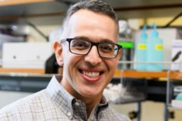
Albert Almada PhD ’13 studies the mechanics of how stem cells rebuild tissues. “Digging deep into the science is what MIT taught me,” he says.
Lillian Eden | Department of Biology
When Albert E. Almada PhD ’13 embarks on a new project, he always considers two criteria instilled in him during his time as a graduate student in the Department of Biology at MIT.
“If you want to make a big discovery, you have to approach it from a unique perspective — a unique angle,” Almada says. “You also have to be willing to dive into the unknown and go to the leading edge of your field.”
This is not without its challenges — but with an innovative spirit, Almada says, one can find ways to apply technologies and approaches to a new area of research where a roadmap doesn’t yet exist.
Now an assistant professor of orthopedic surgery and stem cell biology and regenerative medicine at the Keck School of Medicine of the University of Southern California (USC), Almada studies the mechanics of how stem cells rebuild tissues after trauma and how stem cell principles are dysregulated and drive conditions like degenerative disease and aging, exploring these topics through an evolutionary lens.
He’s also trying to solve a mystery that has intrigued scientists for centuries: Why can some vertebrate species like fish, salamanders, and lizards regenerate entire body parts, but mammals cannot? Almada’s laboratory at USC tackles these critical questions in the musculoskeletal system.
Almada’s fascination with muscle development and regeneration can be traced back to growing up in southern California. Almada’s brother had a degenerative muscle disease called Duchenne muscular dystrophy — and, while Almada grew stronger and stronger, his brother grew weaker and weaker. Last summer, Almada’s brother, unfortunately, lost his battle with his disorder at the age of 41.
“Watching his disease progress in those early years is what inspired me to become a scientist,” Almada recalls. “Sometimes science can be personal.”
Almada went to the University of California at Irvine for his undergraduate degree, majoring in biological sciences. During his summers, he participated in the Undergraduate Research Program (URP) at the Cold Spring Harbor Laboratory and the MIT Summer Research Program-Bio (now the Bernard S. and Sophie G. Gould MIT Summer Research Program in Biology, BSG-MSRP-Bio), where he saw the passion, rigor, and drive that solidified his desire to pursue a PhD.
Despite his interest in clinical applications, skeletal muscle, and regenerative biology, Almada was drawn to the Department of Biology at MIT, which is focused on basic fundamental research.
“I was willing to bet that it all came down to understanding basic cellular processes and things going wrong with the cell and how it interacts with its environment,” he says. “The MIT biology program really helped me define an identity for myself and gave me a template for how to tackle clinical problems from a molecular perspective.”
Almada’s PhD thesis work was based on a curious finding that Phillip Sharp, Institute Professor emeritus, professor emeritus of biology, and intramural faculty at the Koch Institute for Integrative Cancer Research, had made in 2007 — that transcription, the process of copying DNA into a messenger molecule called RNA, can occur in both directions at gene promoters. In one direction, it was long understood that fully formed mRNA is transcribed and can be used as a blueprint to make a protein. The transcription Sharp observed, in the opposite direction, results in a very short RNA that is not used as a gene product blueprint.
Almada’s project dug into what those short RNA molecules are — their structure and sequence, and why they’re not produced the same way that coding messenger RNA is. In two papers published in PNAS and Nature, Almada and colleagues discovered that a balance between splicing and transcription termination signals controls the length of an RNA. This finding has wider implications because toxic RNAs are produced and can build up in several degenerative diseases; being able to splice out or shorten RNAs to remove the harmful segments could be a potential therapeutic treatment.
“That experience convinced me that if I want to make big discoveries, I have to focus on basic science,” he says. “It also gave me the confidence that if I can succeed at MIT, I can succeed just about anywhere and in any field of biology.”
At the time Almada was in graduate school, there was a lot of excitement about transcription factor reprogramming. Transcription factors are the proteins responsible for turning on essential genes that tell a cell what to be and how to behave; a subset of them can even theoretically turn one cell type into another.
Almada began to wonder whether a specialized set of transcription factors instructs stem cells to rebuild tissues after trauma. After MIT, Almada moved on to a postdoctoral position in the lab of Amy Wagers, a leader in muscle stem cell biology at Harvard University, to immerse himself in this problem.
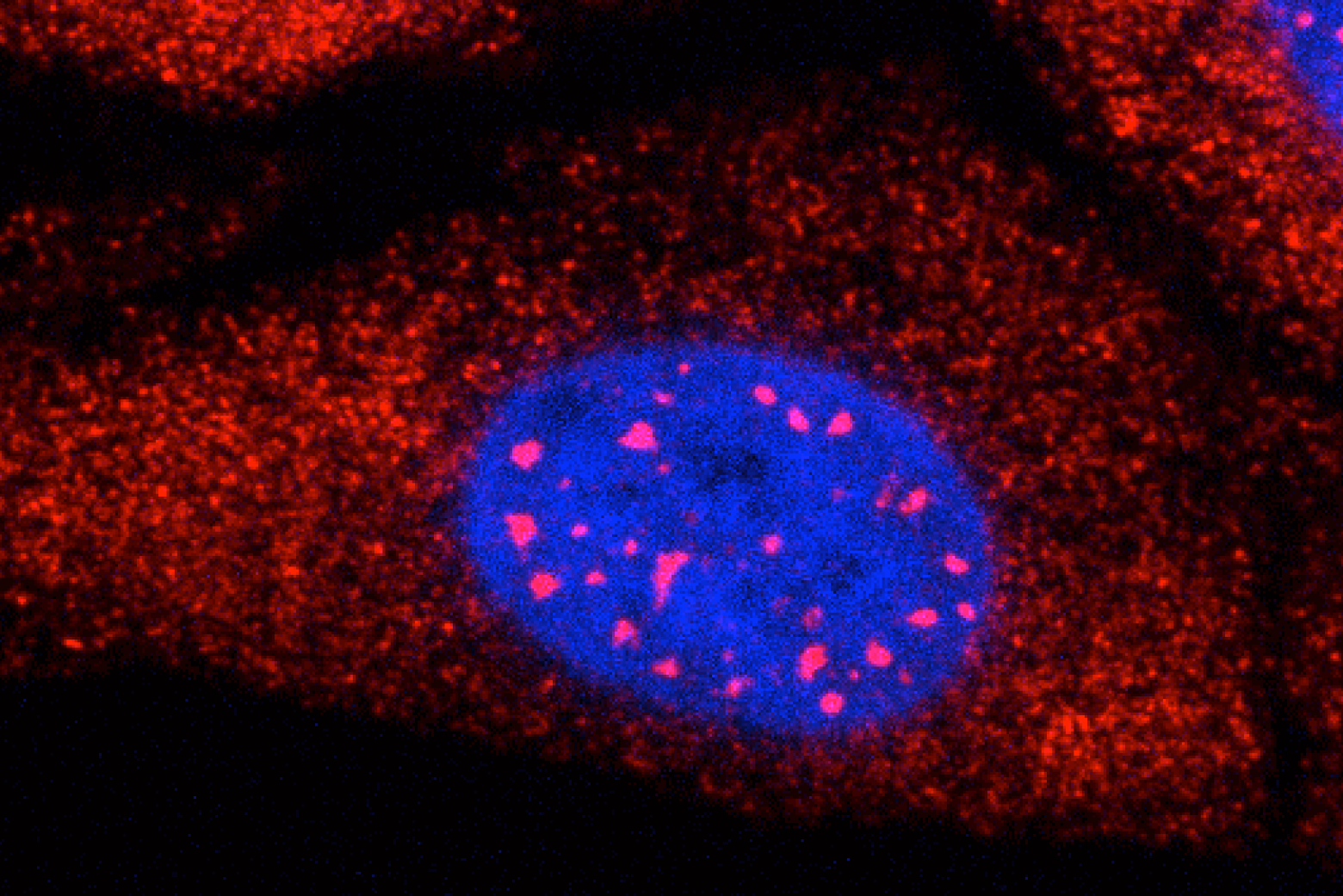
In many tissues in our bodies, a population of stem cells typically exists in an inactive, non-dividing state called quiescence. Once activated, these stem cells interact with their environment, sense damage signals, and turn on programs of proliferation and differentiation, as well as self-renewal, which is critical to maintaining a pool of stem cells in the tissue.
One of the biggest mysteries in the field of regenerative biology is how stem cells transition from dormancy into that activated, highly regenerative state. The body’s ability to turn on stem cells, including those in the skeletal muscle system, declines as we age and is often dysregulated in degenerative diseases — diseases like the one Almada’s brother suffered from.
In a study Almada published in Cell Reports several years ago, he identified a family of transcription factors that work together to turn on a critical regenerative gene program within hours of muscle trauma. This program drives muscle stem cells out of quiescence and speeds up healing.
“Now my lab is studying this regenerative program and its potential dysregulation in aging and degenerative muscle diseases using mouse and human models,” Almada says. “We’re also drawing parallels with super-healing species like salamanders and lizards.”
Recently, Almada has been working on characterizing the molecular and functional properties of stem cells in lizards, attempting to understand how the genes and pathways differ from mammalian stem cells. Lizards can regenerate massive amounts of skeletal muscle from scratch — imagine if human muscle tissue could be regrown as seamlessly as a lizard’s tail can. He is also exploring whether the tail is unique, or if stem cells in other tissues in lizards can regenerate faster and better than the tail, by comparing analogous injuries in a mouse model.
“This is a good example of approaching a problem from a new perspective: We believe we’re going to discover new biology in lizards that we can use to enhance skeletal muscle growth in vulnerable human populations, including those that suffer from deadly muscle disorders,” Almada says.
In just three years of starting his faculty position at USC, his work and approach have already received recognition in academia, with junior faculty awards from the Baxter Foundation and the Glenn Foundation/American Federation of Aging Research. He also received his first RO1 award from the National Institutes of Health with nearly $3 million in funding. Almada and his first graduate student, Alma Zuniga Munoz, were also awarded the HHMI Gilliam Fellowship last summer. Zuniga Munoz is the first to be recognized with this award at USC; fellowship recipients, student and advisor pairs, are selected with the goal of preparing students from underrepresented groups for leadership roles in science.
Almada himself is a second-generation Mexican American and has been involved in mentoring and training throughout his academic career. He was a graduate resident tutor for Spanish House at MIT and currently serves as the chair of the Diversity, Equity, and Inclusion Committee in the Department of Stem Cell Biology and Regenerative Medicine at USC; more than half of his lab members identify as members of the Hispanic community.
“The focus has to be on developing good scientists,” Almada says. “I learned from my past research mentors the importance of putting the needs of your students first and providing a supportive environment for everyone to excel, no matter where they start.”
As a mentor and researcher, Almada knows that no question and no challenge is off limits — foundations he built in Cambridge, where his graduate studies focused on teaching him to think, not just do.
“Digging deep into the science is what MIT taught me,” he says. “I’m now taking all of my knowledge in molecular biology and applying it to translationally oriented questions that I hope will benefit human health and longevity.”