
In 2020, Kathrin “Kat” Kajderowicz’s father passed away from lung cancer. Kajderowicz was in charge of her father’s health care for as long as she can remember. While he suffered from various cardiovascular issues for several years, it wasn’t until the beginning of the Covid-19 pandemic that he was diagnosed with late-stage metastatic small-cell lung cancer. Jumping into a primary caregiver position, she closely monitored the treatments he received from doctors to no avail. “I was frustrated with the many medications he was prescribed without the doctors fully understanding how they interacted with each other,” she says. Even if a single physician had been overseeing his comprehensive treatment plan, she says, they still could not definitively say whether the medication combinations have adverse effects that outweigh any positive impact.
This frustration set her on a scientific journey that has now culminated in her research as a PhD student at MIT’s Department of Brain and Cognitive Sciences (BCS) and the Whitehead Institute for Biomedical Research. “My experience led me to a significant medical problem: How can we eventually shift the medical paradigm to develop treatments that consider not only one specific pathway or problem but contextualize systemic tissue or organ dysfunction?”
To engage with this problem, Kajderowicz studies animals uniquely adapted to handle different stressors and environments, possibly modeling human disease states. “Perhaps we can turn to nature and see how different organisms have adapted to overcome and mitigate similar challenges,” she says.
Kajderowicz now works in Professor Siniša Hrvatin’s lab at Whitehead, where she researches cold tolerance. “I’m interested in exploring the mechanisms underlying cellular cold tolerance in hibernating organisms.” Engineering cold tolerance and stasis has many potential revolutionary future applications. In the near term, her work could improve organ transplantation and cell or tissue preservation. In the longer term, she hopes her work could catalyze a shift in the medical field away from its current crisis-mode approach: “By slowing down bodily processes and disease progression, a lower metabolic state could pave the way for a new class of hypothermic therapies that induce human hibernation-like states for cells, organs, or even whole organisms.”
First-generation student and scientist
Kajderowicz’s clearheaded pursuit of fundamental, large-scale scientific questions has propelled her impressive career as a young scientist. Recently, she was awarded the Paul and Daisy Soros Fellowship for New Americans, recognizing her unique path as the daughter of immigrants from Soviet Poland. Her parents arrived in the United States without having completed higher education degrees, without any savings, knowledge of English, medical insurance, or immigration papers. They worked hard to make a living — her father was a construction worker and her mother a housekeeper — using much of their earnings to become naturalized citizens.
Kajderowicz developed an early interest in a scientific career. “My parents, who didn’t go to college, didn’t push me toward any specific profession,” she says. “This gave me the freedom to explore any field I wanted, and my curiosity naturally led me to science.”
As a teenager, she worked as a golf caddie to help her parents financially. Clients at the golf course assisted her in obtaining internships at biotech and tech companies. Having won Best in Category at the Illinois State Science Fair, Kajderowicz received a substantial scholarship to support her studies at Cornell University, but she continued working to pay for her expenses and tuition. At Cornell, Kajderowicz joined the renowned Lab of Ornithology, where she applied machine-learning techniques to study songbird communication and other behavioral patterns.
Kajderowicz’s journey as a neuroscientist began at Harvard Medical School in Professor Connie Cepko’s lab, where she studied the developmental trajectory of a population of retinal interneurons. “Learning how to identify cell signatures was a fascinating introduction to the complexity of life. But I ultimately realized I wanted to pursue the questions that kept me up at night — both how we process information and how and why these processes change during aging. For me, these are life’s biggest unanswered questions, and I believe neuroscience is the foundation for everything. This led me to MIT’s Department of Brain and Cognitive Sciences.”
Learning from hamsters
Kajderowicz applied and was admitted to over two dozen graduate programs — “but I knew I wanted to go to MIT BCS. That was a no-brainer,” she says. “The department has faculty members in all levels of neuroscience: the cellular and molecular, systems, computational, and cognitive levels. It’s amazing to have all these people under one roof.”
Shortly after starting her graduate work at MIT, Kajderowicz realized she wanted to focus on the cellular level. “I think it’s important first to understand how things work within cells before focusing on function and systems.” She also seeks a translational avenue connecting theory and therapy, bridging the gap between basic science and applied treatment.
Kajderowicz found what she sought at the Whitehead Institute’s Hrvatin Lab and Weissman Lab. “It’s truly unique to have access to two very different communities at MIT. In BCS, I am seen as a biologist, while at the Whitehead Institute, I am more of a neuroscientist. It’s great having folks from different training backgrounds challenging my ideas and work.”
Instead of working directly on how cognition is encoded at the cellular level, Kajderowicz decided to embark on a project that would allow her to figure out how different species survive extreme stressors and environments. She is now developing tools to study cold tolerance across several species on the cellular level.
“Hibernating hamsters can safely endure prolonged durations during which their body temperature drops to 4 degrees [Celsius]. By taking a comparative species approach, I want to identify whether hibernators are uniquely genetically programmed to withstand these conditions or whether non-hibernators don’t activate these genetic pathways,” she says. Next, Kajderowicz hopes to figure out how to transfer or activate cold-protective effects to human cells and, someday, whole humans. While she isn’t directly studying the root of cognition, she hopes her research will help maintain or enhance cognitive functioning throughout aging by pushing the boundaries of the types of medicines and therapeutics available.
Building a scientific community
Kajderowicz’s involvement in the scientific community extends beyond her immediate work. At the height of the pandemic, she initiated a digital platform facilitating conversations on biotechnology trends among researchers, biotech professionals, venture capitalists, and others interested in staying updated on cutting-edge developments. Known as “DNA Deviants,” the community she built consists of several thousand active members on several social media platforms.
“It started with an informal journal club I had with some friends, where we would meet over coffee and discuss new papers. Then, when the pandemic shut down everything, I started a real-time podcast on the Clubhouse app with a friend, discussing emerging biotech trends. Eventually, it became an online journal club, and people just kept joining. We got experts to serendipitously join conversations within their realm of expertise from around the world.” Today, almost a dozen PhD, MD-PhD, and motivated undergraduates worldwide take turns leading conversations with different paper authors.
“It’s been incredibly rewarding to remain connected not only to my work, but also to gain a comprehensive understanding of what’s happening in the world,” Kajderowicz says. “You always need to look beyond your immediate circle.”
Sipping a beer on a warm summer evening, one might not consider that humans and yeast have been inextricably linked for thousands of years; winemaking, baking, and brewing all depend on budding yeast. Outside of baking and fermentation, researchers also use Saccharomyces cerevisiae, classified as a fungus, to study fundamental questions of cell biology.
Budding yeast gets its name from the way it multiplies. A daughter cell forms first as a swelling, protruding growth on the mother cell. The daughter cell projects further and further from the mother cell until it detaches as an independent yeast cell.
How do cells decide on a front and back? How do cells decode concentration gradients of chemical signals to orient in useful directions, or sense and navigate around physical obstacles? New Department of Biology faculty member Daniel “Danny” Lew uses the model yeast S. cerevisiae, and a non-model yeast with an unusual pattern of cell division, to explore these questions.
Q: Why is it useful to study yeast, and how do you approach the questions you hope to answer?
A: Humans and yeast are descended from a common ancestor, and some molecular mechanisms developed by that ancestor have been around for so long that yeast and mammals often use the same mechanisms. Many cells develop a front and migrate or grow in a particular direction, like the axons in our nervous system, using similar molecular mechanisms to those of yeast cells orienting growth towards the bud.
When I started my lab, I was working on cell cycle control, but I’ve always been interested in morphogenesis and the cell biology of how cells change shape and decide to do different things with different parts of themselves. Those mechanisms turn out to be conserved between yeast and humans.
But some things are very different about fungal and animal cells. One of the differences is the cell wall and what fungal cells have to do to deal with the fact that they have a cell wall.
Fungi are inflated by turgor pressure, which pushes their membranes against the rigid cell wall. This means they’ll die if there is any hole in the cell wall, which would be expected to happen often as cells remodel the wall in order to grow. We’re interested in understanding how fungi sense when any weak spots appear in the wall and repair them before those weak spots become dangerous.
Yeast cells, like most fungi, also mate by fusing with a partner. To succeed, they must do the most dangerous thing in the fungal lifecycle: get rid of the cell wall at the point of contact to allow fusion. That means they must be precise about where and when they remove the wall. We’re fascinated to understand how they know it is safe to remove the wall there, and nowhere else.
We take an interdisciplinary approach. We’ve used genetics, biochemistry, cell biology, and computational biology to try and solve problems in the past. There’s a natural progression: observation and genetic approaches tend to be the first line of attack when you know nothing about how something works. As you learn more, you need biochemical approaches and, eventually, computational approaches to understand exactly what mechanism you’re looking at.
I’m also passionate about mentoring, and I love working with trainees and getting them fascinated by the same problems that fascinate me. I’m looking to work with curious trainees who love addressing fundamental problems.
Q: How does yeast decide to orient a certain way—towards a mating partner, for example?
A: We are still working on questions of how cells analyze the surrounding environment to pick a direction. Yeast cells have receptors that sense pheromones that a mating partner releases. What is amazing about that is that these cells are incredibly small, and pheromones are released by several potential partners in the neighborhood. That means yeast cells must interpret a very confusing landscape of pheromone concentrations. It’s not apparent how they manage to orient accurately toward a single partner.
That got me interested in related questions. Suppose the cell is oriented toward something that isn’t a mating partner. The cell seems to recognize that there’s an obstacle in the way, and it can change direction to go around that obstacle. This is how fungi get so good at growing into things that look very solid, like wood, and some fungi can even penetrate Kevlar vests.
If they recognize an obstacle, they have to change directions and go around it. If they recognize a mating partner, they have to stick with that direction and allow the cell wall to get degraded. How do they know they’ve hit an obstacle? How do they know a mating partner is different from an obstacle? These are the questions we’d like to understand.
Q: For the last couple of years, you’ve also been studying a budding yeast that forms multiple buds when it reproduces instead of just one. How did you come across it, and what questions are you hoping to explore?
A: I spent several years trying to figure out why most yeasts make one bud and only one bud, which I think is related to the question of why migrating cells make one and only one front. We had what we thought was a persuasive answer to that, so seeing a yeast completely disobey that and make as many buds as it felt like was a shock, which got me intrigued.
We started working on it because my colleague,Amy Gladfelter, had sampled the waters around Woods Hole, Massachusetts. When she saw this specimen under a microscope, she immediately called me and said, “You have to look at this.”
A question we’re very intrigued by is if the cell makes five, seven, or 12 buds simultaneously, how do they divide the mother cell’s material and growth capacity five, seven, or 12 ways? It looks like all of the buds grow at the same rate and reach about the same size. One of our short-term goals is to check whether all the buds really get to exactly the same size or whether they are born unequal.
And we’re interested in more than just growth rate. What about organelles? Do you give each bud the same number of mitochondria, nuclei, peroxisomes, and vacuoles? That question will inevitably lead to follow-up questions. If each bud has the same number of mitochondria, how does the cell measure mitochondrial inheritance to do that? If they don’t have the same amount, then buds are each born with a different complement and ratio of organelles. What happens to buds if they have very different numbers of organelles?
As far as we can tell, every bud gets at least one nucleus. How the cell ensures that each bud gets a nucleus is a question we’d also very much like to understand.
We have molecular candidates because we know a lot about how model yeasts deliver nuclei, organelles, and growth materials from the mother to the single bud. We can mutate candidate genes and see if similar molecular pathways are involved in the multi-budding yeast and, if so, how they are working.
It turns out that this unconventional yeast has yet to be studied from the point of view of basic cell biology. The other thing that intrigues me is that it’s a poly-extremophile. This yeast can survive under many rather harsh conditions: it’s been isolated in Antarctica, from jet engines, from all kinds of plants, and of course from the ocean as well. An advantage of working with something so ubiquitous is we already know it’s not toxic to us under almost any circumstances. We come into contact with it all the time. If we learn enough about its cell biology to begin to manipulate it, then there are many potential applications, from human health to agriculture.
Despite the proliferation of novel therapies such as immunotherapy or targeted therapies, radiation and chemotherapy remain the frontline treatment for cancer patients. About half of all patients still receive radiation and 60-80 percent receive chemotherapy.
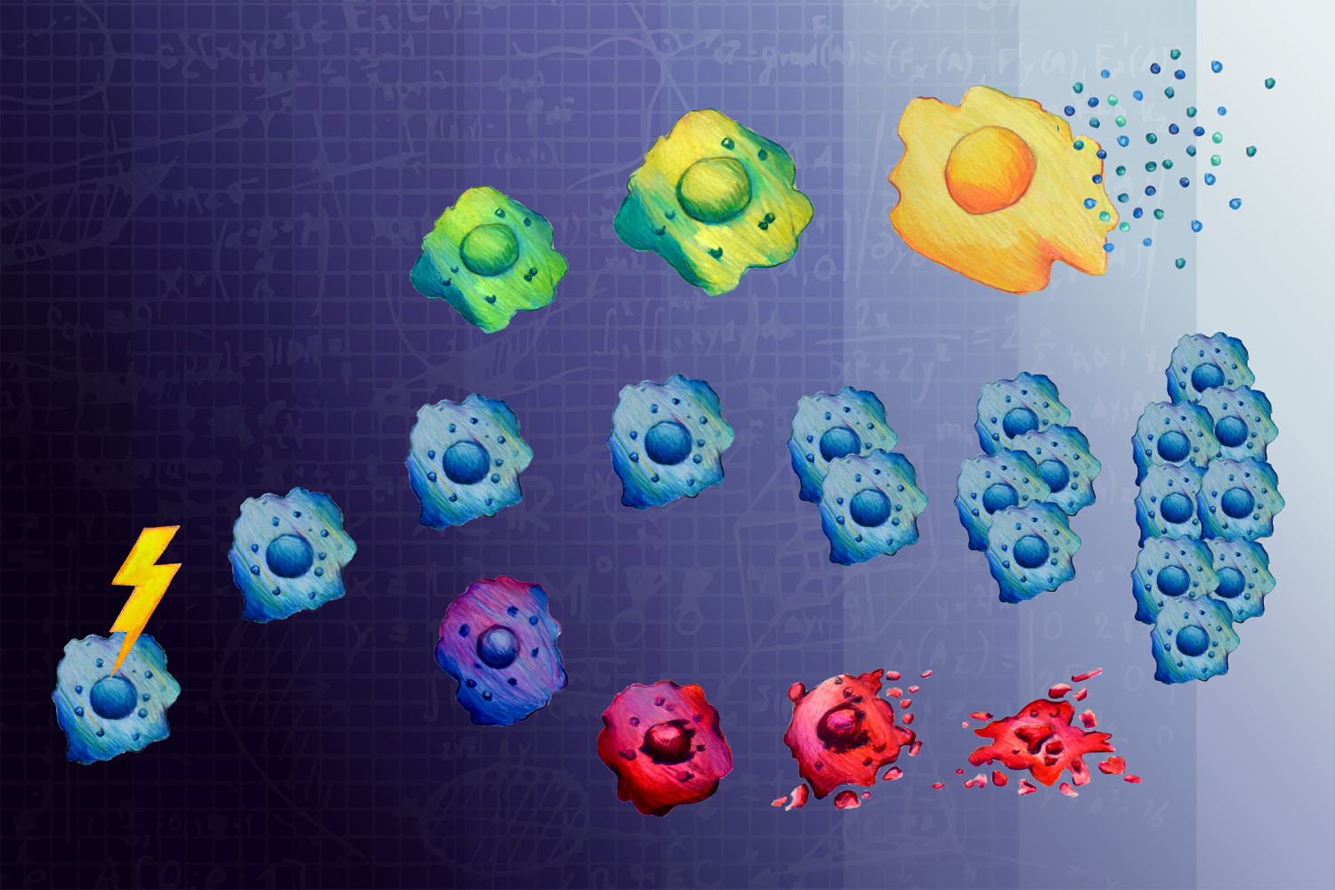
Both radiation and chemotherapy work by damaging DNA, taking advantage of a vulnerability specific to cancer cells. Healthy cells are more likely to survive radiation and chemotherapy since their mechanisms for identifying and repairing DNA damage are intact. In cancer cells, these repair mechanisms are compromised by mutations. When cancer cells cannot adequately respond to the DNA damage caused by radiation and chemotherapy, ideally, they undergo apoptosis or die by other means.
However, there is another fate for cells after DNA damage: senescence — a state where cells survive, but stop dividing. Senescent cells’ DNA has not been damaged enough to induce apoptosis but is too damaged to support cell division. While senescent cancer cells themselves are unable to proliferate and spread, they are bad actors in the fight against cancer because they seem to enable other cancer cells to develop more aggressively. Although a cancer cell’s fate is not apparent until a few days after treatment, the decision to survive, die, or enter senescence is made much earlier. But, precisely when and how that decision is made has not been well understood.
In an open-access study of ovarian and osteosarcoma cancer cells appearing July 19 in Cell Systems, MIT researchers show that cell signaling proteins commonly associated with cell proliferation and apoptosis instead commit cancer cells to senescence within 12 hours of treatment with low doses of certain kinds of chemotherapy.
“When it comes to treating cancer, this study underscores that it’s important not to think too linearly about cell signaling,” says Michael Yaffe, who is a David H. Koch Professor of Science at MIT, the director of the MIT Center for Precision Cancer Medicine, a member of MIT’s Koch Institute for Integrative Cancer Research, and the senior author of the study. “If you assume that a particular treatment will always affect cancer cell signaling in the same way — you may be setting yourself up for many surprises, and treating cancers with the wrong combination of drugs.”
Using a combination of experiments with cancer cells and computational modeling, the team investigated the cell signaling mechanisms that prompt cancer cells to enter senescence after treatment with a commonly used anti-cancer agent. Their efforts singled out two protein kinases and a component of the AP-1 transcription factor complex as highly associated with the induction of senescence after DNA damage, despite the well-established roles for all of these molecules in promoting cell proliferation in cancer.
The researchers treated cancer cells with low and high doses of doxorubicin, a chemotherapy that interferes with the function with topoisomerase II, an enzyme that breaks and then repairs DNA strands during replication to fix tangles and other topological problems.
By measuring the effects of DNA damage on single cells at several time points ranging from six hours to four days after the initial exposure, the team created two datasets. In one dataset, the researchers tracked cell fate over time. For the second set, researchers measured relative cell signaling activity levels across a variety of proteins associated with responses to DNA damage or cellular stress, determination of cell fate, and progress through cell growth and division.
The two datasets were used to build a computational model that identifies correlations between time, dosage, signal, and cell fate. The model identified the activities of the MAP kinases Erk and JNK, and the transcription factor c-Jun as key components of the AP-1 protein likewise understood to involved in the induction of senescence. The researchers then validated these computational findings by showing that inhibition of JNK and Erk after DNA damage successfully prevented cells from entering senescence.
The researchers leveraged JNK and Erk inhibition to pinpoint exactly when cells made the decision to enter senescence. Surprisingly, they found that the decision to enter senescence was made within 12 hours of DNA damage, even though it took days to actually see the senescent cells accumulate. The team also found that with the passage of more time, these MAP kinases took on a different function: promoting the secretion of proinflammatory proteins called cytokines that are responsible for making other cancer cells proliferate and develop resistance to chemotherapy.
“Proteins like cytokines encourage ‘bad behavior’ in neighboring tumor cells that lead to more aggressive cancer progression,” says Tatiana Netterfield, a graduate student in the Yaffe lab and the lead author of the study. “Because of this, it is thought that senescent cells that stay near the tumor for long periods of time are detrimental to treating cancer.”
This study’s findings apply to cancer cells treated with a commonly used type of chemotherapy that stalls DNA replication after repair. But more broadly, the study emphasizes that “when treating cancer, it’s extremely important to understand the molecular characteristics of cancer cells and the contextual factors such as time and dosing that determine cell fate,” explains Netterfield.
The study, however, has more immediate implications for treatments that are already in use. One class of Erk inhibitors, MEK inhibitors, are used in the clinic with the expectation that they will curb cancer growth.
“We must be cautious about administering MEK inhibitors together with chemotherapies,” says Yaffe. “The combination may have the unintended effect of driving cells into proliferation, rather than senescence.”
In future work, the team will perform studies to understand how and why individual cells choose to proliferate instead of enter senescence. Additionally, the team is employing next-generation sequencing to understand which genes c-Jun is regulating in order to push cells toward senescence.
This study was funded, in part, by the Charles and Marjorie Holloway Foundation and the MIT Center for Precision Cancer Medicine.
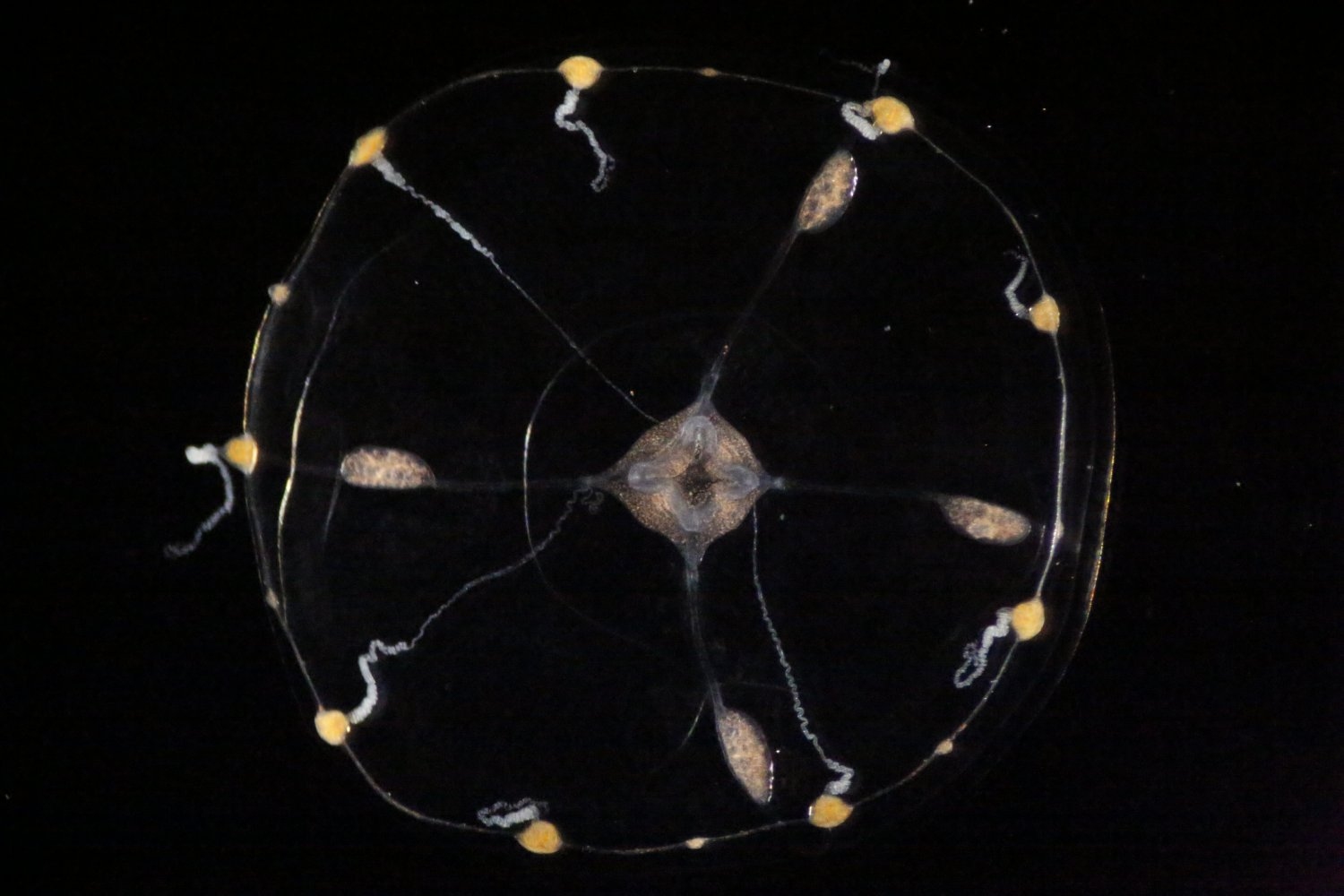
The Clytia hemisphaerica jellyfish is not only a hypnotically graceful swimmer, but also an amazing neuron-manufacturing machine with a remarkable ability to expand and regenerate its nervous system.
Now, thanks to a prestigious Klingenstein-Simons Fellowship Award in Neuroscience, MIT Assistant Professor Brady Weissbourd will study how the tiny, transparent animals use this ability to build, organize, and rebuild a stable, functional, and robust nervous system throughout their lives.
“As we look more broadly across the animal kingdom it is amazing to see how similar the basic biology is of animals that look completely different — even jellyfish have neurons similar to our own that generate their behavior,” says Weissbourd, a faculty member in MIT’s Department of Biology and The Picower Institute for Learning and Memory, whose work to engineer genetic access to C. hemisphaerica in 2021 established it as a new neuroscience model organism. “At the same time, it could be just as important to examine what is different across species, particularly when it comes to some of the incredible capabilities that have evolved.”
Weissbourd is just one of 13 researchers nationally to be recognized with this fellowship, which provides $300,000 over three years. It will enable Weissbourd’s lab to tackle several questions raised by the jellyfish’s prodigious production of neurons. Where does the constant stream of newborn neurons come from, and what guides them to their eventual places in the jellyfish’s mesh-like neural network? How does the jellyfish organize these ever-changing neural populations — for instance, into functional circuits — to enable its various behaviors?
Another question hails from the surprising results of an experiment in which Weissbourd ablated the entire class of the neurons that the jellyfish uses to fold up its umbrella-shaped body — about 10 percent of the 10,000 or so neurons that it has. He found that within a week enough new neurons had taken their place that the folding behavior was restored. Weissbourd’s studies will also seek to determine how the animal can so readily bounce back from the destruction of a whole major neural network and the behavior it produces.
“We were studying the neural control of a particular behavior and stumbled across this shocking observation that the subnetwork that controls this behavior is constantly changing size and can completely regenerate,” Weissbourd says. “We want to understand the mechanisms that allow this network to be so robust, including the ability to rebuild itself from scratch. I’m very grateful to the Klingenstein Fund and the Simons Foundation for supporting our work.”
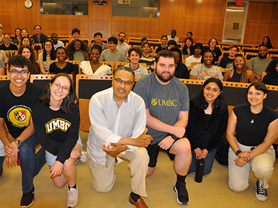
A group of more than 50 — predominantly MSRP-Bio students and alums and current students from the Meyerhoff Scholars program and the University of Maryland, Baltimore County — recently had the pleasure of sitting down for an informal chat at MIT with distinguished educator, author, and mathematician Freeman Hrabowski.
Hrabowski is widely credited for transforming UMBC into a world-renowned, innovative institution while serving as its president from 1992-2022. The educator also ushered in a generation of Black students to earn PhDs in science and engineering, co-founding the Meyerhoff Scholars Program at UMBC. Founded in 1988, the program has become a national model for increasing diversity in STEM. Hrabowski was also a member of the President’s Advisory Commission on Educational Excellence for African Americans during the Obama administration.
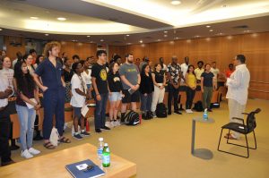
Hrabowski began by quoting poet William Carlos Williams “It is difficult to get the news from poems yet men die miserable every day for lack of what is found there,” and leading a call-and-response recitation of the poem Dreams by Langston Hughes as well as a mantra encouraging students to use their words, actions, and habits to shape their character and their destiny. Afterward, the students asked Hrabowski about his life and experiences.
“The audience of high-achieving students asked terrific, insightful questions reflecting their contemplation of their own paths,” says Biology Department head Amy Keating. “When students spoke up, Hrabowski engaged with them, and their ideas and perspectives were welcomed and respected. By the end of his time with them, almost everyone had their hand up and wanted to contribute to the lively discussion.”
Tobias Coombs, a Meyerhoff Scholars program alumnus and current graduate student in the Spranger Lab, says the event was an example of “classic Freeman Hrabowski”: Hrabowski injected the crowd with excitement and energy. Coombs also remarked that Hrabowski, named by Time as one of the world’s most influential people in 2012, acknowledged to the group that he’s shy, something Hrabowski is still pushing himself to overcome.
“He makes a point of being this down-to-earth person that you feel you can talk to about real issues and have real conversations with,” Coombs says. “He genuinely wants to motivate you to think science and math are cool.”
Before taking questions from the students in attendance, Hrabowski posed one to them: what do you think it takes to be successful in research in STEM? Among the responses were passion, curiosity, and a supportive community. After each response, Hrabowski encouraged a round of applause for each student brave enough to stand and give an answer because “Everybody needs support.”
“The way that you think about yourselves, the language that you use, the way that you interact with each other, and the values that you hold, will be so important. You become like the things that you love,” Hrabowski says.
For his lifetime of accomplishments increasing diversity in STEM, the Howard Hughes Medical Institute recently announced a new program named after Hrabowski. The HHMI Freeman Hrabowski Scholars were selected for their potential to become leaders in their research fields and to foster diverse and inclusive lab environments. The inaugural class of 31 scholars includes MIT Biology faculty members Seychelle Vos, the Robert A. Swanson Career Development Professor of Life Sciences, and Hernandez Moura Silva, an assistant professor and Ragon Institute core member, as well as MIT Biology and Cheeseman lab alumna Kara McKinley, PhD ’16.
Vos and Moura Silva were among the faculty attending the event, and both say Hrabowski was an inspiring guest to have on campus.
“Dr. Hrabowski’s smile, energy, and words are a true force of nature,” Hernandez says. “His words of wisdom showed us that we can all make the impossible possible by bringing a positive attitude to build a strong, supportive, and diverse community. It was such an honor to have him here.”
Biology department undergraduate officer Adam Martin says he noticed the pride in Hrabowski’s eyes when Hrabowski discussed what his trainees and faculty in his programs have accomplished. Biology department graduate officer Mary Gehring said his visit made her remember why she wanted to be a professor: “to help others follow their passions to their full potential.”
Hrabowski reflected on many topics, including the recent Supreme Court ruling on affirmative action. He pointed out that this was not the first time the Supreme Court had ruled on a racially conscious initiative, namely the 1995 decision that a UMBC scholarship program was unconstitutional. To continue the Meyerhoff Scholars Program, which was affected by the Supreme Court decision at the time, Hrabowski worked with Maryland’s Attorney General, found language and methods to encourage broad participation of diverse individuals, and focused on what the program was trying to achieve.
“My message to everyone was ‘where there’s a will, there’s a way.’ If the institution wants to continue to build diversity and broader participation, we can do it,” he says. “What we’re
working to achieve in the Meyerhoff program and in the Freeman Hrabowski Scholars program is to have everybody included.”
Hrabowski also offered advice on more everyday challenges: good students, himself included, can focus too much, forgetting to make time for other important aspects of their lives. He has learned to make time for Tai Chi, acupuncture, and getting his steps in; he encouraged the students similarly to take time for themselves outside work or school.
“When you can have fun and laugh, you’re a much better person. You can be a better thinker if you take care of yourself overall,” he says. “It’s the healthy person who can be most effective.”
As for being intimidated or nervous to talk to a superior, Hrabowski had the room roaring with laughter at his advice: “Just remember they go to the bathroom, too.”
Keating noted that Hrabowski engaged with the audience with energy, compassion, and humor.
She also observed, “No one can hide in Dr. Hrabowski’s classroom.”
“He put students front and center in his presentation, and his emphasis on the joys and importance of learning, knowledge, and achievement inspired us all to go back to the lab and classroom and be our best selves,” Keating says. “He acknowledged that paths in STEM demand much of us, and he encouraged students to have the discipline needed to stay the course while also taking care of themselves.”
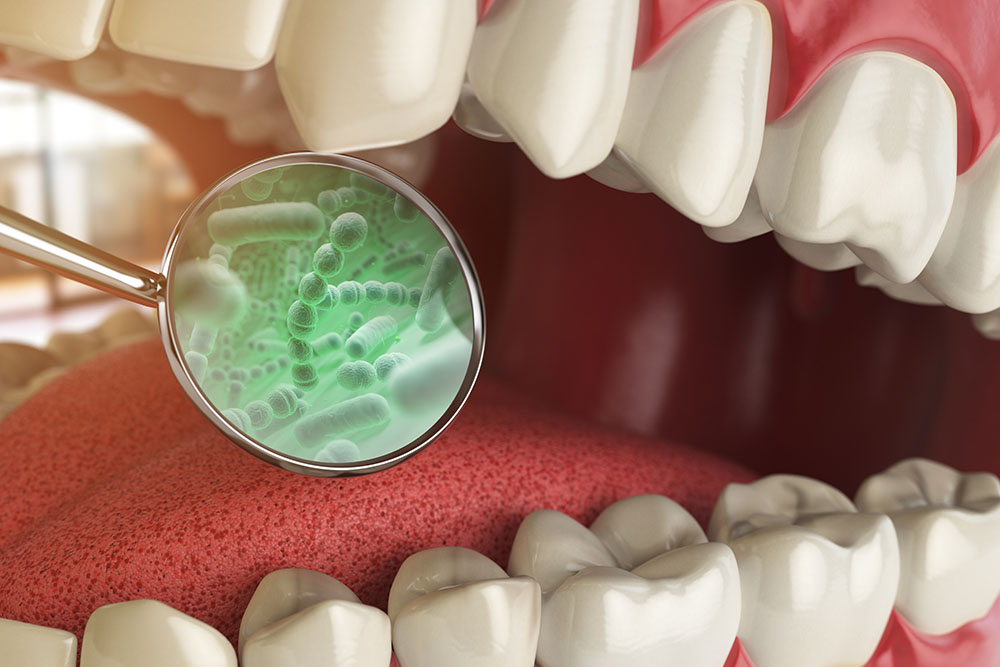
Your mouth is a crucial interface between the outside world and the inside of your body. Everything you breathe, chew, or drink interacts with your oral cavity — the proteins and the microbes, including microbes that can harm us. When things go awry, the result can range from the mild, like bad breath, to the serious, like tooth and gum decay, to more dire effects in the gut and other parts of the body.
Even though the oral microbiome plays a critical role as a front-line defense for human health and disease, we still know very little about the intricacies of host-microbe interactions in the complex physiological environment of the mouth; a better understanding of those interactions is key to developing treatments for human disease.
In a recent study published in PNAS, a team of scientists from MIT and elsewhere revealed that one of the most abundant proteins found in our saliva binds to the surface of select microbes found in the mouth. The findings shed light on how salivary proteins and mucus play a role in maintaining the oral cavity microbiome.
The collaboration involved members of the labs of Barbara Imperiali in the MIT Department of Biology and Laura Kiessling in the MIT Department of Chemistry, as well as the groups of Stefan Ruhl at the University at Buffalo School of Dental Medicine and Catherine Grimes at the University of Delaware.
The work is focused on an abundant oral cavity protein called zymogen granule protein 16 homolog B (ZG16B). Finding ZG16B’s interaction partners and gaining insight into its function were the overarching goals of the project. To accomplish this, Soumi Ghosh, a postdoc in the Imperiali lab, and colleagues engineered ZG16B to add reporter tags such as fluorophores. They called these modified proteins “microbial glycan analysis probes (mGAPs)” because they allowed them to identify ZG16B binding partners using complementary methods. They applied the probes to samples of healthy oral microbiomes to identify target microbes and binding partners.
The results excited them.
“ZG16B didn’t just bind to random bacteria. It was very focused on certain species, including a commensal bacteria called Streptococcus vestibularis,” says Ghosh, who is first author on the paper.
Commensal bacteria are found in a normal healthy microbiome and do not cause disease.
Using the mGAPs, the team showed that ZG16B binds to cell wall polysaccharides of the bacteria, which indicates that ZG16B is a lectin, a carbohydrate-binding protein. In general, lectins are responsible for cell-cell interactions, signaling pathways, and some innate immune responses against pathogens. “This is the first time that it has been proven experimentally that ZG16B acts as a lectin because it binds to the carbohydrates on the cell surface or cell wall of the bacteria,” Ghosh highlights.
ZG16B was also shown to recruit Mucin 7 (MUC7), a salivary glycoprotein in the oral cavity, and together the results suggest that ZG16B may help maintain a healthy balance in the oral microbiome by forming a complex with MUC7 and certain bacteria. The results indicate that ZG16B regulates the bacteria’s abundance by preventing overgrowth through agglutination when the bacteria exceed a certain level of growth.
“ZG16B, therefore, seems to function as a missing link in the system; it binds to different types of glycans — the microbial glycans and the mucin glycans — and ultimately, maintains a healthy balance in our oral cavity,” Ghosh says.
Further work with this probe and samples of oral microbiome from healthy and diseased subjects could also reveal the lectin’s importance for oral health and disease.
Current attention is focused on developing and applying additional mGAPs based on other human lectins, such as those found in serum, liver, and intestine to reveal their binding specificities and their roles in host-microbe interactions.
“The research carried out in this collaboration exemplifies the kind of synergy that made me excited to move to MIT five years ago,” says Kiessling. “I’ve been able to work with outstanding scientists who share my interest in the chemistry and the biology of carbohydrates.”
Kiessling and Imperiali, both senior authors of the paper, came up with the term for the probes they’re creating: “mGAPS to fill in the gaps” in our understanding of the role of lectins in the human microbiome, according to Ghosh.
“If we want to develop therapeutics against bacterial infection, we need a better understanding of host-microbe interactions,” Ghosh says. “The significance of our study is to prove that we can make very good probes for microbial glycans, find out their importance in the front-line defense of the immune system, and, ultimately, come up with a therapeutic approach to disease.”
This research was supported by the National Institute of Health.
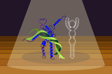
Transcription factors could be the Swiss Army knives of gene regulation; they are versatile proteins containing multiple specialized regions. On one end they have a region that can bind to DNA. On the other end they have a region that can bind to proteins. Transcription factors help to regulate gene expression—turning genes on or off and dialing up or down their level of activity—often in partnership with the proteins that they bind. They anchor themselves and their partner proteins to DNA at binding sites in genetic regulatory sequences, bringing together the components that are needed to make gene expression happen.
Transcription factors are a well-known family of proteins, but new research from Whitehead Institute Member Richard Young and colleagues shows that the picture we have had of them is incomplete. In a paper published in Molecular Cell on July 3, Young and postdocs Ozgur Oksuz and Jonathan Henninger reveal that along with DNA and protein, many transcription factors can also bind RNA. The researchers found that RNA binding keeps transcription factors near their DNA binding sites for longer, helping to fine tune gene expression. This rethinking of how transcription factors work may lead to a better understanding of gene regulation, and may provide new targets for RNA-based therapeutics.
“It’s as if, after carrying around a Swiss Army knife all your life for its blade and scissors, you suddenly realize that the odd, small piece in the back of the knife is a screwdriver,” Young says. “It’s been staring you in the face this whole time, and now that you finally see it, it becomes clear how many more uses there are for the knife than you had realized.”
A few papers, including one from Young’s lab, had previously identified individual transcription factors as being able to bind RNA, but researchers thought that this was a quirk of the specific transcription factors. Instead, Young, Oksuz, Henninger and collaborators have shown that RNA-binding is in fact a common feature present in at least half of transcription factors.
“We show that RNA binding by transcription factors is a general phenomenon,” Oksuz says. “Individual examples in the past were thought to be exceptions to the rule. Other studies dismissed signs of RNA binding in transcription factors as an artifact—an accident of the experiment rather than a real finding. The clues have been there all along, but I think earlier work was so focused on the DNA and protein interactions that they didn’t consider RNA.”
The reason that researchers had not recognized transcription factors’ RNA binding region as such is because it is not a typical RNA binding domain. Typical RNA binding domains form stable structures that researchers can detect or predict with current technologies. Transcription factors do not contain such structures, and so standard searches for RNA binding domains had not identified them in transcription factors.
Young, Oksuz and Henninger got their biggest clue that researchers might be overlooking something from the human immunodeficiency virus (HIV), which produces a transcription factor-like protein called Tat. Tat increases the transcription of HIV’s RNA genome by binding to the virus’ RNA and then recruiting cellular machinery to it. However, Tat does not contain a structured RNA binding site; instead, it binds RNA from a region called an arginine-rich motif (ARM) that is unstructured but has a high affinity for RNA. When the ARM binds to HIV RNA, the two molecules form a more stable structure together.
The researchers wondered if Tat might be more similar to human transcription factors than anyone had realized. They went through the list of transcription factors, and instead of looking for structured RNA binding domains, they looked for ARMs. They found them in abundance; the majority of human transcription factors contain an ARM-like region between their DNA and protein binding regions, and these sequences were conserved across animal species. Further testing confirmed that many transcription factors do in fact use their ARMs to bind RNA.
Next, the researchers tested to see if RNA binding affected the transcription factors’ function. When transcription factors had their ARMs mutated so they couldn’t bind RNA, those transcription factors were less effective in finding their target sites, remaining at those sites and regulating genes. The mutations did not prevent transcription factors from functioning altogether, suggesting that RNA binding contributes to fine-tuning of gene regulation.
Further experiments confirmed the importance of RNA binding to transcription factor function. The researchers mutated the ARM of a transcription factor important to embryonic development, and found that this led to developmental defects in zebrafish. Additionally, they looked through a list of genetic mutations known to contribute to cancer and heritable diseases, and found that a number of these occur in the RNA binding regions of transcription factors. All of these findings point to RNA binding playing an important role in transcription factors’ regulation of gene expression.
They may also provide therapeutic opportunities. The transcription factors studied by the researchers were found to bind RNA molecules that are produced in the regulatory regions of the genome where the transcription factors bind DNA. This set of transcription factors includes factors that can increase or decrease gene expression. “With evidence that RNAs can tune gene expression through their interaction with positive and negative transcription factors,” says Henninger, “we can envision using existing RNA-based technologies to target RNA molecules, potentially increasing or decreasing expression of specific genes in disease settings.”
Ozgur Oksuz, Jonathan E. Henninger, Robert Warneford-Thomson, Ming M. Zheng, Hailey Erb, Adrienne Vancura, Kalon J. Overholt, Susana Wilson Hawken, Salman F. Banani, Richard Lauman, Lauren N. Reich, Anne L. Robertson, Nancy M. Hannett, Tong I. Lee, Leonard I. Zon, Roberto Bonasio, Richard A. Young. “Transcription factors interact with RNA to regulate genes.” Molecular Cell, July 3, 2023. https://doi.org/10.1016/j.molcel.2023.06.012.
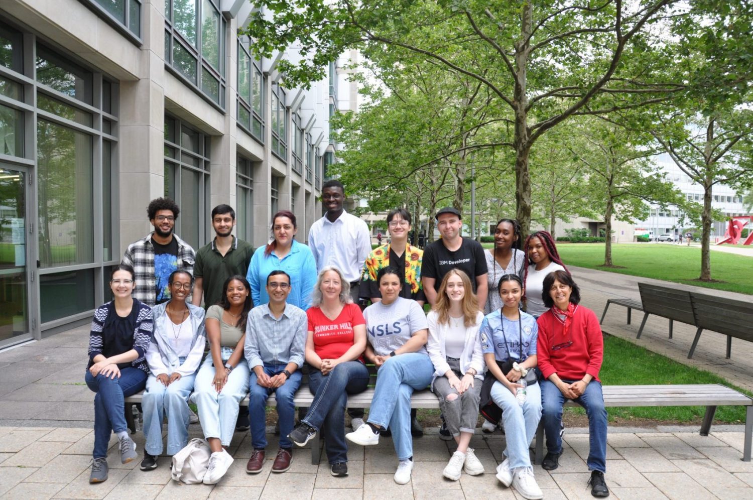
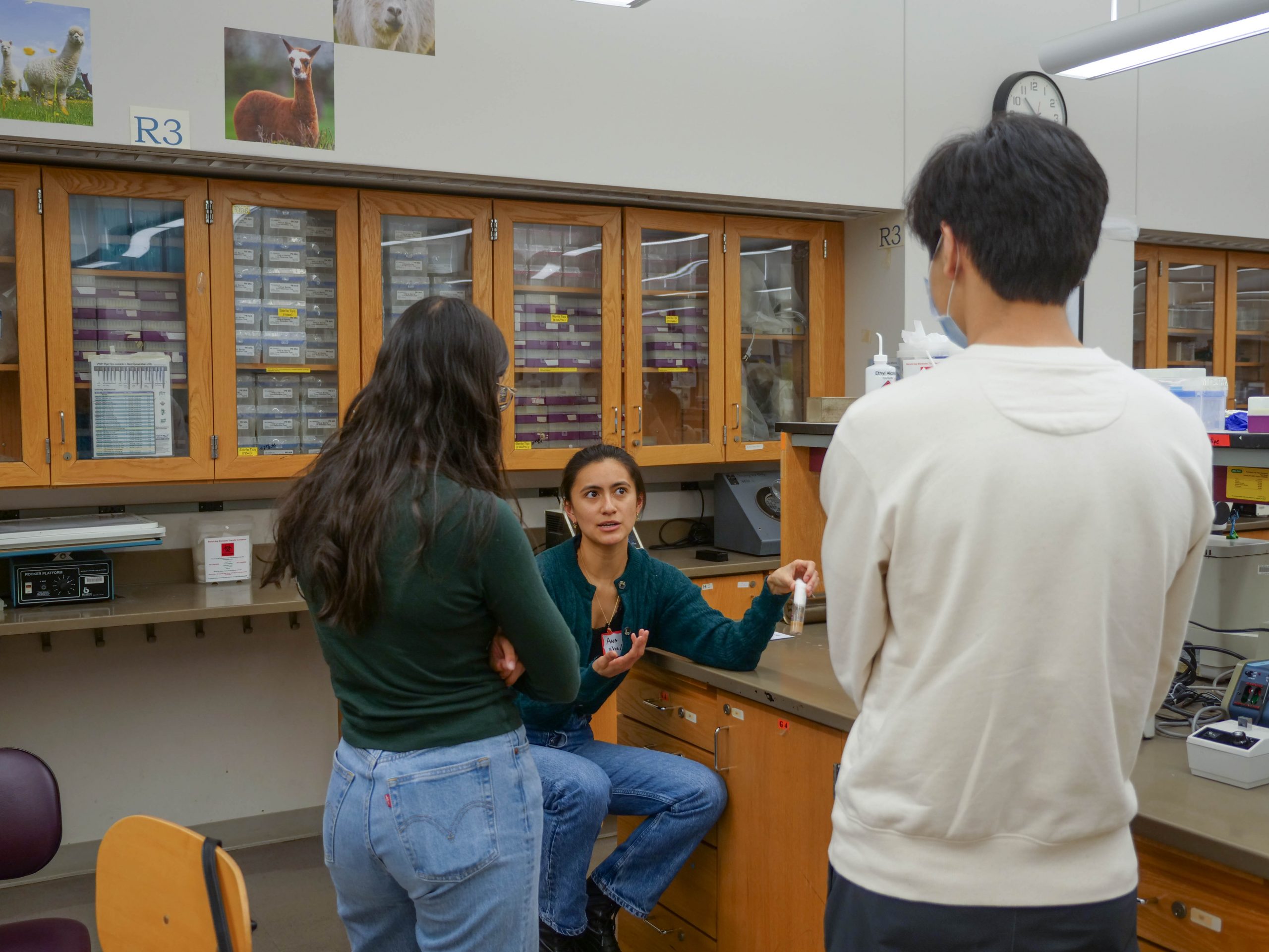
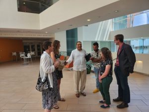
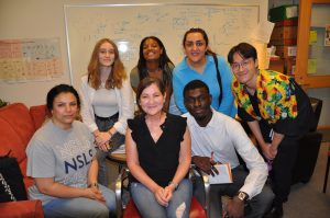
Although many undergraduates may be home for the summer, the halls and labs of MIT are still teeming with activity. On a sunny Thursday in June, 15 students from Bunker Hill Community College (BHCC) got to peek behind the curtain of research at MIT.
The Community College Partnership builds ties with two local community colleges that serve diverse, nontraditional students. The program was first conceived in 2020 as part of the biology department’s participation in #ShutDownSTEM, a day to consider equity and inclusion for marginalized communities and to educate and take action against injustice.
The visit is part of a larger effort to encourage students to pursue research opportunities and careers in research at and beyond MIT; other initiatives include virtual career panels and workshops for students at BHCC and Roxbury Community College. In addition, two community college students perform research as part of MRSP-Bio each summer, thanks to funding from the Packard Foundation acquired by Ankur Jain, Assistant Professor of Biology and Core Member of the Whitehead Institute.
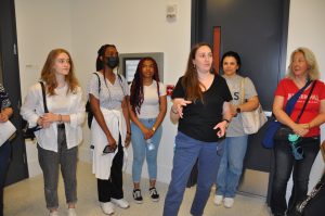
Sarah Sterling, Director of the Cryo-EM facility, loves giving tours because students ask such great questions, and the BHCC students were no exception: they were curious and inquisitive at every stop of their tour. Sterling explained that she chose her position, in part, because she enjoys “facilitating science”—helping researchers use cutting-edge equipment to find answers to their questions.
The students also visited labs in Building 68, Whitehead Institute, and the Picower Institute for Learning and Memory.
Reddien Lab postdoc Thomas Cooke described exploring mechanisms of regeneration in planarians, and Professor Laurie Boyer described the core questions underlying her research on heart development.
“The process of forming tissues and organs works sufficiently well that we’re all here, and we’re all relatively healthy,” Boyer says. “To me, that is remarkable.”
What isn’t well understood, she explains, is how faulty regulation can lead to disease and congenital malformations, and research using model systems can provide answers. For example, creating a model system in a dish can lead to a better understanding of the formation of circuits and molecular players. That, in turn, can lead to therapies or early diagnosis. The lab also works on tools to visualize what is occurring inside cells because “seeing is believing.”
“As scientists, we are not only trying to plan the best experiments possible, but we are also trying to develop new tools that push the boundaries of what we can discover,” she says. “Keeping an eye on the big picture is important because you’re never studying a problem in isolation. You’re studying a biological mechanism that has implications for many different things.”
It was “really eye-opening” to see what’s happening in some of the labs, according to BHCC student Robinson Le. Le is a dancer turned Biology major but had only ever come to campus for breakdancing practice—a skill they showed off to cheering BHCC students during lunch.
Badara Mbengue, another BHCC student, was excited to learn “what everyday life is like in the department.”
“It makes me very happy to see how much this has grown and continues to grow,” says Sheena Vasquez, PhD ‘23, who helped spearhead the initiative.
BHCC alums at MIT also showed students around the labs they are working in and shared their experiences at MIT, including as MSRP-Bio students, Quantitative Methods Workshop students, and as an undergraduate transfer student.
Libby Dunphy, a professor at BHCC, helped arrange the visit. She says the trip was an excellent opportunity for her students, who don’t get much exposure to real research.
“Seeing actual researchers, seeing that they’re real people, and that they’re nice, can help students imagine themselves in this place,” Dunphy says. “The Bunker Hill motto is ‘imagine the possibilities.’ And it’s cheesy, but we’re imagining the possibilities here.”
Boyer also offered advice for pursuing research at this stage in the students’ careers.
“The opportunities are unlimited, and so many people would be happy to support you—but sometimes, you have to ask,” Boyer advises. “Stay ambitious. You should be so proud of yourselves for embarking on this journey.”