Time and again, an unassuming roundworm has illuminated aspects of biology with major consequences for human health.
Jennifer Michalowski | McGovern Institute
December 12, 2025
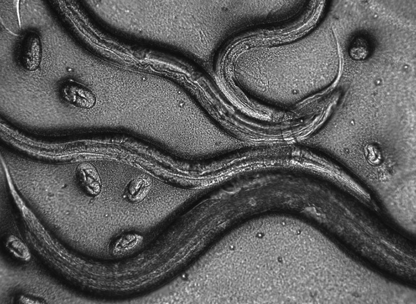
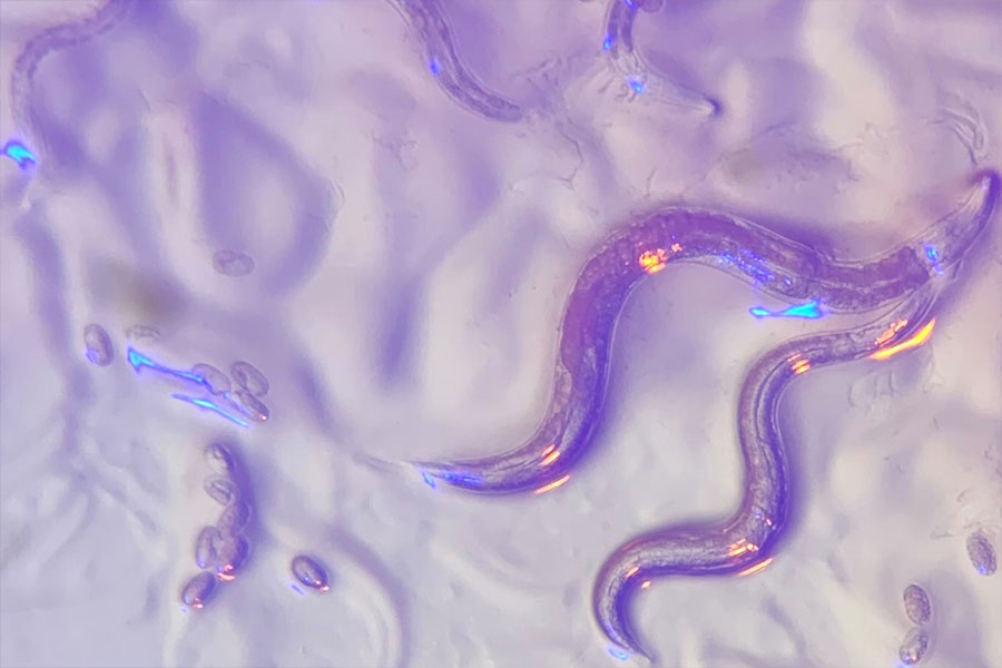
For decades, scientists with big questions about biology have found answers in a tiny worm. That worm–a millimeter-long creature called Caenorhabditis elegans–has helped researchers uncover fundamental features of how cells and organisms work. The impact of that work is enormous: Discoveries made using C. elegans have been recognized with four Nobel prizes and have led to the development of new treatments for human disease.
In a perspective piece published in the November 2025 issue of the journal PNAS, eleven biologists including Robert Horvitz, the David H. Koch (1962) Professor of Biology at MIT, celebrate Nobel Prize-winning advances made through research in C. elegans. The authors discuss how that work has led to advances for human health and highlight how a uniquely collaborative community among worm researchers has fueled the field.
MIT scientists are well represented in that community: The prominent worm biologists who coauthored the PNAS paper include former MIT graduate students Andy Fire and Paul Sternberg, now at Stanford University and the California Institute of Technology, and two past postdoctoral researchers in Horvitz’s lab, University of Massachusetts Medical School professor Victor Ambros and Massachusetts General Hospital investigator Gary Ruvkun. Ann Rougvie at the University of Minnesota is the paper’s corresponding author.
Early worm discoveries
“This tiny worm is beautiful—elegant both in its appearance and in its many contributions to our understanding of the biological universe in which we live,” says Horvitz, who in 2002 was awarded the Nobel Prize in Medicine along with colleagues Sydney Brenner and John Sulston for discoveries that helped explain how genes regulate programmed cell death and organ development. Horvitz is also a member of MIT’s McGovern Institute for Brain Research and Koch Institute for Integrative Cancer Research as well as an investigator at the Howard Hughes Medical Institute.
Those discoveries were among the early successes in C. elegans research, made by pioneering scientists who recognized the power of the microscopic roundworm. C. elegans offers many advantages for researchers: The worms are easy to grow and maintain in labs; their transparent bodies make cells and internal processes readily visible under a microscope; they are cellularly very simple (e.g., they have only 302 nerve cells, compared with about 100 billion in a human) and their genomes can be readily manipulated to study gene function.
Most importantly, many of the molecules and processes that operate in C. elegans have been retained throughout evolution, meaning discoveries made using the worm can have direct relevance to other organisms, including humans. “Many aspects of biology are ancient and evolutionarily conserved,” Horvitz explains. “Such shared mechanisms can be most readily revealed by analyzing organisms that are highly tractable in the laboratory.”
In the 1960s, Brenner, a molecular biologist who was curious about how animals’ nervous systems develop and function, recognized that C. elegans offered unique opportunities to study these processes. Once he began developing the worm into a model for laboratory studies, it did not take long for other biologists to join him to take advantage of the new system.
In the 1970s, the unique features of the worm allowed Sulston to track the transformation of a fertilized egg into an adult animal, tracing the origins of each of the adult worm’s 959 cells. His studies revealed that in every developing worm, cells divide and mature in predictable ways. He also learned that some of the cells created during development do not survive into adulthood and are instead eliminated by a process termed programmed cell death.
By seeking mutations that perturbed the process of programmed cell death, Horvitz and his colleagues identified key regulators of that process, which is sometimes referred to as apoptosis. These regulators, which both promote and oppose apoptosis, turned out to be vital for programmed cell death across the animal kingdom.
In humans, apoptosis shapes developing organs, refines brain circuits, and optimizes other tissue structures. It also modulates our immune systems and eliminates cells that are in danger of becoming cancerous. The human version of CED-9, the anti-apoptotic regulator that Horvitz’s team discovered in worms, is BCL-2. Researchers have shown that activating apoptotic cell death by blocking BCL-2 is an effective treatment for certain blood cancers. Today, researchers are also exploring new ways of treating immune disorders and neurodegenerative disease by manipulating apoptosis pathways.
Collaborative worm community
Horvitz and his colleagues’ discoveries about apoptosis helped demonstrate that understanding C. elegans biology has direct relevance to human biology and disease. Since then, a vibrant and closely connected community of worm biologists—including many who trained in Horvitz’s lab—has continued to carry out impactful work. In their PNAS article, Horvitz and his coauthors highlight that early work, as well as the Nobel Prize-winning work of:
- Andrew Fire and Craig Mello, whose discovery of an RNA-based system of gene silencing led to powerful new tools to manipulate gene activity. The innate process they discovered in worms, known as RNA interference, is now used as the basis of six FDA-approved therapeutics for genetic disorders, silencing faulty genes to stop their harmful effects.
- Martin Chalfie, who used a fluorescent protein made by jellyfish to visualize and track specific cells in C. elegans, helping launch the development of a set of tools that transformed biologists’ ability to observe molecules and processes that are important for both health and disease.
- Victor Ambros and Gary Ruvkun, who discovered a class of molecules called microRNAs that regulate gene activity not just in worms, but in all multicellular organisms. This prize-winning work was started when Ambros and Ruvkun were postdoctoral researchers in Horvitz’s lab. Humans rely on more than 1,000 microRNAs to ensure our genes are used at the right times and places. Disruptions to microRNAs have been linked to neurological disorders, cancer, cardiovascular disease, and autoimmune disease, and researchers are now exploring how these small molecules might be used for diagnosis or treatment.
Horvitz and his coauthors stress that while the worm itself made these discoveries possible, so too did a host of resources that facilitate collaboration within the worm community and enable its scientists to build upon the work of others. Scientists who study C. elegans have embraced this open, collaborative spirit since the field’s earliest days, Horvitz says, citing the Worm Breeder’s Gazette, an early newsletter where scientists shared their observations, methods, and ideas.
Today, scientists who study C. elegans—whether the organism is the centerpiece of their lab or they are looking to supplement studies of other systems—contribute to and rely on online resources like WormAtlas and WormBase, as well as the Caenorhabditis Genetics Center, to share data and genetic tools. Horvitz says these resources have been crucial to his own lab’s work; his team uses them every day.
Just as molecules and processes discovered in C. elegans have pointed researchers toward important pathways in human cells, the worm has also been a vital proving ground for developing methods and approaches later deployed to study more complex organisms. For example, C. elegans, with its 302 neurons, was the first animal for which neuroscientists successfully mapped all of the connections of the nervous system. The resulting wiring diagram, or connectome, has guided countless experiments exploring how neurons work together to process information and control behavior. Informed by both the power and limitations of the C. elegans’ connectome, scientists are now mapping more complex circuitry, such as the 139,000-neuron brain of the fruit fly, whose connectome was completed in 2024.
C. elegans remains a mainstay of biological research, including in neuroscience. Scientists worldwide are using the worm to explore new questions about neural circuits, neurodegeneration, development, and disease. Horvitz’s lab continues to turn to C. elegans to investigate the genes that control animal development and behavior. His team is now using the worm to explore how animals develop a sense of time and transmit that information to their offspring.
Also at MIT, Steven Flavell’s team in the Department of Brain and Cognitive Sciences and the Picower Institute for Learning and Memory is using the worm to investigate how neural connectivity, activity, and modulation integrate internal states, such as hunger, with sensory information, such as the smell of food, to produce sometimes long-lasting behaviors. Flavell is Horvitz’s academic grandson, as Flavell trained with one of Horvitz’s postdoctoral trainees. As new technologies accelerate the pace of scientific discovery, Horvitz and his colleagues are confident that the humble worm will bring more unexpected insights.