Mass. Professor Goes Viral After Putting Crib in Office to Help Grad Student with Infant Daughter
People Magazine
May 20, 2021

A Massachusetts professor is being praised for his kind actions after setting up a crib in his office to help a graduate student who has a 10-month-old child.

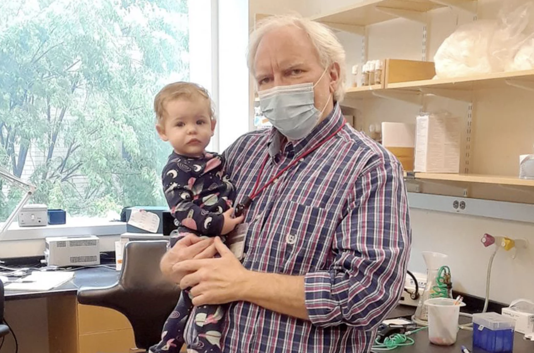
Dr. Troy Littleton, a professor of neuroscience at the Massachusetts Institute of Technology, recently went viral on Twitter after revealing that he had set up a travel crib for a grad student’s daughter.

Good Morning America identified the student as Karen Cunningham and her 10-month-old daughter, Katie.

“My favorite new equipment purchase for the lab – a travel crib to go in my office so my graduate student can bring her little girl to work when necessary and I get to play with her while her mom gets some work done. Win-win!!” Littleton tweeted on May 7 beside a photo of the crib.

“This is what equity and support of women look like!” wrote one user. “This port-a-cot image has made my day. It is so NOT hard if you care to think of inclusive solutions.”

“This is beautiful!” added another person. “We need more workplaces like this that consider caretaking as a healthy & necessary part of an adult work life, not a distraction from it.”

Speaking to GMA, Littleton — who serves as director of MIT’s Molecular and Cellular Neuroscience Graduate Program and runs their research lab — explained that he set up the crib for Cunningham as a late baby shower gift.

“Usually in non-pandemic times we always have baby showers for expectant mothers and fathers where we give them gifts and we weren’t able to do that with Karen because of the pandemic, so this was sort of the lab gift for Karen, 10 months later,” he told the outlet. “It’s always a challenge [being a parent while in graduate school] so anything you can do in a lab to facilitate and help out, we try.”

Added Cunningham: “The first year of being a parent is hard and it’s helpful to have a lot of support. I think during the pandemic parents have been isolated from a lot of their support so [the crib] is definitely an add-on and a really wonderful one.”

Recently, Littleton decided to post the image of the crib after Cunningham brought her daughter to the lab for the first time, not knowing the massive response his tweet would receive, according to GMA.

“The tweet came from just being delighted to be able to see Katie for the first time and to have the opportunity on occasion, when Karen wants to bring her in, to be able to play with her a little bit,” Littleton explained to the outlet. “That was the genesis of the tweet, not from any idea it was going to create this large discussion about the challenges mothers face in the workplace.”

In response to his tweet, which has gained over 117,000 likes and 8,000 retweets, some users pointed out the difficulties that moms face with a work-life balance.

One user even called the moment a “sad substitute for parental leave and daycare,” leading to a discussion about the challenges of paying for child care services.

Though the response was completely unexpected, Littleton told GMA he was pleased with the productive conversations that were taking place on social media.

“I’m glad it had that effect because we need to be solving these issues, both in academia and on a broader level as well,” he shared with the outlet, noting that MIT does currently offer resources for working moms, including a daycare on its campus.

“It’s highlighted that this is a really important issue for our community,” he added.

According to Cunningham, her husband usually watches Katie each day and will continue to do so until she begins daycare in the fall. Still, she said, she appreciates having a backup option with MIT and Littleton.

“There’s the solid, focused six to eight hours of work that you wouldn’t want to bring a baby in for, but then there’s the lab errands that you do here and there and that’s when it’s really useful,” Cunningham explained to GMA. “I can put Katie down and just go do something quick and I can see her and talk to her and she can nap in there. It’s great.”

“One of the reasons I picked MIT was because I got a really positive response from the biology department when I brought up the fact that I was definitely going to want to have a baby during grad school,” she added. “I was thinking about that the whole time.”

While she’s grateful for the support from MIT and Littleton, Cunningham told GMA she hopes other schools, professors and employees will follow their lead.

“It’s really easy to look at the systemic challenges facing parents and moms in our country … and kind of throw up your hands and be like, ‘Well it’s huge. I can’t fix that,'” she explained. “But then these sort of local ways that people in positions of power can protect parents against the systemic things, like what Troy’s been doing in creating a really supportive and inclusive lab, I think that does make a really big difference and it’s great to have an example of that.”

As for Littleton, he said he doesn’t see his gesture as anything out of the ordinary and commended moms everywhere for their ability to balance both work and their personal life.

“I wish people were able to spot the real hero here,” he wrote on May 9 in response to his viral tweet. “It’s the graduate student mom, not me. She’s amazing to do all she has to with her daughter and still keep up her thesis project research. Happy Mother’s Day to all – they deserve it!”

The proteins that package DNA to fit inside cells have another role: tuning gene expression
Raleigh McElvery
May 19, 2021

The DNA inside a single human cell is several meters long — yet it must be condensed to fit inside a space one-tenth the diameter of a hair. That’s like stretching a string from Philadelphia, Pennsylvania to Washington, D.C., and then trying to stuff it into a soccer ball. Imagine then organizing all of this information for each of the body’s 3 trillion cells! The DNA is condensed by proteins called histones that create a spool around which the DNA can wrap itself. How tightly the DNA is wound determines whether it is accessible enough for other proteins to bind to and copy into RNA, toggling gene expression levels up or down.

One specialized type of histone, H2A.Z, is ubiquitous and essential among multicellular organisms. But there have been conflicting reports about how it affects gene expression, especially during embryonic development.

Several years ago, Laurie Boyer’s lab at MIT was the first to show that H2A.Z wraps the DNA located around the start sites of most genes, where the molecular machine RNA polymerase II (RNAPII) binds to copy the DNA into RNA. Boyer’s team demonstrated that removing H2A.Z prevented embryonic cells from turning on genes that are important for forming organs and tissues. But scientists still weren’t sure how H2A.Z exerted its effects.

Now, in a recent Nature Structural and Molecular Biology study, a team from the Boyer lab, led by former postdoc Constantine Mylonas, has revealed how H2A.Z regulates the ability of RNAPII to properly transcribe DNA into the messages that specify all cell types in the body. The researchers found that in embryonic stem cells, H2A.Z serves as a “yellow traffic light,” signaling RNAPII to slow the process of transcribing DNA into RNA. Although there are other proteins that also contribute to RNAPII pausing, H2A.Z establishes a second barrier to transcription that allows gene expression to be tuned in response to developmental signals.

“H2A.Z appears to regulate how fast RNAPII begins to transcribe DNA, and this allows the cell time to respond to important cues that ultimately direct a stem cell to become a brain or heart cell, for example,” says Boyer, a professor of biology and biological engineering. “This connection was a critical missing piece of the puzzle, and explains why H2A.Z is essential for development across all multicellular organisms.”

Illustration of molecules
As RNAPII starts to transcribe a gene, it encounters a cluster of eight histones (a “nucleosome”) including H2A.z, which slows its progression — allowing for tuning of gene expression in response to developmental signals.

According to Boyer, H2A.Z’s role in gene expression has been difficult to pin down because previous approaches only provided static snapshots of how proteins interact with DNA days after loss of the histone. Boyer’s team overcame this shortcoming by leveraging a system that allowed for targeted degradation of H2A.Z within hours. They combined this technique with high-resolution genomic approaches and live cell imaging of RNAPII dynamics using super-resolution microscopy. With help from Ibrahim Cissé’s lab, they were able to visualize RNAPII dynamics in real time at the single molecule level in embryonic stem cells. Upon loss of H2A.Z, they found a remarkable increase in RNAPII movement in the cells, consistent with their genomic results showing a faster release of RNAPII and an increase in transcription in the absence of H2A.Z.

Next, the researchers plan to determine precisely how H2A.Z is targeted to the start sites of genes and how it forms a barrier to RNAPII passage.

Boyer says pinpointing the way histone variants like H2A.Z control gene expression is fundamental to understanding how developmental decisions are made, and will help researchers understand why misregulation of H2A.Z has been linked to diseases such as cancer.

“Emerging evidence indicates that DNA ‘packaging proteins’ like histones directly participate in how RNAPII can read and transcribe DNA,” she explains, “and that crucial connection wasn’t clear before.”

Image credits: courtesy of Laurie Boyer
Top image: Live cell super-resolution imaging showing RNAPII dynamics at a single molecule level in embryonic stem cells. The bright and colored clusters represent RNAPII molecules.

Citation:
“A dual role for H2A. Z. 1 in modulating the dynamics of RNA Polymerase II initiation and elongation.”
Nature Structural & Molecular Biology, online May 10, 2021, DOI: 10.1038/s41594-021-00589-3
Constantine Mylonas, Choongman Lee, Alexander L. Auld, Ibrahim I. Cisse, and Laurie A. Boyer.

Childhood hobbies jump-start a research career

MIT Biology junior Eduardo Canto tinkered with science long before he started studying Treacher Collins syndrome in the Calo lab.

Saima Sidik | Department of Biology
May 19, 2021

In seventh grade, Eduardo Canto wanted a dog. His mom said no, though. She didn’t want to spend her days vacuuming fur. They reached a compromise: Canto was allowed to have pet fish. Soon Canto’s disappointment with his new pets turned to curiosity. While he couldn’t train the fish to sit or roll over, he decided that breeding the fish could be a fun pastime.

An internet search told Canto that some aquarists use dried Indian almond leaves, a traditional Asian herbal remedy, to stimulate fish breeding, although no one is quite sure how the leaves do this. However, finding Indian almond leaves presented a problem for a kid without an Amazon account living far from the tree’s native habitat. On a whim, Canto picked up some similar-looking leaves in a park near his house in Puerto Rico. He knew they weren’t from an Indian almond tree, but he put them in the tank anyhow, just to see what would happen. A few days later, he noticed a collection of eggs attached to the bottom of a leaf!

Canto often took on little experiments like this, which caused his grandfather to predict early on that he would have a scientific career. Eight years after the breeding endeavor, Canto is fulfilling his grandfather’s prediction by studying Course 7 (Biology) at MIT, where he’s currently in his third year of a bachelor’s degree. Once again, fish have come into Canto’s life — he’s working in Eliezer Calo’s lab, where researchers use zebrafish to study a genetic disorder called Treacher Collins syndrome, which causes deformities in eyes, ears, cheekbones, and chins.

Throughout middle school and high school, Canto dipped his toes into many scientific disciplines. School science fairs motivated him to build a dry ice-powered trolley, a solar-powered water heater, and start a vegetable garden.

Sometimes, he admits, his motivation for joining science clubs wasn’t lofty. “I joined the math club because I got to miss a day of school every year for their annual competition,” he says with a laugh. But he also talks excitedly about his early experiments, particularly in biology. “I’ve always loved working with my hands,” he says.

Canto’s father, a medical doctor, encouraged his son’s interest by letting Canto shadow him at work. He also started a molecular biology summer program at Canto’s high school that taught students how to pipette and do simple experiments. By the time Canto applied to college, he was convinced he wanted to study biology, and MIT drew his attention because of its reputation as a top science school with excellent biology teachers. He knew it was the right choice for him when he attended Campus Preview Weekend, and found a large Puerto Rican community ready to welcome him. Even far from the island, he felt at home.

Canto has kept up with his roots since joining MIT by playing on a soccer team for Puerto Rican students. He’s also become part of a new community in a lab run by Eliezer Calo — who is a Puerto Rican himself. The lab is interested in ribosomes, the molecular machines that build proteins. Treacher Collins syndrome arises when cells can’t make ribosomes properly, and Canto wants to understand why that is.

Before Canto joined the Calo lab, the group had already started studying a protein called DDX21 that’s involved in making ribosomes in both humans and zebrafish. When genetic mutations in zebrafish cause DDX21 to go to the wrong part of the cell, the fish develop jaw deformations that mirror Treacher Collins syndrome. The Calo lab thinks cells with mislocalized DDX21 probably don’t produce ribosomes as well as normal cells, but they’re still testing this hypothesis.

Canto wants to probe the relationship between DDX21 and Treacher Collins syndrome further, but fish reproduce slowly, so they’re not ideal organisms for his research. Instead, he’s built a strain of Escherichia coli bacteria that carry DDX21 in place of the equivalent bacterial gene. DDX21 helps these bacteria survive the stress associated with cold temperatures, so without it, the bacteria will die in the cold. Canto hopes to take advantage of this trait by finding small molecules that stop the bacteria from growing at low temperatures — just like a DDX21 mutation would. Studying how these molecules bind DDX21 will help him understand which parts of this protein are important for its function.

The possibility that this work will one day reveal how Treacher Collins syndrome develops in patients is rewarding to Canto, and in fact he hopes helping patients will soon become his life’s focus. He wants to attend medical school, and eventually become a doctor. The human physiology class he took last semester was one of his favorites, even though it was over Zoom due to the COVID-19 pandemic. Becoming a doctor will let him help others while studying topics he finds fascinating. “Medicine is like biology on steroids!” he says.

And who knows — one day after he’s a doctor, maybe he’ll even get that pet he’s always wanted. But unlike Canto’s interest in biology, some of his interests have evolved over time. These days, he prefers cats over dogs.

Photo credit: Saima Sidik
Posted: 5.19.21
Whitehead Institute appoints two new faculty members
Merrill Meadow | Whitehead Institute
May 4, 2021

Whitehead Institute director Ruth Lehmann announced the appointment of two dynamic new Members: Olivia Corradin, currently a Whitehead Fellow, and Sinisa Hrvatin, currently an instructor and postdoctoral fellow at Harvard Medical School. Both will also become assistant professors of biology at Massachusetts Institute of Technology (MIT). Corradin’s joint appointments begin in July 2021, Hrvatin’s in January 2022.

“Both Olivia and Sinisa are creative, collaborative, and highly accomplished early-career scientists,” says Lehmann. “Each has impressed the Whitehead Institute and MIT faculties with their drive, intellect, and their scientific vision. We look forward to their contributions — as researchers, educators, and colleagues — for many years to come.”

Corradin investigates gene variants, small differences in DNA sequence, which can prompt disease-causing changes in gene regulation. During her nearly five years as a Whitehead Fellow, her lab defined the concept of “outside variants,” which helps to explain how genetic variants increase one’s likelihood of developing disease. She also developed a method to identify the cell type affected by a specific disease-linked variant; and then used it to single out oligodendrocytes as one type of brain cell involved in multiple sclerosis. Most recently, Corradin created an approach for defining epigenetic variation — which is caused by factors other than DNA sequence changes — in some individuals with opioid use disorder; this will help researchers’ identify genes associated with the disorder.

Before becoming the Scott Cook and Signe Ostby Fellow at Whitehead Institute in 2016, Corradin earned a PhD at Case Western Reserve University. There her research focused on genetic and epigenetic dysregulation in human disease, and she pioneered approaches to predict gene targets of regulatory DNA sequences associated with variants.

“I’m incredibly excited to be stepping into this new stage at Whitehead Institute and MIT Biology,” Corradin says. “I look forward to continued collaboration and to becoming a part of the rich history that shapes our Institute.”

Hrvatin investigates how organisms enter torpor and hibernation and how their cells adapt and survive in these states. As a postdoctoral research fellow in the lab of Harvard Medical School neurobiologist Michael Greenberg, Hrvatin established an experimental paradigm for studying a hibernation-like behavior in mice — and used this system to discover the neurons that control entry into this state. In addition, he pioneered the Paralleled Enhancer Single Cell Assay platform — a new method to generate cell-type-specific AAV vectors that can be used for targeted human gene therapy, as well as to control defined neuronal cell types across species, including in hibernating animals.

Hrvatin earned a PhD in stem cell and regenerative medicine from Harvard University, where he studied the process of directed differentiation from human embryonic stem cells to pancreatic beta cells. After his graduate work, he served as a postdoctoral associate at MIT in the lab of Daniel Anderson, where he investigated approaches for targeted siRNA delivery to pancreatic beta cells. Hrvatin also founded ReadCube, a startup dedicated to disseminating access to scientific literature and developing reference management tools for research scientists.

“I’ve always been inspired by the exceptional scientists, educators, pioneers, and visionaries at the Whitehead Institute and MIT Biology,” Hrvatin says. “I am absolutely thrilled for the opportunity to learn from and become a part of this extraordinary community.”

These worms’ stem cells are developmental shapeshifters
Eva Frederick | Whitehead Institute
April 20, 2021

Planarians are small water-dwelling worms known for their regenerative capacity. If you chop one into ten pieces, you’ll end up with ten fully-formed worms.

While humans have pools of specialized stem cells that can create our regenerative body parts like hair and skin, these worms owe their regenerative superpowers to a special kind of stem cell called a neoblast. At least some of these cells are “pluripotent,” meaning that they can divide to create almost any cell type in a worm’s body at any time. Neoblasts are actually the only dividing cells in planarians — fully committed cells like those in the eyes or intestines cannot divide again.

“The big question for us is, how does a neoblast go from being able to make anything, to making one particular thing?” says Amelie Raz, a postdoctoral researcher at Whitehead Institute who conducted her graduate research in the lab of Whitehead Institute Member Peter Reddien. “How do they go from being able to make anything in the body to being, say, an intestine cell that’s going to stay an intestine cell until it dies?”

Now, in a paper published online April 20 in the journal Cell Stem Cell, researchers at Whitehead Institute lay out a new model for how these stem cells commit to their fates and go on to create fully differentiated cells. The process of cellular differentiation is often viewed as a hierarchy, with one special stem cell at the top which can take a number of potential paths to arrive at a specialized state. This is generally thought to take place over a series of cell divisions in which each generation’s fate is gradually restricted.

“We’re proposing something happens that is very different from the conventional view,” says senior author Reddien, who is also a professor of biology at Massachusetts Institute of Technology and an investigator with the Howard Hughes Medical Institute. “We think that stem cells can make broad jumps in state without going through a series of fate-restricting divisions. We call it the single-step fate model.”

In the new model, neoblasts that are on a path toward creating skin cells or intestine cells can produce progeny cells that can switch fates to create cells of other types. The work is a step in the long road to understanding these worms’ regenerative capacities, and could possibly inform regenerative medicine approaches far in the future.

“The ability of planarian stem cells to essentially switch their fate is really, really powerful,” says Raz, the first author of the paper. “Obviously this is a long way off, but theoretically the concept of stem cell fate switching could be applied to regenerative medicine, with human stem cell programming.”

Upturning the hierarchy

Neoblasts can be sorted into many “classes.” For example, one class of neoblasts contains all the materials to make skin cells, and others have the necessary toolkit to form the worms’ primitive kidneys or their intestines. According to the hierarchical model, these specialized neoblasts are intermediaries between a pluripotent cell at the top of the hierarchy, and the non-dividing body cells.

“You can imagine that the special cell at the top is a blank slate with no predisposition towards any cell type — it can make anything,” says Raz. “This is how we’ve often imagined development works.”

But Raz, Reddien and Omri Wurtzel, a former postdoc in the Reddien lab now at Tel Aviv University, started to question this assumption after noticing a few mysterious properties of planarian cells.

First of all, researchers have observed in the past that when a planarian is treated with radiation to kill all existing stem cells, a single neoblast can rescue the animal by forming a colony containing many different classes of neoblasts. If, as previous theories suggested, there was a single class of neoblast that gave rise to all these types, Raz and Reddien reasoned that that class should be a common resident in every colony that formed. After creating many of these colonies and analyzing their composition, however, the researchers saw that this was not the case. “For every class we looked at, there were plenty of colonies that lacked that class altogether,” says Reddien. “There was no unique class present in all colonies.”

Another sticking point: the researchers began to realize that, when applying the hierarchy model, the math of planarian cell divisions and potency just didn’t add up. In a prior cell transplantation study, the Reddien lab found that many of the neoblasts they tested were pluripotent —in this study they found that proportion to be larger than what they would expect if only non-specialized neoblasts were pluripotent. “When you add up all the different kinds of specialized neoblasts, it’s at least three quarters of the neoblast population, and almost certainly higher than that.” says Raz. Therefore, the researchers wondered if some specialized neoblasts could be pluripotent as well.

Another study from the Reddien lab showed that skin-specialized neoblasts did not retain skin fate through more than one cell division. Also, in about half of all cell divisions in planarians, the two daughter cells will be different from one another. This raised the possibility that specialized neoblasts can divide asymmetrically as a possible route to stem cells changing fate.

Furthermore, the timeline for regeneration was off — the rate at which planarians were able to regrow body parts didn’t allow for several rounds of fate-restricting divisions.

After conducting experiments to study these different situations, Raz, Wurtzel, and Reddien were able to create a case for their new model of cell differentiation. “What we think is happening is that planarians have a ton of plasticity in their general stem cell population, where individual cells can move in and out of different specialized stages through the process of cell division in order to give rise to what is required,” Raz says.

“This is just the beginning of exploring this process, even though we’ve been studying it for many years,” Reddien says. “Focusing on the model, we’re suggesting that the cells can choose one fate, and then through the process of a division with an asymmetric outcome, one of the daughter cells can now divide again and choose a different fate. That fate switching process might be fundamental to explaining pluripotency.”

Reddien’s lab will continue investigating the mechanisms of neoblast fate specification, including how specialization lines up with the timing of the cell cycle.

“Understanding the structure of cell lineage and how fate choices are made is fundamental to understanding adult stem cell biology, and how in the context of injury and repair, new cells can be brought about,” says Reddien. “Do they have to go through long, complex lineage trajectories? Or can they make big jumps in state from stem cells to the final state? How flexible is that? All of these things have potential implications for understanding stem cell biology broadly, and we hope that the work will highlight some of these mechanisms and provide opportunities to explore general principles in the future.”

Two heads are better than one, but two disciplines are even better

How biologists and mathematicians reached across departmental lines to solve a long-standing problem in electron microscopy

Saima Sidik | Department of Biology
April 19, 2021

MIT’s Hockfield Court is bordered on the west by the ultra-modern Stata Center, with its reflective, silver alcoves that jut off at odd angles, and on the east by Building 68, which is a simple, window-lined, cement rectangle. At first glance, Bonnie Berger’s mathematics lab in the Stata Center and Joey Davis’s biology lab in Building 68 are as different as the buildings that house them. And yet, a recent collaboration between these two labs shows how their disciplines complement each other. The partnership started when Ellen Zhong, a graduate student from the Computational and Systems Biology (CSB) Program, decided to use a computational pattern-recognition tool called a neural network to study the shapes of molecular machines. Three years later, Zhong’s project is letting scientists see patterns that run beneath the surface of their data, and deepening their understanding of the molecules that shape life.

Zhong’s work builds on a technique from the 1970s called cryo-electron microscopy (cryo-EM), which lets researchers take high-resolution images of frozen protein complexes. Over the last decade, better microscopes and cameras have led to a “resolution revolution” in cryo-EM that’s allowed scientists to see individual atoms within proteins. But, as good as these images are, they’re still only static snapshots. In reality, many of these molecular machines are constantly changing shape and composition as cells carry out their normal functions and adjust to new situations.

Along with former Berger lab member Tristan Belper, Zhong devised software called cryoDRGN. The tool uses neural nets to combine hundreds of thousands of cryo-EM images, and shows scientists the full range of three-dimensional conformations that protein complexes can take, letting them reconstruct the proteins’ motion as they carry out cellular functions. Understanding the range of shapes that protein complexes can take helps scientists develop drugs that block viruses from entering cells, study how pests kill crops, and even design custom proteins that can cure disease. COVID-19 vaccines, for example, work partly because they include a mutated version of the virus’s spike protein that’s stuck in its active conformation, so vaccinated people produce antibodies that block the virus from entering human cells. Scientists needed to understand the variety of shapes that spike proteins can take in order to figure out how to force spike into its active conformation.

Two women standing by rock wall
Graduate student Ellen Zhong (right), and her co-advisor, Professor of Mathematics Bonnie Berger (left)

Getting off the computer and into the lab

Zhong’s interest in computational biology goes back to 2011 when, as a chemical engineering undergrad at the University of Virginia, she worked with Professor Michael Shirts to simulate how proteins fold and unfold. After college, Zhong took her skills to a company called D. E. Shaw Research, where, as a Scientific Programmer, she took a computational approach to studying how proteins interact with small molecule drugs.

“The research was very exciting,” Zhong says, “but all based on computer simulations. To really understand biological systems, you need to do experiments.”

This goal of combining computation with experimentation motivated Zhong to join MIT’s CSB PhD program, where students often work with multiple supervisors to blend computational work with bench work. Zhong “rotated” in both the Davis and Berger labs, then decided to combine the Davis lab’s goal of understanding how protein complexes form with the Berger lab’s expertise in machine learning and algorithms. Davis was interested in building up the computational side of his lab, so he welcomed the opportunity to co-supervise a student with Berger, who has a long history of collaborating with biologists.

Davis himself holds a dual bachelor’s degree in computer science and biological engineering, so he’s long believed in the power of combining complementary disciplines. “There are a lot of things you can learn about biology by looking in a microscope,” he says. “But as we start to ask more complicated questions about entire systems, we’re going to require computation to manage the high-dimensional data that come back.”

Before rotating in the Davis lab, Zhong had never performed bench work before — or even touched a pipette. She was fascinated to find how streamlined some very powerful molecular biology techniques can be. Still, Zhong realized that physical limitations mean that biology is much slower when it’s done at the bench instead of on a computer. “With computational research, you can automate experiments and run them super quickly, whereas in the wet lab, you only have two hands, so you can only do one experiment at a time,” she says.

Zhong says that synergizing the two different cultures of the Davis and Berger labs is helping her become a well-rounded, adaptable scientist. Working around experimentalists in the Davis lab has shown her how much labor goes into experimental results, and also helped her to understand the hurdles that scientists face at the bench. In the Berger lab, she enjoys having coworkers who understand the challenges of computer programming.

“The key challenge in collaborating across disciplines is understanding each other’s ‘languages’,” Berger says. “Students like Ellen are fortunate to be learning both biology and computing dialects simultaneously.”

Bringing in the community

Man smiling outside
Zhong’s second co-advisor, Professor Joey Davis

Last spring revealed another reason for biologists to learn computational skills: these tools can be used anywhere there’s a computer and an internet connection. When the COVID-19 pandemic hit, Zhong’s colleagues in the Davis lab had to wind down their bench work for a few months, and many of them filled their time at home by using cryo-EM data that’s freely available online to help Zhong test her cryoDRGN software. The difficulty of understanding another discipline’s language quickly became apparent, and Zhong spent a lot of time teaching her colleagues to be programmers. Seeing the problems that non-programmers ran into when they used cryoDRGN was very informative, Zhong says, and helped her create a more user-friendly interface.

Although the paper announcing cryoDRGN was only recently published, the tool created a stir as soon as Zhong posted her code online, many months prior. The cryoDRGN team thinks this is because leveraging knowledge from two disciplines let them visualize the full range of structures that protein complexes can have, and that’s something researchers have wanted to do for a long time. For example, the cryoDRGN team recently collaborated with researchers from Harvard and Washington University to study locomotion of the single-celled organism Chlamydomonas reinhardtii. The mechanisms they uncovered could shed light on human health conditions, like male infertility, that arise when cells lose the ability to move. The team is also using cryoDRGN to study the structure of the SARS-CoV-2 spike protein, which could help scientists design treatments and vaccines to fight coronaviruses.

Zhong, Berger, and Davis say they’re excited to continue using neural nets to improve cryo-EM analysis, and to extend their computational work to other aspects of biology. Davis cited mass spectrometry as “a ripe area to apply computation.” This technique can complement cryo-EM by showing researchers the identities of proteins, how many of them are bound together, and how cells have modified them.

“Collaborations between disciplines are the future,” Berger says. “Researchers focused on a single discipline can take it only so far with existing techniques. Shining a different lens on the problem is how advances can be made.”

Zhong says it’s not a bad way to spend a PhD, either. Asked what she’d say to incoming graduate students considering interdisciplinary projects, she says: “definitely do it.”

Undergraduate students meld biology and art to forge remote collaborations
April 14, 2021

Taught for the first time in 2013, 7.016 (Introductory Biology) introduces MIT undergraduates to fundamental principles of biochemistry, molecular biology, and genetics. While the class has historically packed over 200 students into a lecture hall, the past two iterations have been held over Zoom due to COVID-19 restrictions. In order to incite collaboration and spur creativity in a remote setting, Professor of Biology and Chemistry, Barbara Imperiali, Associate Professor of Biology, Adam Martin, and MITxBio Instructor, Monika Avello, have infused the homework assignments with some whimsey.

One bonus question on a problem set required students to work together in teams to devise a cartoon of the Statue of Liberty. Inspired by a drawing in Chemical & Engineering News that depicted Lady Liberty clad in chemistry gear, Imperiali, Martin, and Avello asked the students to re-imagine the cartoon with a biology theme instead.

“We wanted to create a light-hearted, fun, and rewarding opportunity for 7.016 students to collaborate and connect with each other in our remote class,” Avello says. “We were totally blown away by how creative and talented the students were. So many of them went above and beyond by modifying the cartoon template we provided to showcase their creativity and artistic skills.”
Below is a sampling of responses from the assignment.
Black and white sketch of the Statue of Liberty
Credit: Gary Nguyen and Sandra Tang

This biology-inspired Statue of Liberty features a crown made of deoxyribose (the 5-carbon sugar in DNA). Per lab protocol, she has also donned safety goggles and disposable gloves. She holds some DNA in one hand and a microscope inspecting a cell in the other.

Cartoon of Statue of Liberty
Credit: Cindy Jie and Dion Sukhram

A DNA sash, hydroxyl eyes, and a crown of SARS-CoV-2 spike proteins highlight some of the details that were central to class discussions this semester. With a textbook in hand and her mitochondria torch held high, this cute comic lifted students’ spirits.

Statue of Liberty and a sunset
Credit: Andrew Emmel and Yoni Haile

In one hand, Lady Liberty is holding a microscope to symbolize discovery. She is holding a petri dish in the other hand, indicating that data are absolute. Her crown is a centrifuge, because the experiment is “king.” And the sunglasses? Those are to show that biology is cool.

Cartoon of Statue of Liberty as a red blood cell
Credit: Joanna Cao and Sarah Wei

Here, Lady Liberty takes the form of a red blood cell, which carries oxygen all over the body. As such, she is holding an oxygen molecule in one hand. In the other hand, she holds carbon dioxide.

Sketch of the Statue of Liberty made of molecules
Credit: Savannah Ashley, Katia Pendowski, and Malia Smith

This cartoon features Lady Liberty standing on the lipid bilayer that constitutes the membrane encircling a cell’s internal components. She is holding a DNA molecule and wearing a dress with deoxyribose molecules, which form the “backbone” of DNA. She is also sporting a crown with adenosine triphosphate (ATP) molecules — providing energy to drive cellular processes — and holding a torch of proteins.

An on-off switch for gene editing
Eva Frederick | Whitehead Institute
April 9, 2021

Now, in a paper published online in Cell on April 9, researchers describe a new gene editing technology called CRISPRoff that allows researchers to control gene expression with high specificity while leaving the sequence of the DNA unchanged. Designed by Whitehead Institute Member Jonathan Weissman, University of California San Francisco assistant professor Luke Gilbert, Weissman lab postdoc James Nuñez and collaborators, the method is stable enough to be inherited through hundreds of cell divisions, and is also fully reversible.

“The big story here is we now have a simple tool that can silence the vast majority of genes,” says Weissman, who is also a professor of biology at MIT and an investigator with the Howard Hughes Medical Institute. “We can do this for multiple genes at the same time without any DNA damage, with great deal of homogeneity, and in a way that can be reversed. It’s a great tool for controlling gene expression.”

The project was partially funded by a 2017 grant from the Defense Advanced Research Projects Agency to create a reversible gene editor. “Fast forward four years [from the initial grant], and CRISPRoff finally works as envisioned in a science fiction way,” says co-senior author Gilbert. “It’s exciting to see it work so well in practice.”

Genetic engineering 2.0

The classic CRISPR-Cas9 system uses a DNA-cutting protein called Cas9 found in bacterial immune systems. The system can be targeted to specific genes in human cells using a single guide RNA, where the Cas9 proteins create tiny breaks in the DNA strand. Then the cell’s existing repair machinery patches up the holes.

Because these methods alter the underlying DNA sequence, they are permanent. Plus, their reliance on “in-house” cellular repair mechanisms means it is hard to limit the outcome to a single desired change. “As beautiful as CRISPR-Cas9 is, it hands off the repair to natural cellular processes, which are complex and multifaceted,” Weissman says. “It’s very hard to control the outcomes.”

That’s where the researchers saw an opportunity for a different kind of gene editor — one that didn’t alter the DNA sequences themselves, but changed the way they were read in the cell.

This sort of modification is what scientists call “epigenetic” — genes may be silenced or activated based on chemical changes to the DNA strand. Problems with a cell’s epigenetics are responsible for many human diseases such as Fragile X syndrome and various cancers, and can be passed down through generations.

Epigenetic gene silencing often works through methylation — the addition of chemical tags to to certain places in the DNA strand — which causes the DNA to become inaccessible to RNA polymerase, the enzyme which reads the genetic information in the DNA sequence into messenger RNA transcripts, which can ultimately be the blueprints for proteins.

Weissman and collaborators had previously created two other epigenetic editors called CRISPRi and CRISPRa — but both of these came with a caveat. In order for them to work in cells, the cells had to be continually expressing artificial proteins to maintain the changes.

“With this new CRISPRoff technology, you can [express a protein briefly] to write a program that’s remembered and carried out indefinitely by the cell,” says Gilbert. “It changes the game so now you’re basically writing a change that is passed down through cell divisions — in some ways we can learn to create a version 2.0 of CRISPR-Cas9 that is safer and just as effective, and can do all these other things as well.”

Building the switch

To build an epigenetic editor that could mimic natural DNA methylation, the researchers created a tiny protein machine that, guided by small RNAs, can tack methyl groups onto specific spots on the strand. These methylated genes are then “silenced,” or turned off, hence the name CRISPRoff.

Because the method does not alter the sequence of the DNA strand, the researchers can reverse the silencing effect using enzymes that remove methyl groups, a method they called CRISPRon.

As they tested CRISPRoff in different conditions, the researchers discovered a few interesting features of the new system. For one thing, they could target the method to the vast majority of genes in the human genome — and it worked not just for the genes themselves, but also for other regions of DNA that control gene expression but do not code for proteins. “That was a huge shock even for us, because we thought it was only going to be applicable for a subset of genes,” says first author Nuñez.

Also, surprisingly to the researchers, CRISPRoff was even able to silence genes that did not have large methylated regions called CpG islands, which had previously been thought necessary to any DNA methylation mechanism.

“What was thought before this work was that the 30 percent of genes that do not have a CpG island were not controlled by DNA methylation,” Gilbert says. “But our work clearly shows that you don’t require a CpG island to turn genes off by methylation. That, to me, was a major surprise.”

CRISPRoff in research and therapy

To investigate the potential of CRISPRoff for practical applications, the scientists tested the method in induced pluripotent stem cells. These are cells that can turn into countless cell types in the body depending on the cocktail of molecules they are exposed to, and thus are powerful models for studying the development and function of particular cell types.

The researchers chose a gene to silence in the stem cells, and then induced them to turn into nerve cells called neurons. When they looked for the same gene in the neurons, they discovered that it had remained silenced in 90 percent of the cells, revealing that cells retain a memory of epigenetic modifications made by the CRISPRoff system even as they change cell type.

They also selected one gene to use as an example of how CRISPRoff might be applied to therapeutics: the gene that codes for Tau protein, which is implicated in Alzheimer’s disease. After testing the method in neurons, they were able to show that using CRISPRoff could be used to turn Tau expression down, although not entirely off.  “What we showed is that this is a viable strategy for silencing Tau and preventing that protein from being expressed,” Weissman says. “The question is, then, how do you deliver this to an adult? And would it really be enough to impact Alzheimer’s? Those are big open questions, especially the latter.”

Even if CRISPRoff does not lead to Alzheimer’s therapies, there are many other conditions it could potentially be applied to. And while delivery to specific tissues remains a challenge for gene editing technologies such as CRISPRoff, “we showed that you can deliver it transiently as a DNA or as an RNA, the same technology that’s the basis of the Moderna and BioNTech coronavirus vaccine,” Weissman says.

Weissman, Gilbert, and collaborators are enthusiastic about the potential of CRISPRoff for research as well.  “Since we now can sort of silence any part of the genome that we want, it’s a great tool for exploring the function of the genome,” Weissman says.

Plus, having a reliable system to alter a cell’s epigenetics could help researchers learn the mechanisms by which epigenetic modifications are passed down through cell divisions. “I think our tool really allows us to begin to study the mechanism of heritability, especially epigenetic heritability, which is a huge question in the biomedical sciences,” Nuñez says.

Spying on enzymes while they perform chemical reactions could help treat gut ailments
Raleigh McElvery
March 26, 2021

Humans breathe oxygen, but many microbes deep within in our gut don’t have access to this precious resource. Instead, they breathe sulfur compounds, releasing hydrogen sulfide in the process. This colorless gas is best-known for its rotten stench, but inside the human colon it has been linked to a thinner mucus barrier, and ailments such as inflammatory bowel disease, Crohn’s disease, ulcerative colitis, and colorectal cancer. In order to develop potential treatments, researchers are probing how microbes create hydrogen sulfide and which molecules they use.

To help further these efforts, Catherine Drennan’s lab and Heather Kulik’s lab at MIT collaborated with Emily Balskus’ lab at Harvard University to investigate the structure and mechanism of an enzyme that’s critical for hydrogen sulfide production: isethionate sulfite-lyase (IslA). The team examined IslA while it was bound to a metabolite that’s readily available in the gut — and revealed how the bacterium Bilophila wadsworthia uses this interaction to help generate the hydrogen sulfide precursor called sulfite. The researchers then compared IslA’s binding behavior to other enzymes in the same family, in order to better understand how these enzymes have evolved to perform challenging chemistry on a wide variety of molecules. Their findings were published on Mar. 26 in the journal Cell Chemical Biology.

“Although abundant, sulfide-producing bacteria are not well understood,” says Drennan, a professor of biology and chemistry and a Howard Hughes Medical Institute investigator. “By characterizing the enzymes in these bacteria that are responsible for sulfur metabolism, we can develop therapeutic strategies to limit production of hydrogen sulfide that can lead to disease.”

Although researchers have been studying bacterial sulfur respiration for decades, IslA was only recently identified. This enzyme breaks the bond between a carbon atom and a sulfur atom in a compound called isethionate, which is a prevalent metabolite in the human body. In doing so, IslA releases the sulfite that bacteria such as B. wadsworthia use to produce hydrogen sulfide.

IslA is a member of a large family of enzymes, known as glycyl radical enzyme (GREs). Scientists can learn a lot from examining the way GREs bind to other molecules, according to Christopher Dawson PhD ’20, the study’s co-first author.

GREs contain a binding site (or “active site”) where they latch onto their respective substrates to perform chemical reactions. “Understanding GREs better will aid in drug design efforts to combat the deleterious effects of some of these enzymes,” Dawson says. “It will also help to engineer enzymes that perform diverse, challenging reactions to expand the toolkit for chemical synthesis.”

To this end, Dawson wanted to compare IslA’s active site — where it binds to isethionate to break the C-S bond — to other enzymes in the GRE family. He used X-ray crystallography to visualize this interaction at the level of individual atoms. The GREs he examined shared similar “barrel-like” structures in their active sites, but used these core features in different ways, depending on the substrates they bound. For instance, isethionate bound higher in IslA’s active site compared to the way other GREs bind their respective substrates. While this aberrant binding behavior is quite unique — even among GREs — another group had found something similar when they elucidated IslA’s structure in a different bacterium. And, the Drennan lab suspects this pattern could be prevalent in other classes of enzymes as well.

Next, Dawson and his colleagues wanted to investigate how IslA goes about cleaving the C-S bond once the enzyme has bound to isethionate. Others had predicted this process would occur via a “migration” reaction. In that scenario, the sulfite leaving group first migrates to another carbon atom and then that C-S bond is cleaved to release it. However, after co-first author Stephania Irwin generated multiple IslA variants, the Kulik lab performed computational analyses, and the researchers completed structural comparisons, the team concluded that migration was not occurring. Instead, IslA appeared to be performing an “elimination” reaction that severed the C-S bond without forming another one via migration.

Now that they know more about IslA — and GREs in general — the researchers hope their insights will aid drug design.

“Understanding how pathogens use enzymes like these to extract sulfite from their hosts and fuel hydrogen sulfide production has very clear therapeutic implications,” Dawson says. “And that’s one of the things I like best about this story.”

Citation
“Molecular Basis of C-S Bond Cleavage in the Glycyl Radical Enzyme Isethionate Sulfite-Lyase”
Cell Chemical Biology, online March 26, 2021,
DOI: 10.1016/j.chembiol.2021.03.001
Christopher D. Dawson, Stephania M. Irwin, Lindsey R. F. Backman, Chip Le, Jennifer X. Wang, Vyshnavi Vennelakanti, Zhongyue Yang, Heather J. Kulik, Catherine L. Drennan, and Emily P. Balskus

Study of synapse strength focuses on ‘active zones’

With new NIH grant, team will learn how neurons build key sites that release neurotransmitters a lot, or a little, to drive nervous system communication

Picower Institute
March 16, 2021

Job descriptions for the thousands of types of neurons in the brain typically include a common function: release chemicals called neurotransmitters to communicate across circuit connections called synapses. In a new study funded by the National Institutes of Health, the lab of MIT Professor Troy Littleton will seek to understand how neurons construct synapses of different strengths, a variety that may be key to the diversity of neural communication.

Littleton, Menicon Professor of Neuroscience in The Picower Institute for Learning and Memory and the Departments of Biology and Brain and Cognitive Sciences at MIT, said the findings could increase scientists’ understanding of how neural circuits develop and change to reflect learning and experience – a phenomenon called plasticity – and might also suggest ways to adjust synaptic strength when it is atypical in disorders such as autism or intellectual disability.

Video from a 2018 Littleton Lab study shows calcium flux (green) indicating the release of glutamate at synapses tagged by the presence of a glutamate receptor (red).

Using neurons that control muscles in the Drosophila fruit fly, the study will focus on “active zones” (AZs), which are tiny neural structures that enable the release of neurotransmitters across each synapse. The flies provide a simple model, Littleton said, that can help elucidate many basic factors affecting AZ strength that are also at play in the neurons of other animals, including mammals.

“Understanding the rules in a simple model like Drosophila that help to define when a synapse is strong or weak allows us to view these principles as fundamental elements of how neurons control synaptic growth and development,” he said. “Depending on which of these factors a neuron modifies or plays around with, it is likely to be able to make synapses stronger or weaker in very different patterns.”

During larval development the neurons build hundreds of AZs. In a 2018 study, Littleton’s lab found that AZs vary widely in their strength: About 10 percent release neurotransmitters as much as 50 times more often than the majority of weaker synapses. The researchers also found that the strongest AZs were typically the ones that had the most time to develop and accumulate their many protein building blocks.

In the new study, which will provide nearly $1.9 million over five years, the team will learn how those active zones get built step by step out of more than a dozen different proteins that arrive at different stages of development. Because some AZs apparently build up bigger and stronger than others, Littleton likens the process to the construction of a variety of houses in a neighborhood—from big four-bedroom homes to little townhomes. The new study, including preliminary work the team has done with the support of the Picower Institute Innovation Fund, will help explain how each kind of structure emerges, in their relative abundance, in the same cell.

In one set of experiments, for instance, his team will study whether the supply of building materials – the various proteins – is a limitation on how many AZs can mature to full strength before development ceases (i.e. maybe they don’t all get enough lumber or nails to fully frame the house in time). The scientists will test that, for instance, with genetic manipulations that change the amount of key proteins produced. By imaging the proteins as they accumulate and by looking in on the same AZs day after day, a technique the lab uses called “intravital imaging,” they can see how changing protein availability changes the construction of AZs in a neuron.

With a house blueprint background a cartoon shows two frames: a few lines and circles arranged over a horizontal bar and then a larger array of lines over the bar with the overall appearance of an erupting fountain or a sprouting plant
A model of active zone construction: Numerous proteins arrive over time during development to ultimately build a structure for releasing neurotransmitters.

In another set of experiments, the team will test whether some AZs are better than others at acquiring the available material supply and putting it to use (i.e. some may have more carpenters than others to make the best use of the available nails and lumber). And to better understand how the construction process might work in longer-lived animals like mammals, where protein materials not only need to be gathered but also maintained and replaced, they will artificially prolong the flies’ larval stage.

In a third set of tests they will examine the case of two types of neurons that each connect to the same fly muscles but exert control in different ways. Though each type works by releasing the same neurotransmitter, called glutamate, “tonic” neurons feature small but constant glutamate release, while the “phasic” cells release stronger, but more occasional, bursts. The study will examine how AZ development differs, for instance, due to differences in gene expression to promote the different function of these otherwise similar cells.

In all, their goal will be to determine how neurons build their different capacities and styles of connection and communication.

In addition to Littleton the research team includes research scientists Yulia Akbergenova and Suresh Jetti, and graduate students Karen Leopold Cunningham and Andrés Crane.