Friends of biology gather to recognize achievements
December 10, 2018

Members of the MIT biology community came together on Nov. 7, 2018, to celebrate the department’s myriad accomplishments at the inaugural Friends of Biology Reception at the Hyatt Regency Hotel along the Charles River. Current students, alumni, faculty, staff, parents, supporters, and industry representatives gathered to recognize the remarkable achievements of the Department of Biology, and to honor the many generous supporters who make it all possible. Guests had the opportunity to mingle with faculty and students, who discussed topics including the importance of basic science and research, and applications of this work in cancer, genetics, immunology, and microbiology.

Alan Grossman, Praecis Professor of Biology, and head of the Department of Biology, opened the program portion of the evening by thanking attendees for their crucial role in enabling innovative biology research at MIT. He highlighted recent progress and accomplishments by biology alumni, students, and faculty.

“It was wonderful to bring many of our friends and alumni together for this event and to be able to share with them some of the exciting work going on in our department,” Grossman said, reflecting on the Friends of Biology gathering. “I’m delighted that we were able to showcase talks by some of our graduate students. I think our core mission of excellence in research, teaching, and service resonated with everyone, and we are tremendously grateful to all those who support our mission.”

Five biology graduate students then spoke about their career paths and described the research taking place in each of their diverse laboratories.

Rebecca Silberman described the work of the Amon lab, which examines basic biological processes such as cell growth and division, and how errors in these processes lead to disease.

Conor McClune shared that in the Laub lab studies are revealing how cells process information and regulate their own behavior.

Steve Sando of the Horvitz lab noted their progress in using nematodes to learn more about fundamental human biology and the development of disease.

Jose Orozco discussed research from the Sabatini lab at the Whitehead Institute, which focuses on growth and metabolism regulation in mammals.

Frances Diehl, a student researcher in the Vander Heiden lab, concluded the evening by detailing her team’s efforts to better understand cell metabolism and its role in diseases like cancer.

Daniel E. Griffin, the development officer for the department and organizer of the event, said the reception had registrants from five countries and 13 states and received such a positive response that it will be held annually as a signature event.

Faculty members in attendance included Robert Horvitz, David H. Koch Professor of Biology; Robert Sauer, Salvador E. Luria Professor of Biology; Amy Keating, professor of biology and biological engineering; Rebecca Lamason, Robert A. Swanson Career Development Assistant Professor; and Gene-Wei Li, assistant professor of biology, as well as Michael Sipser, dean of the School of Science.

Professor of Biology Dennis Kim bids farewell to the department

Since his arrival in 2005, Kim has contributed to the MIT community through his exceptional research and commitment to undergraduate education and advising.

Raleigh McElvery
December 6, 2018

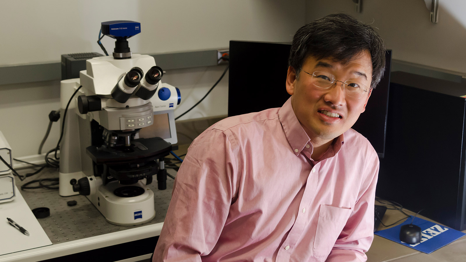
Dennis Kim, the Ivan R. Cottrell Professor of Immunology, is leaving the MIT Department of Biology at the end of this semester to serve as the Chief of the Division of Infectious Diseases at Boston Children’s Hospital.

Kim has been a member of the department for 13 years, serving as the Biology Undergraduate Officer for the past four years and Chair of the Committee on Prehealth Advising for the past six.

“Dennis has made remarkable contributions to our community, including outstanding research and as our Undergraduate Officer,” says Department Head Alan Grossman. “His teaching is exceptional, and his service to the department and MIT has been invaluable. He will be greatly missed.”

Kim became enthralled by basic science as an undergraduate at the University of California, Berkeley. There — in the cold and in the dark — he performed light- and temperature-sensitive experiments under Ken Sauer to understand the mechanism of water oxidation and oxygen evolution in plant photosynthesis. He traces his commitment to undergraduate education back to these formative experiences in the lab. Although he was already planning to attend graduate school, a run-in with a car on his motor scooter and a broken femur ignited an additional passion: human health.

As an MD-PhD student at Harvard Medical School, Kim had the opportunity to explore both avenues. There, under the mentorship of Chris Walsh, he studied enzymatic reaction mechanisms in bacterial cell wall synthesis, cultivating an interest in infectious disease that he pursued during his clinical training.

Later, as a postdoctoral fellow at Massachusetts General Hospital, Kim and his advisor Fred Ausubel, with help from Gary Ruvkun, worked to understand host-microbe interactions with a focus on innate immunity in a simple animal host, the roundworm Caenorhabditis elegans. Together, Kim and colleagues carried out a forward genetic analysis of host defense against pathogen infection in C. elegans, which eventually laid the foundation for his work as an independent investigator.

Kim arrived at the MIT Department of Biology in 2005, and since then has felt continually inspired and supported by the community. For all thirteen years, his lab has been located next door to Nobel Laureate H. Robert Horvitz, who also works in C. elegans.

“Dennis has been my nearest neighbor and closest colleague at MIT for many years,” Horvitz says. “He is a spectacular scientist whose curiosity and demand for rigor have led him to make striking discoveries repeatedly. Our labs have interacted daily, and Dennis has been a wonderful mentor to and role model for the members of my research group. Dennis has been a great friend. I will miss his wisdom and his cheer.”

“It has been an incredible opportunity and privilege to work in the Department of Biology,” Kim says. “MIT Biology promotes freedom to pursue curiosity-driven research, with phenomenal students and fantastic colleagues. And I got to set up my ‘worm’ lab next door to Bob Horvitz’s lab. It’s hard to imagine how anyone could be more fortunate!”

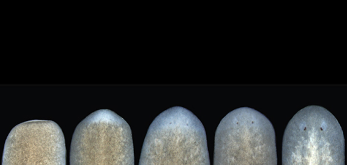
Over the years — and “thanks to some really terrific graduate students” — Kim has discovered molecular pathways governing how C. elegans recognizes and responds to its microbial environment, with a more recent emphasis on understanding how bacterial metabolites can influence host animal behavior. Questions like these are particularly well-suited to C. elegans, given its simplicity, defined nervous system structure, and well-established genetics.

“A picture has started to emerge that reveals how immunity, stress, and physiology are integrated to promote survival of the host organism,” Kim says. “Our findings have implications for understanding how interactions with microbes can affect the physiology of more complex hosts as well.”

In addition to his achievements in the lab, Kim has been a passionate advocate for undergraduate education. He’s been the Department of Biology’s Undergraduate Officer for four years, working at the departmental and institutional levels to develop and implement initiatives related to campus life, educational programs, and the curriculum. He has also chaired the Committee on Prehealth Advising for the past six years and worked to help MIT undergraduates gain admission to competitive medical schools.

“I really enjoyed working in Professor Kim’s lab because he was accessible and always willing to answer questions,” says Sonika Reddy, a former undergraduate researcher in Kim’s lab and currently a student at New York Medical College. “His mentorship was an invaluable part of my education at MIT. He helped me navigate the biology major and decide which courses to take. He also helped me decide what I wanted to do in the future and was an amazing resource as I applied to medical school.”

“The students are really remarkable here at MIT,” Kim says. “Being the Undergraduate Officer and prehealth advisor has allowed me to engage with them on a regular basis and work to improve educational and advising programs based on their feedback. These roles have meant a lot to me over the years.”

During his time at MIT, Kim developed a new subject, 7.26/7.66 (Molecular Basis of Infectious Disease), which provides an overview of viruses, bacterial pathogenesis, and parasites to advanced undergraduates and graduate students, respectively.

Former student Eta Atolia, now an MD-PhD candidate at UCLA-Caltech, says this was one of her favorite classes as an undergraduate. “I already enjoyed the topic, but the elegant way Professor Kim told the story of bacteria, toxins, antibiotics, and drug resistance really made me appreciate the field,” she says. “He also mentored me during the MD-PhD application process. He was very approachable and always available to meet and provide feedback. He introduced me to microbiology, and is a major reason why I am pursuing an MD-PhD now.”

Kim also recently worked with the Biology Undergraduate Committee and colleagues in the Department of Chemistry to develop the new 5-7 (Chemistry and Biology) major.

“I am delighted that there are a number of students who are very enthusiastic about the major and are well-prepared to work at the exciting interface of these traditional disciplines,” he says.

Kim has served at the Institutional level, chairing the Committee on Curricula and serving on the Committee on Nominations, and maintained a part-time clinical instructor appointment at Harvard Medical School.

“When we interviewed Dennis, we were amazed by the versatility and breadth of his research interests,” says Chris Kaiser, the department head who initially hired Kim. “He moves effortlessly from genome wide approaches to incisive pointed tests of mechanism, with interests ranging from innate immunity to neurological sensing and avoidance of bad environments. While running a highly successful basic research lab at MIT, he maintained an enormously important clinical footing in infectious disease at Massachusetts General Hospital. He also developed or redesigned at least five undergraduate classes and energetically ran the Biology Undergraduate program. I will miss having him as an immediate colleague.”

Of his numerous pursuits, Kim has found working with the undergraduates and graduate students to be the most rewarding.

“I’ve really enjoyed watching the scientists and physicians of tomorrow grow and mature,” he says. “To me, being an undergraduate advisor and faculty member requires having an open door for students, and these mentor-mentee relationships have been incredibly gratifying.”

As Kim transitions to his new position as Chief of the Division of Infectious Diseases at Boston Children’s Hospital, it will take three people to fill his shoes as Undergraduate Officer and Prehealth Advisor. Adam Martin and Catherine Drennan have agreed to share the role of Undergraduate Officer, and Matthew Vander Heiden will co-chair the Committee on Prehealth Advising.

“Dennis has left a significant legacy at MIT as the Undergraduate Officer,” Martin says. “He worked with the Department of Chemistry to get the 5-7 major approved, and revised our lab curriculum to increase the flexibility in Course 7. Dennis is really committed to his trainees; I often see him talking with students and postdocs in his lab and he has trained some wonderful and creative students.”

What will Kim miss the most about MIT? That goes without saying: his students.

Decoding patterns and meaning in biological data

Senior Anna Sappington found her perfect balance of “innovative computer science and innovative biology” as a member of the team mapping every cell in the human body.

Raleigh McElvery
December 5, 2018

When Anna Sappington was six years old, her parents gave her a black and white composition notebook. Together, they began jotting down observations to identify the patterns in their wooded backyard near the Chesapeake Bay. How would the harsh winters or the early springs affect the blooming trees? How many bluebirds nested each season and how many eggs would they lay? When would the cicada population cycle peak? Her father, the environmental scientist, taught her to sift through data to uncover the trends. Her mother, the journalist, gave her the words to describe her findings.

But it wasn’t until Sappington competed in the Intel International Science and Engineering Fair her junior year of high school that she probed one tiny niche of the natural world more keenly than she ever had before: the physiology of the water flea. Specifically, she investigated the developmental changes that these minute creatures experienced after being exposed to the antimicrobial compound triclosan, present in many soaps and toothpastes. She was surprised to learn that it required only a low concentration of triclosan (0.5 ppm) to cause developmental defects.

She’d been familiar with the concept of DNA since middle school, but her fellow science fair finalists were delving beyond their observations and into the letters of the genetic code. This gave her a new impetus: to understand how triclosan worked at the level of the genome and epigenome to engender the physical deformities she observed under the microscope. She just needed the proper tools, so she made some calls.

Environmental geneticist and water flea aficionado John Colbourne took an interest, and invited her to his lab at University of Birmingham in the U.K. the following summer so she could learn basic lab techniques. Although her friends and classmates didn’t quite get why she needed to travel to an entirely different country to study an organism they’d never heard of, as she puts it, she had burning scientific questions that needed answers.

“That was the experience that really turned me on to genomics,” says Sappington, now a senior and 6-7 (Computer Science and Molecular Biology) major. “I was finally getting the tools to dig through large amounts of data, using code to find patterns and meaning. I wanted to keep asking ‘why?’ and ‘how?’ all the way down to the molecular level.”

The summer before her freshman year of college, Sappington asked these questions in humans for the time as an intern at the National Human Genome Research Institute (NHGRI). There, she helped create a computational pipeline to identify the genomic changes associated with heightened risk of cardiovascular disease.

She enrolled at MIT the following fall, because she wanted to be around people from every scientific subfield imaginable. When she arrived, the joint major in computer science and biology was still relatively new.

“While a few of the required classes did meld the two, many of them offered training in each separately,” she says. “That approach really appealed to me because I was hoping to develop both skill sets independently. I wanted to learn code and write algorithms that could be applied to any field, and I also loved understanding the biological mechanisms behind different diseases and viruses.”

Before she’d even officially declared her major, Sappington was already running experiments in Sangeeta Bhatia’s lab. There, at the Koch Institute, she studied the effects of HPV infection on gene expression in liver cells. Sappington’s main role was data analysis, striving to determine which genes were amplified in response to disease.Despite their obvious differences, Sappington found the two areas to be more similar than she had initially anticipated. In her Introduction to Algorithms class, she leveraged an arsenal of algorithms with certain outputs, conditions, and run times to decode her problem sets. In Organic Chemistry, she deployed a list of foundational reactions to solve synthesis questions on her exams. “In each case, you have to combine your understanding of these fundamental rules and come up with a creative solution to decipher an unknown,” she says.

One year later, Sappington moved to Aviv Regev’s lab at the Broad Institute. There, she learned computational techniques for decoding protein interaction networks. After a year, she began working on an international project called the Human Cell Atlas as a member of the Regev and the Sanes lab collaboration.

“The overarching mission is to create a reference map of all human cells,” Sappington explains. “We want to add a layer of functional understanding on top of what we know about the genome, to understand how different cell types differ and how they interact to impact disease. This kind of endeavor has never been undertaken on such a large scale before, so it’s incredibly exciting.”

Even within a single cell type — say, retinal cells — there are about six main cell categories, each of which splinter into as many as 40 subtypes with distinct molecular profiles and roles.

Beyond the biological challenges that go along with trying to distinguish all these cell types, there are numerous computational hurdles as well. Sappington enjoys these the most — grappling with how best to analyze the gene expression of a single cell separated from its tissue of origin.

“Since you’re only working with single cells rather than entire groups of cells from a tissue, the data that you get are much more sparse,” she says. “You have to sequence a lot of individual cells and build up lots of statistical power before you can be confident that a given cell is expressing specific genes. Coming up with models to determine what constitutes a cell type — and map cell types between time points or between species — are broad problems in computer science that we’re now applying to this very specific type of data.”

Although she’s been at the Broad since her sophomore year, Sappington has supplemented her MIT research experiences with summer studies elsewhere: another stint at the NHGRI and an Amgen Scholars fellowship in Japan. She’s especially excited because her first co-authored paper will soon be published. As she puts it, she’s finally found her ideal balance of “innovative computer science and innovative biology.”

But Sappington’s time at MIT has been defined by more than just lab work. She is the co-president of the Biology Undergraduate Student Association, which serves as a liaison between the Department of Biology and the wider community. She’s also a member of MedLinks, a volunteer at the Massachusetts General Hospital Department of Radiology, former managing director of TechX, and a performer for several campus dance troupes. In 2018, Sappington earned the prestigious Barry Goldwater Scholarship Award, alongside fellow 6-7 major Meena Chakraborty.

She was recently awarded the Marshall Scholarship, which will fund her master’s degrees in machine learning at University College London and oncology at the University of Cambridge beginning in the fall of 2019. After two years, she plans to start her MD-PhD. That way, she can become a practicing physician without having to give up her computer science research.

Her advice to prospective students: “When you get to MIT, just explore. Try different academic disciplines, different extracurriculars, and talk to as many people as you can. The campus is full of passionate individuals in every field imaginable, whether that’s computer science or political science.”

Posted 12.5.18
Uncovering the “must-haves” of tissue regeneration
Nicole Davis | Whitehead Institute
November 27, 2018

Cambridge, MA.  – The ability to regrow missing or damaged body parts is one of the great marvels of modern biology. In an effort to lay bare the biological underpinnings of this phenomenon, scientists at Whitehead Institute have begun to define the core features that are required for regeneration in flatworms. Their research, which appears online November 27 in Cell Reports, reveals that a set of cellular and molecular responses — previously thought to be essential for regeneration following amputations and other major injuries — is in fact dispensable.

“This is a real surprise,” said senior author Peter Reddien, a Member of Whitehead Institute, professor of biology at Massachusetts Institute of Technology, and investigator with the Howard Hughes Medical Institute. “These responses are broad, prominent attributes of tissue regeneration and repair and, a reasonable bet was that they function to bring about regeneration.”

About eight years ago, Reddien and his team described a set of biological activities that are triggered by injuries that remove tissue. Whereas a cut or a scrape removes little if any tissue, more damaging injuries, like amputations, cause significant tissue loss. That missing tissue must be regenerated to ensure the organism retains its proper anatomical proportions.

A series of cellular and molecular activities — known collectively as the missing tissue response — were believed to enable this regeneration to occur. They include the sustained action of genes that respond to injury, a period of intense cell division in areas surrounding the wound, and a general increase in cell death throughout the body. “This happens prominently, not only in planarians but also in other organisms capable of regeneration, so we suspected that the missing tissue response must play a very fundamental role in regeneration,” recalled Reddien.

What types of injuries require the missing tissue response for repair, and what is the function of the missing tissue response in regeneration? Graduate student and first author Aneesha Tewari, Reddien and colleagues, including Sarah Stern and Isaac Oderberg, set out to uncover the answers. This work forms the basis of their latest Cell Reports study.

The researchers harnessed an earlier discovery that a gene known as follistatin is required for the missing tissue response in flatworms (known as planarians). By using molecular tools to inhibit this gene, they could block the missing tissue response and observe what happens under various wound conditions, ranging from minimal (the removal of an eye, for example) to moderate (the removal of the pharynx or part of the head) to significant tissue loss (the removal of a complete side of the body). Remarkably, in every case, the missing tissue was regenerated, albeit much more slowly than it would be otherwise.

“These results tell us that what the missing tissue response is really doing is simply pushing the foot down on the gas pedal — basically accelerating the process of regeneration,” explained Reddien. “If you can’t accelerate, you’ll still get there, it just takes longer.”

Tewari, Reddien, and their colleagues also cracked a thorny mystery surrounding the missing tissue response. Although their results show that it is not required across a wide range of injuries, there is one lingering instance in which regeneration failed to occur when they blocked the missing tissue response: head amputation.

“This was a big puzzle,” said Tewari. “It left us wondering whether or not we could generalize our findings to all types of wounds — is there something special about the head that makes it uniquely dependent on the missing tissue response?”

The answer, it turns out, is no. When follistatin is blocked, a key signaling protein, called Wnt1, kicks into overdrive. And when that happens, the tissue destined to form the head does not receive the positional cues it needs to properly regrow, which means regeneration fails to proceed. But, when both the missing tissue response and Wnt1 are blocked, the head does indeed regenerate, the team uncovered.

Taken together, the researchers’ findings begin to clarify what is essential for regeneration to take place and what is not. “Our study greatly simplifies the picture of what it takes to regenerate,” said Reddien. “And that’s an important step along the path towards dissecting the central elements of regeneration in animals that do regenerate well, like flatworms, and then applying that knowledge to understand what the limits might be in those animals that don’t regenerate as well, like humans.”

This research was supported by the NIH (R01GM080639), the National Science Foundation, the Eleanor Schwartz Charitable Foundation, and the Howard Hughes Medical Institute.

***
Peter Reddien’s primary affiliation is with Whitehead Institute for Biomedical Research, where his laboratory is located and all his research is conducted. He is also a Howard Hughes Medical Institute Investigator and a professor of biology at Massachusetts Institute of Technology.
***
Full citation:
Cell Reports,  Vol. 25, Is. 9, P2577-2590.E3, November 27, 2018, DOI:https://doi.org/10.1016/j.celrep.2018.11.004
“Cellular and molecular responses unique to major injury are dispensable for planarian regeneration”
Aneesha G. Tewari (1,2), Sarah R. Stern (1,2), Isaac M. Oderberg (1,2,4), and Peter W. Reddien (1,2,3)
 1.Whitehead Institute for Biomedical Research, Cambridge, MA 02142, USA
2. Department of Biology, Massachusetts Institute of Technology, Cambridge, MA 02139, USA
3. Howard Hughes Medical Institute, Chevy Chase, MD 20815, USA
4. Brigham and Women’s Hospital, Harvard Medical School, Boston, MA 02115, USA
Computer model offers more control over protein design

New approach generates a wider variety of protein sequences optimized to bind to drug targets.

Anne Trafton | MIT News Office
October 15, 2018

Designing synthetic proteins that can act as drugs for cancer or other diseases can be a tedious process: It generally involves creating a library of millions of proteins, then screening the library to find proteins that bind the correct target.

MIT biologists have now come up with a more refined approach in which they use computer modeling to predict how different protein sequences will interact with the target. This strategy generates a larger number of candidates and also offers greater control over a variety of protein traits, says Amy Keating, a professor of biology, a member of the Koch Institute, and the leader of the research team.

“Our method gives you a much bigger playing field where you can select solutions that are very different from one another and are going to have different strengths and liabilities,” she says. “Our hope is that we can provide a broader range of possible solutions to increase the throughput of those initial hits into useful, functional molecules.”

In a paper appearing in the Proceedings of the National Academy of Sciences the week of Oct. 15, Keating and her colleagues used this approach to generate several peptides that can target different members of a protein family called Bcl-2, which help to drive cancer growth.

Recent PhD recipients Justin Jenson and Vincent Xue are the lead authors of the paper. Other authors are postdoc Tirtha Mandal, former lab technician Lindsey Stretz, and former postdoc Lothar Reich.

Modeling interactions

Protein drugs, also called biopharmaceuticals, are a rapidly growing class of drugs that hold promise for treating a wide range of diseases. The usual method for identifying such drugs is to screen millions of proteins, either randomly chosen or selected by creating variants of protein sequences already shown to be promising candidates. This involves engineering viruses or yeast to produce each of the proteins, then exposing them to the target to see which ones bind the best.

“That is the standard approach: Either completely randomly, or with some prior knowledge, design a library of proteins, and then go fishing in the library to pull out the most promising members,” Keating says.

While that method works well, it usually produces proteins that are optimized for only a single trait: how well it binds to the target. It does not allow for any control over other features that could be useful, such as traits that contribute to a protein’s ability to get into cells or its tendency to provoke an immune response.

“There’s no obvious way to do that kind of thing — specify a positively charged peptide, for example — using the brute force library screening,” Keating says.

Another desirable feature is the ability to identify proteins that bind tightly to their target but not to similar targets, which helps to ensure that drugs do not have unintended side effects. The standard approach does allow researchers to do this, but the experiments become more cumbersome, Keating says.

The new strategy involves first creating a computer model that can relate peptide sequences to their binding affinity for the target protein. To create this model, the researchers first chose about 10,000 peptides, each 23 amino acids in length and helical in structure, and tested their binding to three different members of the Bcl-2 family. They intentionally chose some sequences they already knew would bind well, plus others they knew would not, so the model could incorporate data about a range of binding abilities.

From this set of data, the model can produce a “landscape” of how each peptide sequence interacts with each target. The researchers can then use the model to predict how other sequences will interact with the targets, and generate peptides that meet the desired criteria.

Using this model, the researchers produced 36 peptides that were predicted to tightly bind one family member but not the other two. All of the candidates performed extremely well when the researchers tested them experimentally, so they tried a more difficult problem: identifying proteins that bind to two of the members but not the third. Many of these proteins were also successful.

“This approach represents a shift from posing a very specific problem and then designing an experiment to solve it, to investing some work up front to generate this landscape of how sequence is related to function, capturing the landscape in a model, and then being able to explore it at will for multiple properties,” Keating says.

Sagar Khare, an associate professor of chemistry and chemical biology at Rutgers University, says the new approach is impressive in its ability to discriminate between closely related protein targets.

“Selectivity of drugs is critical for minimizing off-target effects, and often selectivity is very difficult to encode because there are so many similar-looking molecular competitors that will also bind the drug apart from the intended target. This work shows how to encode this selectivity in the design itself,” says Khare, who was not involved in the research. “Applications in the development of therapeutic peptides will almost certainly ensue.”

Selective drugs

Members of the Bcl-2 protein family play an important role in regulating programmed cell death. Dysregulation of these proteins can inhibit cell death, helping tumors to grow unchecked, so many drug companies have been working on developing drugs that target this protein family. For such drugs to be effective, it may be important for them to target just one of the proteins, because disrupting all of them could cause harmful side effects in healthy cells.

“In many cases, cancer cells seem to be using just one or two members of the family to promote cell survival,” Keating says. “In general, it is acknowledged that having a panel of selective agents would be much better than a crude tool that just knocked them all out.”

The researchers have filed for patents on the peptides they identified in this study, and they hope that they will be further tested as possible drugs. Keating’s lab is now working on applying this new modeling approach to other protein targets. This kind of modeling could be useful for not only developing potential drugs, but also generating proteins for use in agricultural or energy applications, she says.

The research was funded by the National Institute of General Medical Sciences, National Science Foundation Graduate Fellowships, and the National Institutes of Health.

Plant characteristics shaped by parental conflict
Greta Friar | Whitehead Institute
November 19, 2018

CAMBRIDGE, Mass. – Different subpopulations of a plant species can have distinct traits, varying in size, seed count, coloration, and so on. The primary source of this variation is genes: different versions of a gene can lead to different traits. However, genes are not the only determinant of such traits, and researchers are learning more about another contributor: epigenetics. Epigenetic factors are things that regulate genes, altering their expression, and like genes they can be inherited from generation to generation, even though they are independent of the actual DNA sequences of the genes.

One epigenetic mechanism is DNA methylation, in which the addition of chemical tags called methyl groups can turn genes on or off. Genes that share the identical DNA sequence but have different patterns of methylation are called epialleles. Several studies have shown that epialleles, like different versions of genes, can cause differences in traits between plant subpopulations, or strains, but whether genetic factors are also at play can be difficult to determine.

The lab of Whitehead Member Mary Gehring, who is also an associate professor at Massachusetts Institute of Technology, has described evidence that epialleles alone can lead to different heritable traits in plants. In research published online November 5 in the journal PLoS Genetics, Gehring, along with co-first authors and former lab members Daniela Pignatta and Katherine Novitzky, showed that altering the methylation state of the gene HDG3 in different strains of the plant Arabidopsis thaliana was enough to cause changes in seed weight and in the timing of certain aspects of seed development.

In plants, methylation states of genes change most frequently during seed development, when genes are switched on or off to progress development of the organism. This period is also when a conflict of interest arises in the genome of each seed between the parts inherited from its mother and father. The mother plants produce seeds fertilized by different fathers at the same time. It’s in the mother’s interest to give an equal share of nutrients to each seed—to have many smaller seeds. But it’s in the father’s interest for its seed to get the most nutrients and grow larger. This conflict plays out through an epigenetic mechanism called imprinting, in which, through differential methylation between the father’s and mother’s copies of a gene, one parent’s copy is silenced in the offspring so that only the other parent’s version of the gene is expressed.

The gene HDG3 is imprinted in one strain of Arabidopsis so that only the father’s copy is expressed. Gehring and her team found that when the strain loses its paternal imprinting, the timing of seed development is affected and the plant ends up with smaller seeds. This is consistent with the theory of imprinting: When the father’s genes have the advantage, the seeds are larger than when both parents’ genes are equally expressed.

Other experiments tested the effect of either activating or silencing HDG3 by methylation in a variety of scenarios, both in a separate strain of Arabidopsis in which the gene starts off silenced, as well as in crosses between the two strains. The researchers found that altering the methylation state of the gene was sufficient to affect seed size and the timing of seed development. In the crosses, these traits depended on whether the paternal copy of the gene came from the strain in which HDG3 was normally silenced or the strain in which it was normally activated.

Altogether these experiments demonstrate a link between changes in methylation state and differences in seed development and size. This suggests that epialleles in natural populations function much like variations in genes, creating heritable traits that differ within the larger population.

This work was funded by the National Science Foundation (NSF grant 1453459).

***

Mary Gehring’s primary affiliation is with Whitehead Institute for Biomedical Research, where her laboratory is located and all her research is conducted. She is also an associate professor of biology at Massachusetts Institute of Technology.
***
Full citation:
“A variably imprinted epiallele impacts seed development”
PLoS Genetics, online November 5 2018, https://doi.org/10.1371/journal.pgen.1007469
Daniela Pignatta (1,3), Katherine Novitzky (1,3), P. R. V. Satyaki (1), Mary Gehring (1,2)
1. Whitehead Institute for Biomedical Research, Cambridge, MA, United States of America
2. Department of Biology, Massachusetts Institute of Technology, Cambridge, MA, United States of America
3. These authors contributed equally to this work.
Heart-healthy plant chemistry
Greta Friar | Whitehead Institute
October 29, 2018

Plants have been a rich source of medicines for thousands of years. Compounds such as artemisinin, for example, used to treat malaria, and morphine, a pain reliever, are mainstay therapeutics derived from plants. However, several roadblocks in plant chemistry research have prevented scientists from tapping into the full potential of plant-based medicinal compounds, thwarting drug discovery and development. Researchers typically screen for molecules of interest by breaking the plant into very small pieces, using biochemistry to test the activity of the pieces, and isolating the molecules responsible for the activity. It is often difficult, however, to pick the right compound responsible for a medicinal effect out of the plant mixture, or to identify the genes responsible for producing it.

The Chinese wolfberry plant (Lycium barbarum), also known as goji berry, has been used in traditional Chinese herbal medicine for millennia to treat symptoms such as high blood pressure. Researchers had identified small protein-like molecules called lyciumins, produced by the goji berry, as the source of its antihypertensive properties but little else was known about the molecules.

In research published online October 29 in the journal Proceedings of the National Academy of Sciences, Whitehead Institute Member Jing-Ke Weng and postdoctoral researcher Roland Kersten describe an approach to speed up the process of identifying plant chemistry that they used to investigate lyciumins. The approach capitalizes on the growing number of plants that have had their genomes sequenced. The wealth of genomic data available enabled Kersten to identify the gene that is associated with lyciumin production in goji berries by searching for a DNA sequence that matched the sequences of the lyciumins. Once Kersten found the matching precursor gene in goji berries he inserted it into a tobacco plant, which began producing lyciumins, confirming that he had found the right gene.

Kersten then hunted for lyciumin-producing genes in other plant genomes using a common feature of the genes that he had identified as a search query. He discovered more than one hundred unknown lyciumins in everything from potatoes to beets to soybeans.

Having sped up the gene discovery stage, Kersten used gene expression techniques to likewise speed up the molecule production stage. Being able to quickly produce large quantities of a drug candidate is necessary for testing and manufacturing the drug. Kersten edited the lyciumin precursor genes to make more copies of the molecule and then inserted the edited genes into the tobacco plant to mass produce lyciumins up to 40 times faster than the original plants. Kersten was also able to edit the lyciumins’ DNA sequences to alter the molecules’ structure, creating new varieties of lyciumins not found in nature. Together, these results allow for the future creation of a lyciumin library, a valuable repository for drug discovery research. Millions of different lyciumins can be grown in tobacco and tested for their efficacy as antihypertensive drugs or in other potential agrochemical and pharmaceutical applications.

Weng and Kersten’s approach leverages the recent explosion in plant genomics to uncover important medicinal compounds in plants and reveal the secrets of plants used in traditional global medicine for generations. For Kersten, the research was also an exciting demonstration of just how much undiscovered chemistry lies waiting to be tapped in even the best-studied crop plants.

This work was supported by grants from the Thome Foundation, the Pew Scholars Program in the Biomedical Sciences, the Searle Scholars Program, and the Family Larsson Rosenquist Foundation.

***
Jing-Ke Weng’s primary affiliation is with Whitehead Institute for Biomedical Research, where his laboratory is located and all his research is conducted. He is also an assistant professor of biology at Massachusetts Institute of Technology.
***
Full citation:
“Gene-guided discovery and engineering of branched cyclic peptides in plants”
PNAS, online on October 29.
Roland D. Kersten (1), Jing-Ke Weng (1,2)
1. Whitehead Institute for Biomedical Research, 455 Main Street, Cambridge, MA, United States
2. Department of Biology, Massachusetts Institute of Technology, Cambridge, MA, United States
Activating a new understanding of gene regulation
Greta Friar | Whitehead Institute
November 15, 2018

CAMBRIDGE, Mass. – Regulation of gene expression — turning genes on or off, increasing or decreasing their expression — is critical for defining cell identity during development and coordinating cellular activity throughout the cell’s lifetime. The common model of gene regulation imagines the nucleus of the cell as a large space in which molecules involved in DNA transcription float around seemingly at random until they stumble across a DNA sequence or other transcriptional machinery to which they can bind — a haphazard approach. However, this paradigm is being upended as over the last few years researchers have discovered that rather than being amorphous spaces dependent upon fortuitous collisions, cells actually compartmentalize their processes into discrete membraneless structures in order to congregate relevant molecules, thereby better coordinating their interactions. Research from the lab of Whitehead Member Richard Young and others earlier this year reported that such compartmentalization is a crucial, previously unobserved aspect of gene regulation.[1]

The latest research from Young’s lab, published online November 15 in the journal Cell, delves further into how such compartmentalization helps orchestrate transcriptional regulation by revealing the role of the activation domain, a part of transcription factors previously shrouded in mystery. One side of transcription factors, containing the DNA binding domain, binds to a region of DNA near a gene. The other end, called the activation domain, then captures molecules that impact gene expression, anchoring that transcriptional machinery near the gene.

This most recent work reveals that activation domains do their job by meshing with other transcription proteins to form liquid droplets near the genes they regulate. The process by which the molecules form a distinct liquid compartment within the environment of the cell — like oil refusing to mix with vinegar in a salad dressing — is called phase separation.

Such an evolved understanding of gene regulation has enormous implications for medicine and drug discovery, as errors in gene regulation are key components of many diseases, including cancers. The new model could help illuminate how diseases coopt regulatory mechanisms and how therapeutic interventions might remedy such dysregulation. Transcription factors have traditionally been hard to target therapeutically, and the incomplete understanding of their structure and function may have been part of the reason.

“Transcriptional regulation is important for every human function, from cell differentiation to development to cell maintenance,” says Ann Boija, co-first author and postdoctoral researcher in Young’s lab. “Despite that fact the structure and function of the activation domain on the transcription factors have been poorly understood.”

Most proteins settle into defined three-dimensional structures and can only bind with other molecules that fit them perfectly, in a specific orientation, like a key in a lock. The activation domains of transcription factor proteins, however, contain what are known as intrinsically disordered regions, which behave more like strands of cooked spaghetti, tangling at random into flexible shapes. This disorder allows the molecules to bind at many points, creating a dynamic network of loose connections that appears to precipitate phase separation.

“I have taught regulatory biology for decades using inspiration from lock and key structures. They are elegant, and easy to visualize and model, but they don’t tell the whole story. Phase separation was the missing piece,” says Young, who is also a professor of biology at MIT.

In experiments with a variety of transcription factors, Boija and co-first author Isaac Klein, a postdoctoral researcher in Young’s lab and medical oncology fellow at the Dana-Farber Cancer Institute, found that the transcription factors meshed with Mediator, a molecule that helps activate genes, and phase separated into droplets, and that this process was associated with gene activation. The transcription factors they investigated included OCT4, which is important for maintaining the state of embryonic stem cells; the estrogen receptor (ER), which plays a role in breast cancer; and GCN4, a well-studied model transcription factor in yeast.

The discovery has implications for many diseases, such as cancer, in which cancer genes may use phase separated droplets to help ramp up their expression. New therapeutic approaches could focus on dissolving the droplets, and drug discovery can incorporate testing of how the drug — or target molecule — behaves inside versus outside of the droplets. This new model of how transcription factors function is not only rewriting the understanding of transcriptional regulation, it is opening up new paths for drug discovery and therapeutic approaches.“We found a link between gene activation and phase separation across a broad spectrum of contexts,” Klein says, suggesting that this mechanism is a common feature of transcriptional regulation.

The work was supported by the National Institutes of Health (NIH grants GM123511, GM117370, T32CA009172, T32GM08759), the National Science Foundation (NSF grant PHY1743900), Swedish Research Council (VR 2017-00372), Damon Runyon Cancer Research Foundation (2309-17), Hope Funds for Cancer Research, Cancer Research Institute, and Netherlands Organisation for Scientific Research (NWO).

***
Richard Young’s primary affiliation is with Whitehead Institute for Biomedical Research, where his laboratory is located and all his research is conducted. He is also a professor of biology at Massachusetts Institute of Technology.
***
Full citation:
“Transcription factors activate genes through the phase separation capacity of their activation domains”
Cell, online November 15, DOI: 10.1016/j.cell.2018.10.042
Ann Boija (1,7), Isaac A. Klein (1,2,7), Benjamin R. Sabari (1), Alessandra Dall’Agnese (1), Eliot L. Coffey (1,3), Alicia V. Zamudio (1,3), Charles H. Li (1), Krishna Shrinivas (4,5), John C. Manteiga (1,3), Nancy M. Hannett (1), Brian J. Abraham (1), Lena K. Afeyan (1,3), Yang E. Guo (1), Jenna K. Rimel (6), Charli B. Fant (6), Jurian Schuijers (1), Tong Ihn Lee (1), Dylan J. Taatjes (6), and Richard A. Young (1,3)
  1. Whitehead Institute for Biomedical Research, Cambridge, MA 02142, USA
  2. Department of Medical Oncology, Dana-Farber Cancer Institute, Harvard Medical School, Boston, MA 02215, USA
  3. Department of Biology
  4. Department of Chemical Engineering
  5. Institute of Medical Engineering and Science, Massachusetts Institute of Technology, Cambridge, MA, 02139, USA
  6. Department of Biochemistry, University of Colorado, Boulder, CO 80303, USA
  7. These authors contributed equally
[1] Sabari et al., “Coactivator condensation at super-enhancers links phase separation and gene control,”
Science, June 21, 2018; Cho et al., “Mediator and RNA polymerase II clusters associate in transcription-dependent condensates,” Science, June 21 2018.
A Long Legacy In RNA Discovery
Nicole Giese Rura | Whitehead Institute
October 25, 2018

On January 1, 1994, Whitehead Institute Fellow David Bartel officially opened his lab. Since then, his lab has achieved numerous milestones as a trailblazer in RNA research. Initially the lab studied the ability of RNA to catalyze chemical reactions, including reactions that would have been required early in the evolution of life. In 1999, which was several years after Bartel had become a Whitehead Institute Member, the lab also began to study the ability of RNA to trigger gene silencing.  As part of the field’s vanguard, they made early insights into the roles that small RNAs play during RNA-guided gene regulation and helped discover the abundance of microRNAs (miRNAs), small but critical pieces of RNA that had been largely overlooked until the Bartel lab and others detected their widespread existence in the early 2000s. Since then, the lab has made many discoveries that helped define miRNAs and how plants and animals make these short snippets of RNA and then use them to regulate genes.

To celebrate his lab’s silver anniversary, Bartel, who is also a professor of biology at Massachusetts Institute of Technology and an investigator with the Howard Hughes Medical Institute (HHMI), recently hosted a reunion for all current and former lab members, key collaborators, and their families. About 70 attended the scientific portion, and approximately 130 closed the symposium with dinner at the New England Aquarium. Far-flung participants came from the west coast of the United States and Canada, as well as from France, but the longest trip was made by former Bartel graduate student Huili Guo, her husband, and nine-month old daughter, who traveled from Singapore, where Guo is an independent fellow at the Institute of Molecular and Cell Biology.

True to his quest to expand knowledge, Bartel suggested that the scientific talks presented during the two-day event focus on recent findings and new research directions. The result struck a chord with Bartel’s graduate advisor, Jack Szostak. A professor at Harvard University, and an HHMI investigator, Szostak commented that as a leader of a lab, joy comes not only from seeing biology advance, but also from watching the biologists who have been in the lab—to see what they can do and where they go.

From Szostak to Margaret Glasner (Bartel’s second graduate student, who is now an associate professor at Texas A&M) to current lab members, the speakers during the scientific portion spanned Bartel’s career in science. These speakers presented their own work, which provided insight into how ideas planted and tended in Bartel’s lab sprouted and matured into innovative lines of research and labs. Many who passed through his lab remain focused on catalytic RNAs, miRNAs or other types of RNA, but others have branched off into different areas, such as investigating protein function, studying chromosome architecture, developing therapies for treating human disease, and working with (and in some cases founding) biotechnology firms.

Lab members—current and former alike—credit Bartel and their time in his lab as transformative, educational, and key to their future careers. But for some, being in Bartel’s lab had very important, unexpected benefits. Ulrich Müller (a former postdoc in the Bartel lab and currently an associate professor of chemistry and biochemistry at University of California, San Diego) met his wife, who was in the lab of Whitehead Institute Founding Member Robert Weinberg, at a Whitehead Institute social event on the second floor.

Laura Resteghini, who has been Bartel’s administrative lab manager since late 2000 and with Bartel was the primary organizing force behind the reunion, says that the occasion was two days of stellar research talks, reminiscing with old friends, and making new ones. But most importantly, the reunion celebrated Bartel’s rich history of scientific mentorship.

For Bartel, the reunion was not only an opportunity for everyone reconnect with friends and colleagues—as well as their families—but also for establishing new connections that could form the foundation for the next 25 years of innovative research.


***

David Bartel’s primary affiliation is with Whitehead Institute for Biomedical Research, where his laboratory is located and all his research is conducted. He is also a professor of biology at Massachusetts Institute of Technology and investigator with the Howard Hughes Medical Institute.

How Many Evolutionary Events Can It Take To Screw in Nature’s Lightbulb?

Firefly genomics reveals independent evolution of bioluminescence in beetles

Lisa Girard | Whitehead Institute
October 16, 2018

Cambridge, MA — Researchers at Whitehead Institute and collaborators from fourteen other institutions around the world have shed light on the evolutionary origins of luciferase, the key enzyme behind the glow of fireflies and other bioluminescent beetles. By sequencing the genomes of two American and Japanese firefly species that diverged approximately 100 million years ago, along with a more evolutionarily distant bioluminescent Caribbean click beetle, the team discovered that luciferase appears to have arisen independently in fireflies and click beetles. Examining the genes flanking that encoding the luciferase gene, suggests an evolutionary path along which the luciferase gene arose from duplications and divergences of CoA ligase genes involved in fat metabolism. As described online October 16 in the journal eLife, these findings provide fundamental insights into how enzymes can evolve, potentially inform strategies to help protect bioluminescent beetles from a shifting climate and habitat, and could extend the utility of luciferase, which has also been harnessed for biomedical and agricultural research, as a laboratory tool.

Throughout much of the world, the silent flash of a firefly on a warm evening can only mean one thing-Summer has arrived. But fireflies don’t just signal summer, their glow serves as a mating signal to other fireflies, and is even a warning that they are chemically defended, having a noxious taste capable of repelling the boldest of predators.

Belying its grandeur, the chemistry of firefly bioluminescence is relatively straightforward. Their light is produced by a specialized firefly enzyme, luciferase, that breaks down a molecule called luciferin, producing light in the process. Luciferase has become a mainstay tool in the laboratory. Scientists can fuse their gene of interest to luciferase and assay for gene expression by measuring the intensity of the glow after luciferin is added.

Beyond fireflies, there are other bioluminescent beetles (despite their name, fireflies are actually beetles), including certain tropical click-beetles. Perplexingly, these diverse bioluminescent beetles use very similar luciferase enzymes and luciferin molecules, but have an unrelated anatomy of their light-producing organs (also known as lanterns), making it unclear if their bioluminescence evolved from a common luminous ancestor, or if their special glow evolved independently.

Since fireflies and bioluminescent click-beetles are not model organisms like mice or fruit flies for which there is a wealth of genetic information, Jing-Ke Weng, Whitehead Institute Member and assistant professor of biology at Massachusetts Institute of Technology (MIT), along with a  graduate student in Weng’s lab, Tim Fallon, and Cornell postdoctoral researcher, Sarah Lower, began their investigations by sequencing the genome of the American Big Dipper firefly, Photinus pyralisNamed for its distinctive swooping “J” flash , this common inhabitant of meadows and suburban lawns has been called the “All-American firefly”. Due to its abundance and ease of identification, it was also the firefly of choice for scientific study, and is the species from which luciferin and luciferase were first characterized. Wanting to start their work quickly and make their progress and data available to others in the firefly community, Whitehead Institute researchers and collaborators crowdsourced funds to sequence the Big Dipper firefly.

The Big Dipper genome sequence, they discovered, revealed interesting insights into the origin of the luciferase gene. Examining the genes flanking that encoding luciferase, they found a cluster, or tandem repeat, of fatty acid CoA ligase genes with the luciferase gene sitting in the middle of this cluster. Sequence similarity and proximity between the luciferase and fatty acid CoA ligase genes suggested an evolutionary path along which the luciferase gene was produced from tandem duplication and divergence of an ancestral fatty acid CoA ligase gene.

“When the luciferase gene was cloned, people knew it was similar to the fatty acid CoA ligase gene in sequence, and hypothesized that it must be related to that ancestry. But what we uncovered from the luciferase gene locus is a tandem repeat of five genes, four are still the fatty acid CoA ligases, but then luciferase evolved right in the middle we believe from divergence of one of these duplications,” says Weng.

The Big Dipper sequencing provided important insights into the origin of luciferase and additional factors involved in bioluminescence, but in order to gain additional insights into the evolution of bioluminescence, the researchers set out to sequence two additional species that they hoped would provide the additional context to help them triangulate on some answers.

The bioluminescent click beetle, Ignelater luminosus, is related to the firefly, but on another branch of the tree of life entirely. Instead of producing light at its tail, it has two lanterns behind its head.

“We thought that sequencing the click beetle would provide insights into the evolution of bioluminescence as well as perhaps into how these animals could acquire very similar traits in terms of their biochemistry, but not in terms of their development,” says Fallon.

The third species they selected to sequence was a Japanese aquatic firefly (Aquatica lateralis), known in Japan as the Heike firefly.  Heike and the Big Dipper diverged from one another over 100 million years ago (to give you a sense of how far this is, it is older than the evolutionary distance between humans and rodents).

The researchers analyzed genomic data from the Japanese aquatic firefly and saw a similar arrangement around the luciferase gene locus as they had in the Big Dipper genome, suggesting that luciferase arose from a common ancestral event in both firefly species. The structure around the luciferase locus in the click beetle, however, was entirely absent, suggesting that luciferase arose through a different event.  Taken together, by sequencing and analyzing data from the genomes of two firefly species that diverged approximately 100 million years ago, along with a more evolutionarily distant bioluminescent click beetle, the team discovered that luciferase appears to have evolved independently in both fireflies and click beetles.

“Having the genome allowed us to understand how the evolution of luciferase happened. Before sequencing, we knew there were five genes cloned, including luciferase, in firefly. By sequencing the genomes we actually uncovered those genomic loci where those initial gene duplication events occured,” says Lower.

In addition to the origins of luciferase, these findings also provided the researchers with insights into the evolution of the light organs.

“Since our findings suggest that luciferase originated independently in both lineages, we can infer that anything that came after luciferase, for example the light organs, or other things dependent on luciferase should also be independent,” says Fallon.

Discovering how bioluminescence arose, as well as other complex traits, can be studied now that genomic information is available. The information can also inform strategies to protect fireflies, whose populations in many parts of the world are diminishing. In addition to adding tools to help reveal a constituent parts list that could allow researchers to optimize bioluminescence as a tool, these findings reveal important insights into the evolution of bioluminescence as well as genomic evolution more broadly.

“Luciferase is a perfect example of how to build a new enzyme, duplication of a related progenitor gene followed by mutation and selection,” says Weng. “And one of the most exciting parts of this study was that by examining the evolutionary scars in the genomes we studied we could actually see it happen.”

* * *
Jing-Ke Weng’s primary affiliation is with Whitehead Institute for Biomedical Research, where his laboratory is located and all his research is conducted. He is also an assistant professor of biology at Massachusetts Institute of Technology.
* * *
Full citation:
“Firefly genomes illuminate parallel origins of bioluminescence in beetles”
eLife, online on October 9, 2018. doi: 10.7554/eLife.36495
Timothy R. Fallon (1,2,*), Sarah E. Lower (3,*), Ching-Ho Chang (4) , Manabu Bessho-Uehara (5,6), Gavin J. Martin (7), Adam J. Bewick (8) , Megan Behringer (9) , Humberto J. Debat (10), Isaac Wong (4) , John C. Day (11), Anton Suvorov (7) , Christian J. Silva (4,12), Kathrin F. Stanger-Hall1 (3), David W. Hall (8) , Robert J. Schmitz (8), David R. Nelson (14), Sara M. Lewis (15), Shuji Shigenobu (16), Seth M. Bybee (7) , Amanda M. Larracuente (4), Yuichi Oba (5), and Jing-Ke Weng (1,2)
1. Whitehead Institute for Biomedical Research, Cambridge, Massachusetts 02142, USA.
2.Department of Biology, Massachusetts Institute of Technology, Cambridge, Massachusetts 02139, USA.
3. Department of Molecular Biology & Genetics, Cornell University, Ithaca, New York 14850, USA.
4. Department of Biology, University of Rochester, Rochester, New York 14627, USA.
5. Department of Environmental Biology, Chubu University, Kasugai, Aichi 487-8501, Japan.
6. Graduate School of Bioagricultural Sciences, Nagoya University, Nagoya, Aichi 464-8601, Japan.
7. Department of Biology, Brigham Young University, Provo, Utah 84602, USA.
8. Department of Genetics, University of Georgia, Athens, Georgia 30602, USA.
9. Biodesign Center for Mechanisms of Evolution, Arizona State University, Tempe, Arizona 85287, USA.
10. Center of Agronomic Research National Institute of Agricultural Technology, Córdoba, Argentina.
11. Centre for Ecology and Hydrology (CEH) Wallingford, Wallingford, Oxfordshire, UK.
12. Department of Plant Sciences, University of California Davis, Davis, California, USA.
13. Department of Plant Biology, University of Georgia, Athens, Georgia 30602, USA.
14. Department of Microbiology Immunology and Biochemistry, University of Tennessee  HSC, Memphis 38163, USA.
15. Department of Biology, Tufts University, Medford, Massachusetts 02155, USA.
16. NIBB Core Research Facilities, National Institute for Basic Biology, Okazaki 444-8585, Japan.