Start signal for sex cell creation
Greta Friar | Whitehead Institute
February 27, 2019

Cambridge, MA — Cells can divide and multiply in two ways: mitosis, in which the cell replicates itself, creating two copies identical to the original; or meiosis, in which the cell shuffles its DNA and divides twice, creating four genetically unique cells, each with half of the original cell’s number of chromosomes. In mammals, these latter cells become eggs and sperm.

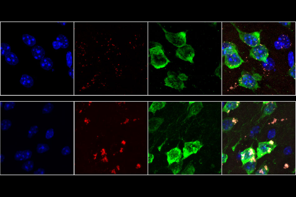
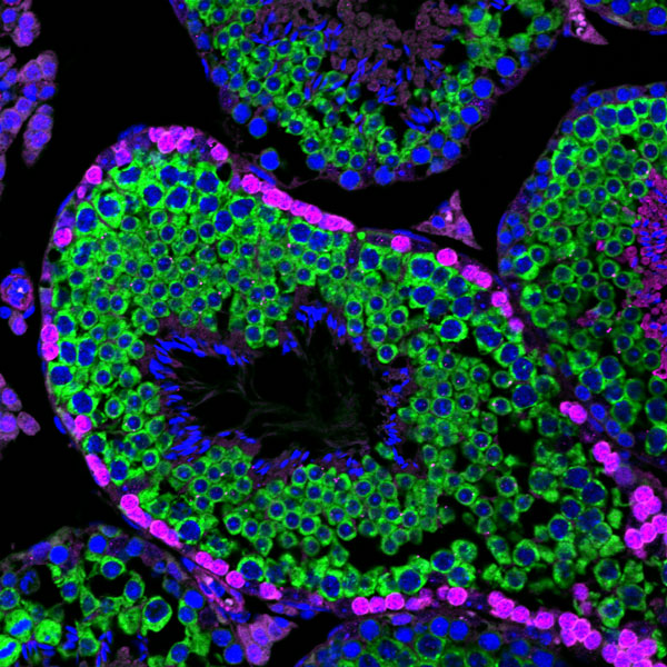
How do germ line cells, the repository of cells that create eggs and sperm, know when to stop replicating themselves and undergo meiosis? Researchers had been aware that a protein called STRA8, which is only active in germ line cells, was involved in initiating meiosis, but they did not know how. New research from Whitehead Member and Institute Director David Page, also a professor of biology at Massachusetts Institute of Technology and an investigator with Howard Hughes Medical Institute; Mina Kojima, formerly a Massachusetts Institute of Technology graduate student and now a postdoctoral researcher at Yale; and visiting scientist Dirk de Rooij has revealed that in mice, STRA8 initiates meiosis by activating and amplifying a network of thousands of genes. This network includes genes involved in the early stages of meiosis, DNA replication, and other cell division processes. The research was published in eLife on February 27, 2019.

In the past, researchers have had difficulty collecting enough cells on the cusp of meiosis to investigate STRA8’s role. In mammals, germ line cells are inside the body, difficult to access, and they begin meiosis in staggered fashion so few cells are at the same stage during an extraction. Researchers in Page’s lab had previously come up with an approach to solve this problem using developmental synchronization, manipulating the cells’ exposure to the chemical that triggers their development in order to prompt all of the cells to begin meiosis simultaneously. Once the cells were synced up, first author Kojima could get a large enough sample to observe patterns in gene expression leading up to and during meiosis, and to figure out where STRA8 is binding.

She found that STRA8 binds to the regulatory portions of DNA called promoter regions, which initiate or increase transcription of adjacent genes, of most critical meiosis genes. With some exceptions, STRA8 does not switch genes from off to on. Rather, genes in the STRA8-regulated network are already expressed at low levels and STRA8 binding massively ramps up their production. The researchers posit that meiosis is then initiated once the genes reach a threshold of expression. This finding sheds light on instances in previous studies in which researchers found meiosis-related genes active in cells not yet undergoing meiosis.

The researchers were surprised to find that STRA8 also amplifies many genes involved in mitosis. However, they suggest that the meiosis-specific genes activated by STRA8 take precedence in determining which of the two cell-cycle processes the cell will undergo. STRA8 regulates certain critical genes, such as Meioc and Ythdc2, which help to establish a meiosis-specific cell-cycle program.

This research enriches our understanding of the process of sexual reproduction. Identifying the expansive STRA8-regulated network has elucidated the start of meiosis: the moment a cell commits to recombining and dividing, relinquishing its genetic identity for the chance to create something — or someone — new.

This work was supported by the National Science Foundation and the Howard Hughes Medical Institute.


Written by Greta Friar

***

David Page’s primary affiliation is with Whitehead Institute for Biomedical Research, where his laboratory is located and all his research is conducted. He is also a Howard Hughes Medical Institute Investigator and a Professor of Biology at the Massachusetts Institute of Technology.

***

Full citation:

“Amplification of a broad transcriptional program by a common factor triggers the meiotic cell cycle in mice”

eLife, February 27, 2019, https://doi.org/10.7554/eLife.43738

Mina L. Kojima (1,2), Dirk G. de Rooij (1), and David C. Page (1,2,3)

1. Whitehead Institute, 455 Main Street, Cambridge, MA 02142, USA

2. Department of Biology, Massachusetts Institute of Technology, Cambridge, MA 02142, USA

3. Howard Hughes Medical Institute, Whitehead Institute, Cambridge, MA 02142, USA

Pumping up red blood cell production
Greta Friar | Whitehead Institute
February 28, 2019

Cambridge, MA — Red blood cells are the most plentiful cell type in our blood and play a vital role transporting oxygen around our body and waste carbon dioxide to the lungs. Injuries that cause significant blood loss prod the body to secrete a one-two punch of signals – stress steroids and erythropoietin (EPO) – that stimulates red blood cell production in the bone marrow. These signals help immature cells along the path to becoming mature red blood cells. In a healthy individual, as much as half of their blood volume can be replenished within a week. Despite its importance, scientists are still working to unravel many aspects of red blood cell production. In a paper published online February 28 in the journal Developmental Cell, Whitehead Institute researchers describe work that refines our understanding of how stress steroids, in particular glucocorticoids, increase red blood cell production and how early red blood cell progenitors progress to the next stage of maturation toward mature red blood cells.

These findings are especially important for patients with certain types of anemia that do not respond to clinical use of EPO to stimulate the final stages of red cell formation, such as Diamond-Blackfan anemia (DBA). In this rare genetic disorder usually diagnosed in infants and toddlers, the bone marrow does not produce enough of early red blood cell progenitors, called burst forming unit-erythroids (BFU-Es), that respond to glucocorticoids. In both healthy people and DBA patients, these BFU-Es divide several times and mature before developing into colony forming unit-erythroids (CFU-Es) that that, stimulated by EPO, repeatedly divide and produce immature red blood cells that are released from the bone marrow into the blood. But the lack of BFU-Es in DBA patients means that the glucocorticoid signal has a limited target, and the cascade of cell divisions that should result in plentiful red blood cells is contracted and instead produces an insufficient amount.

One of the standard treatments for DBA is boosting red blood cell production with high doses of synthetic glucocorticoids, such as prednisone or prednisolone. But the mechanisms behind these drugs and their normal counterparts are not well understood. By deciphering the mechanisms by which glucocorticoids stimulate red cell formation, scientists may be able identify other ways to stoke CFU-E production – and ultimately red blood cell production – without synthetic glucocorticoids and the harsh side effects that their long-term use can cause, such as poor growth in children, brittle bones, muscle weakness, diabetes, and eye problems.

For more than two decades, Whitehead Institute Founding Member Harvey Lodish, has investigated glucocorticoids’ effects on red blood cell production. In his lab’s most recent paper, co-first authors and postdocs Hojun Li and Anirudh Natarajan, describe their research, which helps decipher how BFU-Es progress through their maturation process.

For more than 30 years, scientists have thought that glucocorticoids bestowed BFU-Es with a stem cell-like ability to divide until an unknown switch flipped and the cells matured to the CFU-E stage. By looking at gene expression in individual BFU-Es from normal mice, Li and Natarajan determined that the developmental progression from BFU-E to CFU-E is instead a smooth continuum. They also found that in mice glucocorticoids exert the greatest effect on the BFU-Es at the beginning of the developmental continuum by slowing their developmental progression without affecting their cell division rate. In other words glucocorticoids are able to effectively compensate for a decreased number of BFU-Es by allowing those that do exist, while still immature, to divide more times, producing in mice up to 14 times more CFU-Es than BFU-Es lacking exposure to glucocorticoids.

Li and Natarajan’s work reveals previously unknown aspects of the mechanism by which glucocorticoids stimulate red blood cell production. With this better understanding, scientists are one step closer toward pinpointing more targeted approaches to treat certain anemias such as DBA.

This work was supported by the National Institutes of Health (NIH grants DK06834813 and HL032262-25) and the American Society of Hematology and was performed with the assistance of Whitehead Institute’s Fluorescence Activated Cell Scanning (FACS) Facility and Genome Technology Core facility. Styliani Markoulaki, head of the Whitehead Genetically Engineered Models Center, and M. Inmaculada Barrasa of Bioinformatics and Research Computing (BaRC) are also co-authors of the paper.


Written by Nicole Giese Rura

***

Harvey Lodish’s primary affiliation is with Whitehead Institute for Biomedical Research, where his laboratory is located and all his research is conducted. He is also a professor of biology and a professor of biological engineering at Massachusetts Institute of Technology (MIT). Lodish serves as a paid consultant and owns equity in Rubius, a biotech company that seeks to exploit the use of modified red blood cells for therapeutic applications.

***

Citation:

“Rate of Progression through a Continuum of Transit-Amplifying Progenitor Cell States Regulates Blood Cell Production”

Developmental Cell, online February 28, 2019, https://doi.org/10.1016/j.devcel.2019.01.026

Hojun Li*, Anirudh Natarajan*, Jideofor Ezike, M. Inmaculada Barrasa, Yenthanh Le, Zoë A. Feder, Huan Yang, Clement Ma, Styliani Markoulaki, and Harvey F. Lodish.

*These authors contributed equally

Nedivi named to new professorship
Picower Institute
February 8, 2019

Elly Nedivi, a professor in the Picower Institute and the Departments of Brain and Cognitive Sciences and Biology, has been named the inaugural William R. (1964) & Linda R. Young Professor of Neuroscience, the MIT School of Science announced.

Nedivi, an MIT faculty member since 1998, studies the cellular mechanisms that underlie activity-dependent plasticity in the developing and adult brain through studies of neuronal structural dynamics, identification of the participating genes, and characterization of the proteins they encode.

Her work to identify “candidate plasticity genes” has yielded many insights, including elucidating the neuronal and synaptic function of two previously unknown CPGs: CPG2 and CPG15. In a study published earlier this year, her lab showed that the protein CPG2 is significantly less abundant in the brains of people with bipolar disorder and showed how specific mutations in the SYNE1 gene that encodes CPG2 undermine the protein’s expression and its function in neurons, potentially contributing to disease.

Motivated by the large number of CPGs that affect neuronal structure, her lab has also been collaborating with that of Peter So’s in MIT’s Department of Mechanical Engineering to develop multi-color two photon microscopy for large volume, high resolution imaging of dendritic arbor and synaptic structural dynamics in vivo. Nedivi’s lab was the first to show unambiguous evidence of dendritic arbor remodeling in the adult brain, and identify inhibitory connections as the most plastic component of experience-dependent circuit rearrangements.

Nedivi thanked the Youngs for their support of neuroscience research at MIT.

“I recently met the Youngs, and share their view that study of the brain and mind is an area of science with tremendous potential to improve people’s lives,” she said. “I respect their wish to give back to MIT, and am deeply honored to be named the inaugural William R. (1964) & Linda R. Young Professor of Neuroscience.”

Origin story

Junior Leah McKinney practiced kitchen microbiology on her ranch in Nevada before exploring the intricacies of DNA replication initiation in bacteria at MIT Biology.

Raleigh McElvery
February 6, 2019

Leah McKinney grew up on a 50,000-acre cattle ranch in Nevada — vaccinating sheep, roping calves, digging for fossils, and occasionally hauling home old bovine femurs. She saddled horses, treated sick lambs, and helped ewes struggling to give birth. One Christmas, she even asked Santa for a fetal pig. “He delivered,” McKinney, now a junior in Course 7, recalls with a laugh.

When she was 12 years old, she saved up enough birthday money to purchase a microscope. Even though she permanently dyed the kitchen sink a distinct shade of blue while making slides, her parents (who both hold degrees in animal science) didn’t mind. They even let her grow bacteria in the heater closet and tally them on the kitchen counter — all in the name of the elementary school science fair.

“They were always encouraging my weird scientific endeavors,” she says. “I think my love for science, and microbiology specifically, came out of my agricultural upbringing.”

She grew to appreciate basic science because it allowed her to study the fundamental mechanisms behind key biological processes. She arrived at MIT in 2016 determined to major in Biology, and hasn’t wavered in her decision. Although she relishes the subject matter, she initially feared the classes would be tedious and memorization-heavy.

“I was quite happy to learn that’s not the case here,” she says. “MIT Biology values problem-solving over rote memorization, and encourages you to take the information you’ve learned in class and apply it to interesting problems. And that mindset extends from the classroom into the lab.”

One of the things that drew McKinney to MIT was the institute’s Undergraduate Research Opportunities Program (UROP), which allows students to join labs and collaborate with faculty as early as their first year. She recalls that, while other universities touted similar opportunities, MIT placed theirs front and center.

“I’d heard that all you had to do was email a professor and ask to join the lab, but I didn’t believe it — that just seemed way too easy,” McKinney says. “But when I was looking for a UROP, I just emailed my current principal investigator to set up a time to talk, and now I’ve been in his lab for over a year.”

McKinney is part of Department Head Alan Grossman’s lab, which investigates the molecular mechanisms and regulation underlying basic cellular processes in bacteria. The entire group works with the rod-shaped Bacillus subtilis, but some members study horizontal gene transfer while others focus on DNA replication and gene expression. McKinney and her graduate student mentor Mary Anderson are in this second category, examining a protein called DnaA that is required to initiate DNA replication and also modulates the expression of several genes.

In order to successfully grow and reproduce, a bacterium must first replicate its single chromosome before dividing into two identical daughter cells. DnaA is responsible for beginning DNA replication in all bacteria. It binds to the origin of replication on the chromosome, unwinds some of the nearby DNA, and recruits the other proteins needed to copy the chromosome.

This operation is highly regulated to ensure that each daughter cell receives only a single chromosome. B. subtilis controls replication via several proteins, including YabA. When YabA binds to DnaA, it prevents replication from ever getting started.

Since DnaA also serves as a transcription factor — binding to other DNA sequences called promoters to increase or decrease expression of certain genes, including its own gene dnaA — YabA may also impact DnaA’s gene targets. McKinney hopes to eventually determine exactly how.

While McKinney discovers something new about her bacteria each time she conducts a successful experiment, she learns almost as much when her tests go awry. “I’ve had to practice a lot of troubleshooting,” she says, “and that’s not something you can learn in class. But everyone in the lab is incredibly friendly and always willing to answer questions or give advice.”

As a teaching assistant for the lab class 7.02 (Introduction to Experimental Biology and Communication), McKinney had the chance to help other students conduct experiments, answering their questions and grading their lab notebooks. She took 7.02 last spring, but says it’s been enlightening to experience the class through a different lens. She adds: “I definitely understand the material more deeply than I did before.”

In addition to TAing, McKinney teaches an SAT preparatory program run by MIT students. “At first, standing up and talking in front of a 20-person section was rather terrifying, but it’s become so much easier,” she says. “The experience has been really good for me.”

After she graduates, McKinney knows she wants to go to graduate school — likely for microbiology — but beyond that, nothing is concrete. She is sure of one thing, though: joining the Grossman lab was one of the best decisions she’s made at MIT.

She advises all current and prospective students to do a UROP. “Find something you’re really interested in,” she says. “It’s okay not to know a lot coming in; you’re going to learn so much, including topics and techniques you won’t learn in class. And don’t be too disappointed when things don’t work; that’s just part of the process. And when you finally get something to work that you’ve been troubleshooting for a while, the feeling is absolutely amazing.”

Posted 2.5.19
Puzzling over Pollen

Graduate student Joe Jacobowitz analyzes new enzymes that could reveal key insights into plant reproduction.

Raleigh McElvery
January 24, 2019

Every morning, fifth-year graduate student Joe Jacobowitz takes the elevator to the seventh floor of the Whitehead Institute, passes the soil bins, “false winter” fridges, and toasty growing chambers, and enters his favorite workspace: the greenhouse. There, among the myriad of tall, stout, grass-like, and blooming plants, he attends to his organism of choice, Arabidopsis thaliana. With four simple, white petals interrupted by protruding, yellow stamens, “it looks like something that would grow in the cracks of a sidewalk,” Jacobowitz says. While you or I might pass by it and not think twice, Jacobowitz and the Weng lab hold that Arabidopsis could reveal key insights into pollen development, in particular which enzymes are critical for plant reproduction.

Jacobowitz became fascinated by enzymes as a biochemistry major at Brandeis University, studying the evolution of a single enzyme found in the deadliest form of malaria. After arriving at MIT Biology for graduate school and joining Jing-Ke Weng’s team at the Whitehead, Jacobowitz shifted his focus from biochemistry and biophysics to plant development. His work investigating the pollen-bearing chamber known as the anther represents just one facet of the Weng lab — which probes the origin and evolution of plant metabolism, as well as the small molecules plants produce to interact with their environments.

Above his lab desk, next to hand-drawn sketches and photos of friends, Jacobowitz has taped intricate microscopy images detailing the many complex stages of anther development. The pollen grains inside this structure contain the plant’s male gametes, which are transferred via wind and passersby to the female part, the pistil, of another flower. In the case of Arabidopsis, a single flower can self-pollinate and reproduce on its own, generating seeds and engendering the next generation. As the pollen grains mature, they become coated in a tough outer layer made of the material sporopollenin. This polymer, Jacobowitz explains, has helped sculpt the terrestrial ecosystem we know today.

Nearly 500 million years ago, the first plants migrated from sea to land, and eventually developed this durable coating to protect their delicate pollen grains from the stresses of living above water, such as UV radiation and desiccation. Today, researchers understand the basic sequence of events required for pollen development, but it’s been historically difficult to identify the genes involved — or even break down the resilient sporopollenin to determine its composition. In December of 2018, Weng lab postdoc Fu-Shuang Li and his team became the first to report the successful degradation of this virtually indestructible material and determine its chemical structure.

“Now that we have a better grasp of what this pollen coating looks like at a molecular level,” says co-author Jacobowitz, “we can improve our understanding of the genes that are already known to produce the pollen wall, and make predictions about new enzymes that also likely contribute.”

Jacobowitz aims to pinpoint which enzymes add certain chemical groups to sporopollenin, as well as the molecular players required for anther development. As he puts it, the general premise of his current project is to “examine genes that no one has looked at before.”

Jacobowitz spent almost a year sifting through online databases to compile a list of enzymes that could potentially play a critical role in anther development. He ordered knockout lines that eliminated each enzyme one at a time, and watched as the plants matured.

At first, nothing happened. Jacobowitz was simply rearing a bunch of normal plants. But then it occurred to him that perhaps nature had built in some redundancy, allowing plants to survive these genetic errors. If one enzyme was incapacitated, another might compensate for the loss and assume its function so development could proceed as usual.

“Even though my screens were pretty unsuccessful at first, I still enjoyed the entire process,” he recalls. “That’s when I started to realize that I really like genetics. There’s always this possibility that you’ll stumble upon a new gene, or a new function of a known gene, that no one ever suspected. That was the opposite of my undergraduate experience in biochemistry, where we drilled down into the intimate details of a single, well-studied enzyme.”With this in mind, Jacobowitz crossed two knockouts together and created a double mutant, simultaneously erasing what he suspected were two relatively similar enzymes. This time, he saw an effect — the walls of the anther began to swell, invading the space containing the pollen and preventing the grains from developing properly. He’d made a sterile plant, indicating that these two enzymes (encoded by the PRX9 and PRX40 genes, respectively) were critical for pollen development

Post-MIT, Jacobowitz is considering pursing a postdoc in genetics. He’s open to studying any organism, so plants aren’t off the table just yet.

“As humans, we rely heavily on plant-based medicines and agricultural products,” he says. “In today’s changing climate, it’s especially important recognize our dependence on plants, and put necessary resources into understanding the basic principles governing their reproductive cycle.” In fact, our own lives could depend on it.

Posted 1.24.19
A tough case cracked
Greta Friar | Whitehead Institute
December 17, 2018

CAMBRIDGE, Mass. — For hundreds of millions of years, plants thrived in the Earth’s oceans, safe from harsh conditions found on land, such as drought and UV radiation. Then, roughly 450 million years ago, plants found a way to make the move to land: They evolved spores—small reproductive cells—and eventually pollen grains with tough, protective outer walls that could withstand the harsh conditions in the terrestrial environment until they could germinate and grow into a plant or fertilize an ovule. A key component of the walls is sporopollenin, a durable polymer — a large molecule made up of many small subunits — that is absent in algae but remains ubiquitous in all land plants to this day.

Understanding the molecular composition of polymers found in nature is a fundamental pursuit of biology, with a long history tracing back to the early days of elucidating DNA and protein structures. However, the very toughness that makes sporopollenin so important for all land plants also makes it tough for researchers to study. It is extremely inert, resistant to reacting with other chemicals, including the ones researchers typically use to determine the structures of other plant biopolymers, such as polysaccharides, lignin, and natural rubber. Consequently, scientists have struggled for decades to figure out exactly what the sporopollenin polymer is made of. Now, in an article published in the journal Nature Plants on December 17, Whitehead Institute Member Jing-Ke Weng and first author and Weng lab postdoc Fu-Shuang Li, together with collaborators Professor Mei Hong and graduate student Pyae Phyo from the Massachusetts Institute of Technology (MIT) Department of Chemistry, have used innovative chemical degradation methods and state-of-the-art nuclear magnetic resonance (NMR) spectroscopy to determine the chemical structure of sporopollenin.

“Plants could not have colonized the land if they had not developed a way to withstand harsh environments,” says Weng, who is also an assistant professor of biology at MIT. “Sporopollenin helped make the terrestrial ecosystem as we know it possible.”

In addition to solving a longstanding puzzle in plant chemistry, identifying the structure of sporopollenin opens the door for its potential use in a host of other applications. Sporopollenin’s inertness is a desirable attribute to replicate in the development of, for example, medical implants such as stents, which prop open clogged arteries, to prevent negative interactions between the device and the body. It could also be a good model for durable paints and coatings, such as those used on boats, where its inertness would prevent reactions with compounds in the water and so protect the ship’s hull from environmental degradation.

Finding the shape and composition of sporopollenin was not a simple task. The first challenge was getting enough of the material to study, as pollen amounts that can be collected from most plants are minute. However, pollen from the pitch pine, Pinus rigida, is sold in bulk in China as a topping for rice cakes, so Weng used an unconventional sample collection method: He asked his parents in China to ship him copious quantities of pitch pine pollen.

A common approach to determine a complex plant polymer’s structure is to dissolve it in solutions with specific chemical compounds that will break it apart into smaller and smaller pieces from which the complete structure can be deduced. But since sporopollenin is inert and does not react with the researchers’ usual cadre of chemicals, figuring out how to break down the molecule was a key challenge.

In order to crack this problem — and make the sporopollenin dissolve more easily — Li used a specially designed grinder known as the high-energy ball mill to physically shear the tiny pollen coat into even finer pieces. Then he began testing different chemical mixtures to find ones that could break apart the sporopollenin polymer into more accessible fragments. The big breakthrough came when he tried a chemical degradation process called thioacidolysis, an acid catalyzed reaction with a pinch of a special sulfur-containing compound. This allowed Li to consistently break down 50% of the total sporopollenin polymer into small pieces, with the structure of each of these pieces resolved one by one.

To help complete the puzzle, the researchers collaborated with Mei Hong’s group in MIT’s Department of Chemistry and used magic-angle-spinning solid-state NMR spectroscopy, which can determine the chemical structures of insoluble compounds by having them interact with magnetic fields. This investigation narrowed the possible structures for sporopollenin. Combined with more chemical degradation tests to verify certain possibilities and eliminate others, it ultimately led to the complete structure.

With the structure of sporopollenin in hand, the researchers were then able to identify aspects of this unique polymer that make it such a good protective wall for spores and pollen.

A key finding was that sporopollenin molecules contain two types of cross-linkages, esters and acetals, that act like chemical clips, binding the chains of the molecule together. Other known plant polymers have only one main type of cross-link, and this unique characteristic likely provides the extreme chemical inertness of sporopollenin. Ester bonds are resistant to mildly acidic conditions, while acetals are resistant to basic conditions, meaning the molecule won’t break down in either type of environment in the wild or in the lab.

Other components of sporopollenin that the researchers found include multiple molecules known to provide UV protection, as well as fatty acids, which are water resistant and may protect spores and pollen from drought or other changes in water availability.

The researchers are now looking for differences in sporopollenin between species. Pine is not a flowering plant, but the majority of plants of interest to agriculture and medicine are, so Weng and Li are investigating how sporopollenin may have changed with the evolution of the flowering plants.

“Since I was a student, inspired by the magnificent discovery of the structure of DNA, I have been driven to discover the fundamental forms of things in nature,” Weng says. “It has been so rewarding to illuminate the structure of this crucial biopolymer in plants.”

This work was supported by the Pew Scholar Program in the Biomedical Sciences and the Searle Scholars Program, and the U.S. Department of Energy (# DE-SC0001090).

***

Jing-Ke Weng’s primary affiliation is with Whitehead Institute for Biomedical Research, where his laboratory is located and all his research is conducted. He is also an assistant professor of biology at Massachusetts Institute of Technology.

***

Full citation:
“The molecular structure of plant sporopollenin”
Nature Plants, December 17, 2018, DOI: 10.1038/s41477-018-0330-7
Fu-Shuang Li (1), Pyae Phyo (2), Joseph Jacobowitz (1,3), Mei Hong (2), Jing-Ke Weng (1,3)
1. Whitehead Institute for Biomedical Research, Cambridge, Massachusetts 02142, United States.
2. Department of Chemistry, Massachusetts Institute of Technology, Cambridge, Massachusetts 02139, United States.
3. Department of Biology, Massachusetts Institute of Technology, Cambridge, Massachusetts 02139, United States.
Biologists discover an unusual hallmark of aging in neurons

Snippets of RNA that accumulate in brain cells could interfere with normal function.

Anne Trafton | MIT News Office
November 27, 2018

As we age, neurons in our brains can become damaged by free radicals. MIT biologists have now discovered that this type of damage, known as oxidative stress, produces an unusual pileup of short snippets of RNA in some neurons.

This RNA buildup, which the researchers believe may be a marker of neurodegenerative diseases, can reduce protein production. The researchers observed this phenomenon in both mouse and human brains, especially in a part of the brain called the striatum — a site involved in diseases such as Parkinson’s and Huntington’s.

“The brain is very metabolically active, and over time, that causes oxidative damage, but it affects some neurons more than others,” says Christopher Burge, an MIT professor of biology. “This phenomenon appears to be a previously unrecognized consequence of oxidative stress, which impacts hundreds of genes and may influence translation and RNA regulation globally.”

Burge and Myriam Heiman, the Latham Family Career Development Associate Professor of Brain and Cognitive Sciences, are the senior authors of the paper, which appears in the Nov. 27 issue of Cell Reports. Peter Sudmant, a former MIT postdoc, is the lead author of the paper, and postdoc Hyeseung Lee and former postdoc Daniel Dominguez are also authors.

A mysterious finding

For this study, the researchers used a technique developed by Heiman that allows them to isolate and sequence messenger RNA from specific types of cells. Messenger RNA carries protein-building instructions to cell organelles called ribosomes, which read the mRNA and translate the instructions into proteins by stringing together amino acids in the correct sequence.

Heiman’s technique involves tagging ribosomes from a specific type of cells with green fluorescent protein, so that when a tissue sample is analyzed, researchers can use the fluorescent tag to isolate and sequence RNA from only those cells. This allows them to determine which proteins are being produced by different types of cells.

“This is particularly useful in the nervous system where you’ve got different types of neurons and glia closely intertwined together, if you want to isolate the mRNAs from one particular cell type,” Burge says.

In separate groups of mice, the researchers tagged ribosomes from either D1 or D2 spiny projection neurons, which make up 95 percent of the neurons found in the striatum. They labeled these cells in younger mice (6 weeks old) and 2-year-old mice, which are roughly equivalent to humans in their 70s or 80s.

The researchers had planned to look for gene expression differences between those two cell types, and to explore how they were affected by age. “These two types of neurons are implicated in several neurodegenerative diseases that are aging-related, so it is important to understand how normal aging changes their cellular and molecular properties,” says Heiman, who is a member of MIT’s Picower Institute for Learning and Memory and the Broad Institute of MIT and Harvard.

To the researchers’ surprise, a mysterious result emerged — in D1 neurons from aged mice (but not neurons from young mice or D2 neurons from aged mice), they found hundreds of genes that expressed only a short fragment of the original mRNA sequence. These snippets, known as 3’ untranslated regions (UTRs), were stuck to ribosomes, preventing the ribosomes from assembling normal proteins. “While these RNAs have been observed before, the magnitude and age-associated cell-type specificity was really unprecedented,” says Sudmant.

The 3’ UTR snippets appeared to originate from about 400 genes with a wide variety of functions. Meanwhile, many other genes were totally unaffected.

“There are some genes that are completely normal, even in aged D1 neurons. There’s a gene-specific aspect to this phenomenon that is quite interesting and mysterious,” Burge says.

The findings led the researchers to explore a possible role for oxidative stress in this 3’ UTR accumulation. Neurons burn a great deal of energy, which can produce free radicals as byproducts. Unlike many other cell types, neurons do not get replaced, so they are believed to be susceptible to accumulated damage from these radicals over time.

The MIT team found that the activation of oxidative stress response pathways was higher in D1 neurons compared to D2 neurons, suggesting that they are indeed undergoing more oxidative damage. The researchers propose a model for the production of isolated 3′ UTRs involving an enzyme called ABCE1, which normally separates ribosomes from mRNA after translation is finished. This enzyme contains iron-sulfur clusters that can be damaged by free radicals, making it less effective at removing ribosomes, which then get stuck on the mRNA. This leads to cleavage of the RNA by a mechanism that operates upstream of stalled ribosomes.

“Sending neural signals takes a lot of energy,” Burge says. “Over time, that causes oxidative damage, and in our model one of the proteins that eventually gets damaged is ABCE1, and that triggers the production of 3’ UTRs.”

RNA buildup

The researchers also found the same accumulation in most parts of the human brain, including the frontal cortex, which is very metabolically active. They did not see it in most other types of human tissue, with the exception of liver tissue, which is exposed to high levels of potentially toxic molecules.

In human brain tissue, the researchers found that the amount of 3’ UTRs gradually increased with age, which fits their proposed model of gradual damage by oxidative stress. The researchers’ findings and model suggest that the production of these 3′ UTRs involves the destruction of normal mRNAs, reducing the amount of protein produced from the affected genes.  This buildup of 3′ UTRs with ribosomes stuck to them can also block ribosomes from producing other proteins.

It remains to be seen exactly what effect this would have on those neurons, Burge says, but it is possible that this kind of cellular damage could combine with genetic and environmental factors to produce a general decline in cognitive ability or even neurodegenerative conditions such as Parkinson’s disease. In future studies, the researchers hope to further explore the causes and consequences of the accumulation of 3’ UTRs.

The research was funded by the National Institutes of Health and the JPB Foundation.

How returning to a prior context briefly heightens memory recall
Picower Institute
December 11, 2018

Whether it’s the pleasant experience of returning to one’s childhood home over the holidays or the unease of revisiting a site that proved unpleasant, we often find that when we return to a context where an episode first happened, specific and vivid memories can come flooding back. In a new study in Neuron, scientists in MIT’s Picower Institute for Learning and Memory report the discovery of a mechanism the brain may be employing to make that phenomenon occur.

“Suppose you are driving home in the evening and encounter a beautiful orange twilight in the sky, which reminds you of the great vacation you had a few summers ago at a Caribbean island,” said study senior author Susumu Tonegawa, Picower Professor of Neuroscience at MIT. “This initial recall could be a general recall of the vacation. But moments later, you may get reminded of details of some specific events or situations that took place during the vacation which you had not been thinking about.”

At the heart of that second stage of recall, where specific details are suddenly vividly available, is a change in the electrical excitability of “engram cells,” or the ensemble of neurons that together encode a memory through the specific pattern of their connection. In the new study Tonegawa’s lab, led by postdoc Michele Pignatelli and former member Tomas Ryan, now at Trinity College Dublin, showed that after mice formed a memory in a context, the engram cells encoding that memory in a brain region called the hippocampus would temporarily become much more electrically excitable if the mice were placed back in the same context again. So for instance, if they were given a little shock in a specific context one day, then the engram cells would be much more excitable for about an hour after they were put back in that same context the next day.

The specific change in the engram cells’ electrical properties has some direct implications for learning and behavior that hadn’t been appreciated before. Importantly, during that hour after returning to the initial context, because of the engrams’ elevated excitability, mice proved better able to learn from a shock in that context and better able to distinguish between that and distinct contexts even if they shared some similar cues. The increase in excitability therefore allowed them both to learn to avoid places where danger happened very recently and to continue to function normally in places that happen to have some irrelevant resemblance. And because the effect was short-lived, it didn’t oblige them to remain overly attuned for very long.

“The short-term reactivation increases the future recognition capability of specific cues,” Pignatelli and Tonegawa’s team wrote. “Engram cell excitability may be crucial for survival by facilitating rapid adaptive behavior without permanently altering the fundamental nature of the long-term engram.”

Tonegawa added that “while the survival interpretation may be an evolutionary origin of this multi-step episodic memory recall” it likely also applies to positive episodic memories, like the vacation sunset experience, just as much.