Forging a new understanding of metal-containing proteins

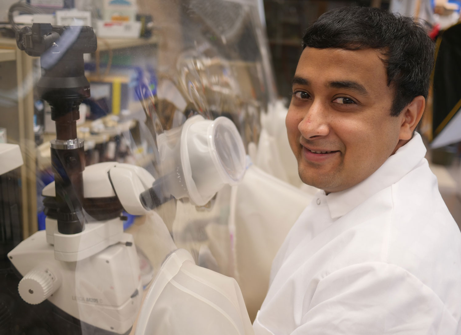
Graduate student Rohan Jonnalagadda analyzes the 3D shapes of iron-containing enzymes to parse their role in cellular processes.

Raleigh McElvery
August 27, 2019

Raised in a computer-savvy family well-versed in software and information technology, Rohan Jonnalagadda had a strong desire to “decode” the world around him. But his kind of code, the genetic one, consists of four repeating letters: A, T, C, and G. “Just like a computer runs on software, I wanted to investigate the code behind the molecular hardware that gives rise to life,” he says. Now a sixth-year graduate student in the Drennan lab, he works to decrypt the structure of metal-containing proteins, in order to determine the roles they play in vital cellular reactions.

When Jonnalagadda was an undergraduate biochemistry major at the University of California, Berkeley, it became clear to him that the genetic code was more than just a string of letters; it also serves as the blueprint for all the proteins in the entire organism. These proteins fold into complex 3D structures, which ultimately beget function.

At UC Berkeley, he joined a lab studying the iron-containing protein Heme-Nitric Oxide/Oxygen (H-NOX) that senses nitric oxide gas in bacterial and eukaryotic cells. When H-NOX binds to nitric oxide, it must change its 3D shape in the process. Jonnalagadda used a technique known as X-ray crystallography to freeze H-NOX in various stages of this conformational change to determine how it binds the gas molecules.

“I think we sometimes ignore the fact that we need trace metals in order to survive,” he says. “I was interested in continuing to think about what different metals could do in the cell. And using metals opens up a whole new world of chemical reactions that you generally don’t learn about in class.”By the time he graduated and began his PhD at MIT Biology, Jonnalagadda had been using X-ray crystallography for over two years. Today, as a member of Catherine Drennan’s lab, he continues to leverage this same method to parse the structure of additional metal-containing proteins.

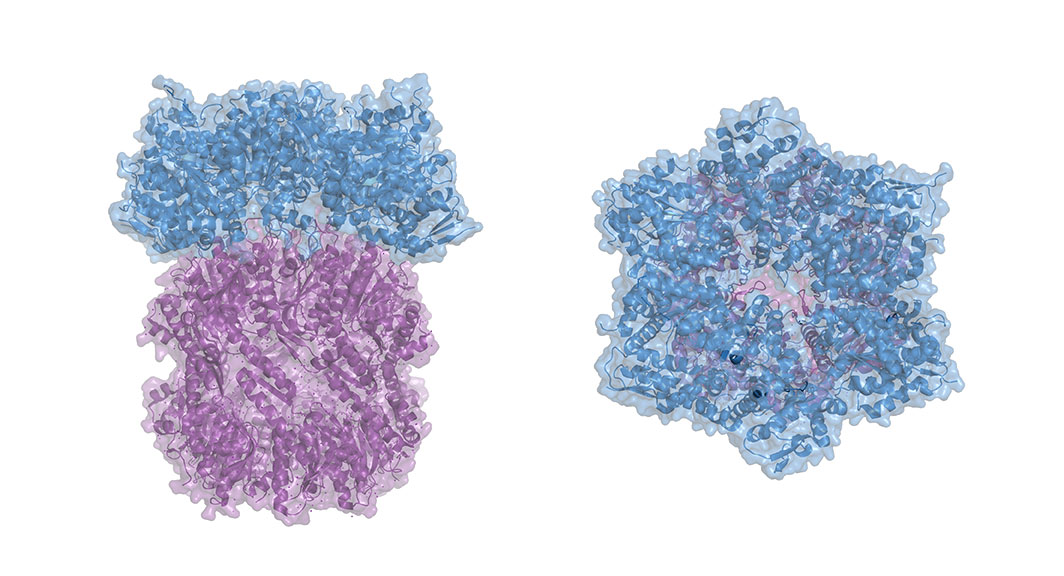
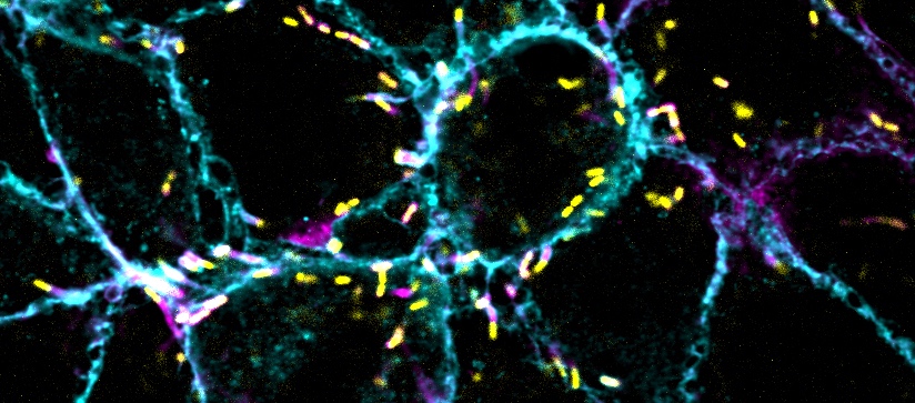
In fact, the two projects that he’s devoted most of his time to over the past five years involve reactions that he’d never even heard of before he arrived at MIT. The focus of his first undertaking was the iron-containing enzyme ribonucleotide reductase (RNR), which helps generate deoxyribonucleotides, the building blocks of DNA.

Jonnalagadda aims to understand how this enzyme is regulated to ensure the cell maintains the proper amount of each type of deoxyribonucleotide, in order to properly replicate and repair its genome. If those ratios are incorrect, the cell could experience detrimental stress.

Because the enzyme is regulated differently in humans than it is in bacteria, scientists hope to one day create antibiotics that target the bacterial RNR while leaving the human RNR unscathed. Jonnalagadda works with the human version, devising an assay that will allow him to better assess the differences between the two enzymes. RNR is notoriously difficult to work with, and so Jonnalagadda has spent much of his time developing ways to purify it so it remains stable.

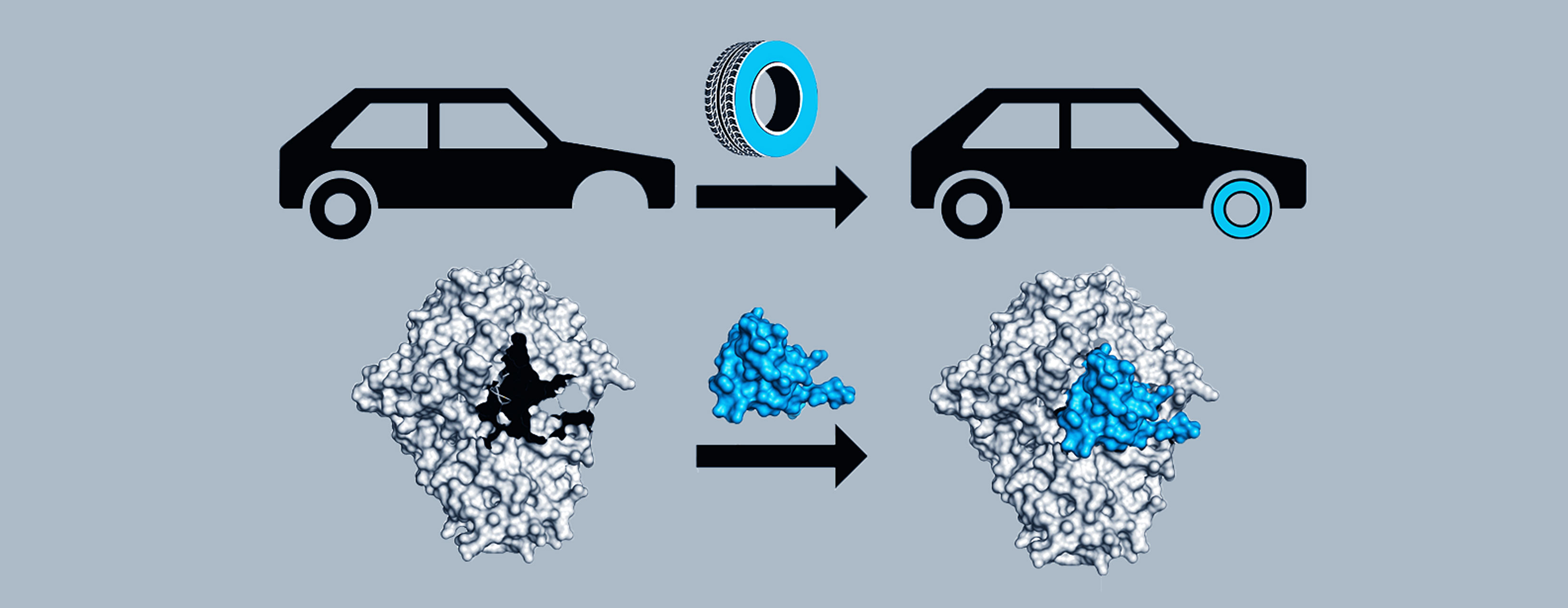
His second project is a collaboration with researchers at his alma mater, UC Berkeley, investigating isonitriles — compounds containing a carbon atom tripled bonded to a nitrogen atom. Because isonitriles are used to make drugs like antibiotics, scientists have a keen interest in exploring new ways to produce them. The team discovered that one bacterium, Streptomyces coeruleorubidus, had a novel and mysterious way of synthesizing these compounds. Jonnalagadda wants to know exactly how these particular bacteria do it.

He is using X-ray crystallography to determine the structure of the iron-containing enzyme ScoE in S. coeruleorubidus, which is responsible for forming the carbon-nitrogen triple bond characteristic of isonitriles.

“It’s exciting to be working on a protein that’s only just been discovered,” he says. “There’s just so much more to learn about its fundamental biological function. I think that’s why basic research is so appealing to me; you never know where the work will take you, or the impacts it could have on human health later on.”

Extending the frontiers of any discipline requires some guesswork and metaphorical bushwhacking, and Jonnalagadda has learned almost as much from his failed experiments as he has from his successful ones. “I’m proud that I’ve been able to use what I’ve learned about experimental design to help others in my lab when they have questions,” he says.

As he considers life post-graduation, he hopes to use the biochemical and structural techniques he’s mastered over the years to secure a job in industry.

“Being part of a department with such broad and wide-ranging research interests has made it easy to see that my work doesn’t exist in a vacuum,” he says. “It connects to many different aspects of biology.”

Photo credit: Raleigh McElvery
Posted 8.23.19
BRAIN grant will fund new tools to study astrocytes
Picower Institute
August 27, 2019
Multi “-omics” approach uncovers the riches of traditional global medicine
Greta Friar | Whitehead Institute
July 22, 2019

Cambridge, MA — Kava (Piper methysticum) is a plant native to the Polynesian islands that people there have used in a calming drink of the same name in religious and cultural rituals for thousands of years. The tradition of cultivating kava and drinking it during important gatherings is a cultural cornerstone shared throughout much of Polynesia, though the specific customs — and the strains of kava — vary from island to island. Over the last few decades, kava has been gaining interest outside of the islands for its pain relief and anti-anxiety properties as a potentially attractive alternative to drugs like opioids and benzodiazepines because kavalactones, the molecules of medicinal interest in kava, use slightly different mechanisms to affect the central nervous system and appear to be non-addictive. Kava bars have been springing up around the United States, kava supplements and teas lining the shelves at stores like Walmart, and sports figures including former and current NFL players in need of safe pain relief are touting its benefits.

This growing usage suggests that there would be a sizeable market for kavalactone based medical therapies, but there are roadblocks to development: for one, kava is hard to cultivate, especially outside of the tropics. Kava takes years to reach maturity, and as a domesticated species that no longer produces seeds it can only be propagated using cuttings. This can make it difficult for researchers to get a large enough quantity of kavalactones for investigations or clinical trials. New research from Whitehead Institute Member and associate professor of biology at MIT Jing-Ke Weng and postdoctoral researcher Tomáš Pluskal, published online in Nature Plants on July 22, describes a way to solve that problem, as well as to create kavalactone variants not found in nature that may be more effective or safe as therapeutics.

“We’re combining historical knowledge of this plant’s medicinal properties, established through centuries of traditional usage, with modern research tools in order to potentially develop new drugs,” Pluskal says.

Weng’s lab has shown that if researchers figure out the genes behind a desirable natural molecule—in this case, kavalactones—they can clone those genes, insert them into species like yeast or bacteria that grow quickly and are easier to maintain in a variety of environments than a temperamental tropical plant, and then get these microbial bio-factories to mass produce the molecule. In order to achieve this, first Weng and Pluskal had to solve a complicated puzzle: how does kava produce kavalactones? There is no direct kavalactone gene; complex metabolites like kavalactones are created through a series of steps using intermediate molecules. Cells can combine these intermediates, snip out parts of them, and add bits onto them to create the final molecule—most of which is done with the help of enzymes, cells’ chemical reaction catalysts. So, in order to recreate kavalactone production, the researchers had to identify the complete pathway plants use to synthesize it, including the genes for all of the enzymes involved.

The researchers could not use genetic sequencing or common gene editing tools to identify the enzymes because the kava genome is huge; it has 130 chromosomes compared to humans’ 46. Instead they turned to other methods, including sequencing the plant’s RNA to survey the genes expressed, to identify the biosynthetic pathway for kavalactones.

“It’s like you have a lot of LEGO pieces scattered on the floor,” Weng says, “and you have to find the ones that fit together to build a certain object.”

Weng and Pluskal had a good starting point: they recognized that kavalactones had a similar structural backbone to chalcones, metabolites shared by all land plants. They hypothesized that one of the enzymes involved in producing kavalactones must be related to the one involved in producing chalcones, chalcone synthase (CHS). They looked for genes encoding similar enzymes and found two synthases that had evolved from an older CHS gene. These synthases, which they call PmSPS1 and PmSPS2, help to shape the basic scaffolding of kavalactones molecules.

Then, with some trial and error, Pluskal found the genes encoding a number of the tailoring enzymes that modify and add to the molecules’ backbone to create a variety of specific kavalactones. In order to test that he had identified the right enzymes, Pluskal cloned the relevant genes and confirmed that the enzymes they encode produced the expected molecules. The team also identified key enzymes in the biosynthetic pathway of flavokavains, molecules in kava that are structurally related to kavalactones and have been shown in studies to have anti-cancer properties.

Once the researchers had their kavalactone genes, they inserted them into bacteria and yeast to begin producing the molecules. This proof of concept for their microbial bio-factory model demonstrated that using microbes could provide a more efficient and scalable production vehicle for kavalactones. The model could also allow for the production of novel molecules engineered by combining kava genes with other genes so the microbes would produce modified kavalactones. This could allow researchers to optimize the molecules for efficiency and safety as therapeutics.

“There’s a very urgent need for therapies to treat mental disorders, and for safer pain relief options,” Weng says. “Our model eliminates several of the bottlenecks in drug development from plants by increasing access to natural medicinal molecules and allowing for the creation of new-to-nature molecules.”

Kava is only one of many plants around the world containing unique molecules that could be of great medicinal value. Weng and Pluskal hope that their model—combining the use of drug discovery from plants used in traditional medicine, genomics, synthetic biology, and microbial mass production—will be used to better harness the great diversity of plant chemistry around the world in order to help patients in need.


This work was supported by grants from the Smith Family Foundation, Edward N. and Della L. Thome Memorial Foundation, the Family Larsson-Rosenquist Foundation, and the National Science Foundation (CHE-1709616). T.P. is a Simons Foundation Fellow of the Helen Hay Whitney Foundation. J.K.W is supported by the Beckman Young Investigator Program, Pew Scholars Program in the Biomedical Sciences (grant number 27345), and the Searle Scholars Program (grant number 15-SSP-162).


Written by Greta Friar


***

Jing-Ke Weng’s primary affiliation is with Whitehead Institute for Biomedical Research, where his laboratory is located and all his research is conducted. He is also an associate professor of biology at Massachusetts Institute of Technology.

***


Full citation:

“The biosynthetic origin of psychoactive kavalactones in kava”

Nature Plants, online July 22, 2019, doi: 10.1038/s41477-019-0474-0

Tomáš Pluskal (1), Michael P. Torrens-Spence (1), Timothy R. Fallon (1,2), Andrea De Abreu (1,2), Cindy H. Shi (1,2), and Jing-Ke Weng (1,2)

1. Whitehead Institute for Biomedical Research, 455 Main Street, Cambridge, MA 02142 USA.

2. Department of Biology, Massachusetts Institute of Technology, Cambridge, MA 02139 USA.

Bacteria use “spare part” proteins to repair damage and survive inhospitable conditions
Saima Sidik
July 17, 2019

Millennia ago, before the evolution of multicellular life, bacteria developed a group of proteins called glycyl radical enzymes to help them turn food into cellular energy. Glycyl radical enzymes functioned efficiently for millions of years until bacteria encountered a new hurdle: oxygen build-up in the atmosphere. Glycyl radicals are easily damaged by oxygen, and bacteria needed a way to continue to process nutrients under these new conditions.

In a recent study published in Journal of Biological Inorganic Chemistry, Sarah Bowman et al. from MIT provide structural evidence for how bacteria use “spare part” proteins to repair glycyl radical enzymes by replacing their damaged portions.

“Instead of degrading the whole protein and building a new one, bacteria make a small spare part protein that can bind to the glycyl radical enzyme and restore its function,” says Lindsey Backman, a graduate student in the MIT Department of Chemistry and co-author on the study.

Senior author Catherine Drennan, an MIT professor of biology and chemistry and a Howard Hughes Medical Institute Investigator, is leading the effort to characterize a glycyl radical enzyme called pyruvate formate lyase, or PFL, and its corresponding spare part protein, YfiD. Using a technique called nuclear magnetic resonance spectroscopy, her lab examined the shape of YfiD. Previous work revealed that upon oxygen exposure, part of PFL is cleaved off, leaving the enzyme unable to perform its chemistry. Then, using in silico modeling, the Drennan lab showed that YfiD fits neatly into the hole in oxygen-damaged PFL, replacing the missing piece.

“If you have a car and you get a flat tire, you don’t buy a new car, you just change the tire,” Backman says.

Although YfiD is one of only two spare part proteins that have been identified, Drennan suspects there are many more. It’s easy for researchers to mistake spare parts for the portions of proteins that they replace, and Drennan thinks that scientists have probably overlooked spare parts when analyzing the complex milieu of proteins found in cells.

Although identifying spare parts is technically challenging, Drennan thinks the applications for synthetic biology make it well worth the effort. Specifically, she’s interested in bacteria that live in the deep sea where there’s very little oxygen and use other glycyl radical enzymes to degrade hydrocarbons. Because they can break down hydrocarbons, these bacteria could be valuable tools for cleaning up oil spills. Oil spills occur on the ocean’s surface where there’s a lot of oxygen, so spare parts might be necessary to stabilize oil-degrading proteins under these conditions.

“The question always becomes, ‘what about the oxygen sensitivity?’” Drennan says, referring to these oil-degrading proteins. “What if we expressed a spare part to make them more stable?”

Backman began working on spare part proteins as an undergraduate participating in the MIT Summer Research Program, or MSRP, as did several of the study’s other authors, and Drennan says that this project’s success highlights the valuable role these students play in MIT labs. Now a full-time graduate student, Backman has teamed up with Drennan lab postdoc and co-author Mary Andorfer, and together they plan to continue characterizing the interaction between YfiD and PFL. They think their findings may be the first step in showing that spare parts are a common protein repair mechanism, and that characterizing them will add a new tool to the synthetic biology toolbox.

Citation:
“Solution structure and biochemical characterization of a spare part protein that restores activity to an oxygen-damaged glycyl radical enzyme”
JBIC Journal of Biological Inorganic Chemistry, online June 26, 2019, DOI: 10.1007/s00775-019-01681-2.
Sarah E. J. Bowman, Lindsey R. F. Backman, Rebekah E. Bjork, Mary C. Andorfer, Santiago Yori, Alessio Caruso, Collin M. Stultz, and Catherine L. Drennan.

Researchers determine what makes some proteins “slippery” enough to evade destruction
Raleigh McElvery
July 10, 2019

All cells must balance generating new proteins with eliminating excess or damaged ones by way of powerful degradation machines — which, much like wood chippers, chew up proteins and spit them out. But, these proteins are often folded into intricate structures, and must be unfurled before they can be fed into these degradation machines, broken into tiny bits, and ultimately recycled. In bacteria, a molecular motor known as ClpX must grip the end of the ill-fated protein and apply force to straighten it. However, until now, researchers weren’t sure precisely how ClpX gripped its target tightly enough to accomplish this task.

There had been evidence to suggest that some amino acids — the chemical building blocks that comprise proteins — are “slippery” and thus more difficult to grip. In a new study published in eLife, researchers at the MIT Department of Biology examined each amino acid’s individual contribution to grip. By parsing the physical basis for this molecular interaction, they hope to better understand how some proteins evade destruction.

“Previous studies had shown that small amino acids were notoriously hard to grip, but no one really understood why,” says Tristan Bell, graduate student and first author on the paper. “It’s like watching a game of tug-of-war and knowing that a person’s hands are important for pulling on the rope, but having no idea what allows the hands to get a good grip on the rope.”

ClpX, he explains, is roughly shaped like donut with loops protruding into the center hole. These loops grip the target protein, jamming it against the surface of ClpX, and unfolding it so it can be threaded through the hole and shredded.

The researchers engineered proteins with tails comprised of various amino acid combinations, and measured how well ClpX could grip them, both in bacteria and in test tubes. They determined that ClpX can only grip between six and eight amino acids at a time, and that only a handful of the 20 possible amino acids could actually be “well-gripped.” When ClpX was able to grasp multiple amino acids simultaneously, its grip strength increased.

“We think that somehow the charge is preventing ClpX from making strong contacts with the target protein, preventing it from achieving a stable grip state,” Bell says.Just like in previous experiments, large amino acids appeared easier to grip than small ones, “similar to the way a knotted rope is easier to grasp than a smooth, slippery one,” Bell says. But, regardless of size, amino acids that carried electric charge seemed to be more slippery.

The team thinks that proteins with slippery tails might have an evolutionary advantage, because they are harder to grip and therefore less likely to be degraded.

Invaders like viruses have been known to insert a slippery sequence into certain proteins to prevent the host cell from destroying them and thus promoting replication. Even healthy cells produce proteins with strategically placed slippery sequences, which allow a portion of the protein to break away from the degradation machinery unscathed. In the bacteria Caulobacter crescentus, this planned breakage actually produces a version of one protein that’s needed for DNA replication.

“Next,” Bell says, “we’re hoping to look across entire proteomes in different organisms to find more proteins that escape destruction.”

“Tristan’s experiments and results reveal some of the molecular determinants of grip in the bacterial degradation machines we study,” says Bob Sauer, the Salvador E. Luria Professor of Biology and senior author on the study. “Many of the rules he discovered apply to related machines that function in all biological organisms, including humans, emphasizing the common evolution of these machines.”

Citation:
“Interactions between a subset of substrate side chains and AAA+ motor pore loops determine grip during protein unfolding”
eLife, online June 28, 2019, DOI: 10.7554/eLife.46808
Tristan A. Bell, Tania A. Baker, and Robert T. Sauer.

Researchers identify important proteins hijacked by pathogens during cell-to-cell spread
Raleigh McElvery
July 9, 2019

Listeria monocytogenes, the food-borne bacterium responsible for listeriosis, can creep from one cell to the next, stealthily evading the immune system. This strategy of cell-to-cell spread allows them to infect many different cell types, and can spur complications like meningitis. Yet the molecular details of this spread remain a mystery.

In a paper recently published in Molecular Biology of the Cell, researchers from the MIT Department of Biology, University of California, Berkeley, and Chan Zuckerberg Biohub are beginning to piece together the elusive means by which Listeria moves from one cell to the next. This mode of transport, the scientists suggest, looks a lot like trans-endocytosis, a process that healthy, uninfected cells use to exchange organelles and various cytoplasmic components. In fact, the two processes are so similar that Listeria may be co-opting the host cell’s trans-endocytosis machinery for its own devices.

Although the particulars of trans-endocytosis are poorly understood, the process permits neighboring cells to exchange materials via membrane-bound compartments called vacuoles, which release their cargo upon reaching their final destination.

Much like trans-endocytosis, cell-to-cell spread relies on vacuoles to ferry Listeria. First, the pathogen commandeers the host cell’s own machinery to assemble a tail of proteins that allows it to rocket around inside the cell and ram against both the membrane of the host and that of the adjacent cell. The resulting protrusion is then somehow engulfed into a double-membrane vacuole, and the bacteria burst through their containment to begin the process anew in the recipient cell.

“There’s been a lot of work looking at Listeria cell-to-cell spread,” says Rebecca Lamason, the Robert A. Swanson (1969) Career Development Assistant Professor in the MIT Department of Biology and senior author on the study. “But we still don’t really understand the molecular mechanisms that allow the bacteria to manipulate the membrane to promote engulfment. Depending on what we uncover, we might also be able to apply that information to better grasp how an uninfected cell regulates trans-endocytosis.”

Lamason and her team anticipated that the same proteins implicated in trans-endocytosis would also be involved in Listeria cell-to-cell spread, which would indicate that the pathogen was appropriating these proteins for its own purposes. The researchers made a list of 115 host genes of interest, and then used an RNAi screen to identify just 22 that are critical for cell-to-cell spread.

They were excited to find that, of those 22 genes, several are also implicated in endocytosis, which suggests Listeria is using a similar strategy. These include genes encoding caveolin proteins that control membrane trafficking and remodeling, as well as another protein called PACSIN2 that interacts with caveolins to regulate protrusion engulfment.

Now that the researchers have pinpointed these key proteins, the next step is to determine how they work together in order to promote cell-to-cell spread — especially since the protrusions created by Listeria are much larger than those required for trans-endocytosis.

“As we drill down even deeper into the molecular mechanisms, it will be interesting to see where trans-endocytosis and cell-to-cell spread differ, and where they are similar,” Lamason says. “Our hope is that investigating the mechanisms of bacterial spread will reveal fundamental insights into host intercellular communication.”

Citation:
“RNAi screen reveals a role for PACSIN2 and caveolins during bacterial cell-to-cell spread”
Molecular Biology of the Cell, online June 26, 2019, DOI: 10.1091/mbc.E19-04-0197
Allen G. Sanderlin, Cassandra Vondrak, Arianna J. Scricco, Indro Fedrigo, Vida Ahyong, and Rebecca L. Lamason

Unusual labmates: Lighting up the lab
July 7, 2019

Unusual Labmates is a series that explores some of the more unusual models used for research at Whitehead Institute. From rare plants to luminescent beetles to regenerative starfish and worms, these organisms and their unusual traits provide insights into the underlying biology and incredible diversity of living things.

Massachusetts Institute of Technology (MIT) graduate student Tim Fallon is standing in a field in New Jersey, holding a net and waiting for the last glimmers of sunlight to disappear. As the trees surrounding the field fade into shadow, Fallon watches the ground intently. The air is still and then, hovering above the grass, a small point of light appears. It floats through the air in a concave upwards arc and winks out. Soon, more lights follow suit. These are fireflies, unassuming beetles by day, but at night they put on a dazzling luminescent display in the hope of attracting a mate. Fallon sweeps his net through the air, capturing some of them. He has traveled to New Jersey with other members of Whitehead Institute Member and associate professor of biology at MIT Jing-Ke Weng’s lab to collect specimens of Photinus pyralis, the big dipper firefly, in order to sequence a firefly genome for the very first time.

Fireflies have been around for more than one hundred million years, and in that time have diverged into more than 2,000 species and spread to every continent except Antarctica. The beetles (despite their name, fireflies are actually beetles) are widely known, and often beloved, for their enchanting courtship rituals, but they have also piqued the interest of scientists, who have harnessed the gene behind their light emitting capability for use in research. However, for all of fireflies’ appeal, they are a difficult animal to work with in the lab, and much of their biology remains shrouded in mystery. In the hopes of improving that situation, Weng, Fallon, and collaborators—including Sarah Lower, assistant professor of biology at Bucknell University, and Yuichi Oba, professor at Chubu University in Japan—have been investigating fireflies, primarily by sequencing and analyzing their genomes. This research is providing insights into the evolution of fireflies’ light-producing ability, the biomolecular pathways the fireflies use to luminesce, and could perhaps even inform how the use of firefly-based luminescence in research can be improved.

The chemistry of light

Fireflies produce light using two main ingredients: an enzyme called luciferase and the small molecule luciferin. Luciferase facilitates a chemical reaction that oxidizes the luciferin, and one product of the reaction is light. In the 1980s, researchers recreated the process in plants and plant cells by cloning the firefly gene responsible for the luciferase enzyme from big dipper fireflies and then inserting it into the genomes of their specimens in the lab.[1] When they injected luciferin into the specimens, they began to glow – just like fireflies. Researchers now use this approach in both plant and animal models to track various aspects of biology. They can link the luminescence to a trait or process, and then measure the level of light emitted. For example, researchers can fuse the luciferase gene to a gene of interest such that the two genes will be expressed as one. Then they introduce luciferin to the system and measure the light output using very sensitive equipment that can sense minute changes. The more light that is emitted, the higher the activity level of both luciferase and the gene of interest. This is a widely used assay that, Fallon says, every biologist uses or at least learns about during their training. Luciferase-luciferin has many applications, and along with tracking gene expression it has also been used to track cancer metastasis, monitor medical treatment efficacy, and check for microbial contamination—including on space vehicles such as the Mars Curiosity Rover.[2] The extreme sensitivity of luciferin-luciferase tests makes them an attractive choice in many experiments.

In spite of the widespread use of firefly luminescence, a lot about it still isn’t known, especially when it comes to luciferin. While the gene encoding luciferase has been identified, luciferin is thought to be created through a process involving multiple genes, and the complete set of those genes is unknown, as are the steps involved in the production of luciferin: the intermediate molecules produced and how they are modified to reach the final product. Although Weng’s lab generally studies plants and focuses on understanding how plants evolved biochemical pathways to produce unique small molecules with traits of interest, particularly those with medicinal value, when Fallon joined the lab, he convinced Weng that investigating the unknowns of the small molecule luciferin was a similar and suitable project.

Fireflies in the wild

When Fallon joined the Weng lab, there were not a lot of research tools available for investigating fireflies, starting with the lack of a sequenced genome. Only a handful of firefly genes had even ever been identified. Weng and his collaborators crowdfunded the money to sequence the first firefly genome, and then received funding to sequence a second firefly species and a bioluminescent click beetle, providing a wealth of new data to explore.

The lack of tools for firefly research was due in part to how difficult it is to rear fireflies in the lab, which makes them tricky animals to study. Fireflies are very sensitive to changes in their environments, and in the lab it’s difficult to mimic the right vegetation, climate, seasonal shifts, and other factors that the beetles rely on to time their metamorphoses and thrive.

Unpredictable environmental changes are becoming a common challenge for fireflies beyond the lab as well, due to the impact of humans on their habitats. Fireflies’ sensitivity to these changes may be causing their disappearance. Anecdotally, many people have fond memories of seeing fireflies every summer when they were younger and can attest to the absence of fireflies in those same places now. A large citizen science research project called Firefly Watch is currently underway to figure out the extent to which firefly populations are declining across the United States (U.S.).[3] And in a case indicative of a larger problem, conservation groups recently submitted an urgent petition to recognize the Bethany Beach firefly, whose key habitat in Delaware is at risk due to human development, as the first endangered firefly species in the U.S.[4]

A significant manmade disruption to the fireflies’ habitats is light pollution. Fireflies find each other by signaling with light, but their lanterns are no match for electricity; a firefly trying to outshine a streetlamp might as well be facing off with the sun. For species that evolved faint glows to flash in the dead of night, in the dark of forests, finding a place where their light can be seen is getting harder and harder. Big dipper fireflies have fared better than many others species. They’re large, with bright lanterns, and they tend to flash at twilight, so they are used to competing with some ambient light. Unsurprisingly, big dipper fireflies remain abundant in the wild, including in and near cities. They can be spotted all over the eastern and midwestern U.S., where they are easily identifiable thanks to the distinctive J-shape—resembling the curve of a dipper—that they make as they flash.

Big dipper fireflies have been important in biology research—it is from them that the firefly luciferase gene was first cloned—and so they were the first species that Fallon and his collaborators chose for their genome sequencing project. However, big dipper fireflies fare no better than other firefly species in a lab setting. No one has ever successfully reared big dipper fireflies through a full lifecycle (from egg to egg) in the lab, Fallon says. So, in spite of the ease of collecting big dipper fireflies in the eastern U.S., in order to get a population of fireflies going in the lab, Fallon had to look elsewhere: to Japan.

In Japan, fireflies are beloved. Watching them is a popular summer pastime, and they are celebrated in myths, songs, and other media. The two dominant species are both aquatic in their larval stages: the heike-botaru (Aquatica lateralis), which mostly lives in flooded rice paddy fields, and the larger and brighter genji-botaru (Luciola cruciata), which lives in streams and rivers. Both species need clean bodies of water to survive, and so their numbers have diminished in cities—but unlike in America, Japan has not allowed fireflies to fade away quietly into the night. As fireflies have grown scarcer, breeding centers have begun rearing the insects in large numbers, using big facilities that can recreate the fireflies’ natural habitat on a scale not possible in a U.S. lab. Fireflies are sometimes bred for conservation purposes, such as an effort to bolster the heike population in the water by the Imperial Palace in Tokyo. The fireflies are also bred for spectacle, released during summer festivals and firefly-watching events in the city to recreate the lost experience of glow-filled nights. The Fussa firefly festival has drawn large crowds for more than fifty years.

Rearing insects is also a relatively common pastime in Japan. When Fallon was looking for a type of firefly that would be a good addition to the genome sequencing project and could survive in lab conditions, he learned of the heike, which are kept in captivity and used in research in Japan. Although heike are still very sensitive to small changes in their environments, they have been demonstrated to survive for multiple generations indoors, unlike the big dipper firefly. The larvae are aquatic, and maintaining a controlled aquarium is also easier than a terrarium, Fallon says. Fallon received his fireflies from a collaborator in Japan, firefly expert Dr. Yuichi Oba, a professor at Chubu University. Oba works with Haruyoshi Ikeya, a high school teacher in Yokohama, Japan who cracked the code of rearing heike fireflies indoors several decades ago; the population from which Fallon received his specimens has been lab-bred since its original capture in 1990. Fallon received tips from Oba on how to successfully rear the fireflies—though not all of these could be followed, due to stricter regulations for how to keep the fireflies contained in the U.S., where the United States Department of Agriculture considers them a potential plant pest.

Fireflies in the lab

Fallon rears the fireflies in a small room separate from the main space of the Weng lab. The room is tightly packed with supplies and various aquariums: Fallon must move the firefly specimens between environments as they go through their lifecycle. There is tinfoil over the windows so when Fallon switches the lights off the room becomes a darkroom, where he can observe the fireflies flashing.

Maintaining a lab population serves a number of purposes. First of all, collecting fireflies is time-consuming, limited by season, and in the case of foreign species, involves further transportation and regulatory roadblocks. When collecting specimens in the wild, it’s easy to find adults, but harder to get access to every stage of the lifecycle, especially eggs and pupae, the way you can in the lab. Having a lab population also ensures that you are working with one species, whereas when collecting in the wild it can be easy to get a mixed-up batch—one field may contain a dozen similar-looking species—and molecular biology research requires species-specific material. Overall, having a lab population means access to live specimens whenever you need them: whenever the researchers have a new research question to answer or a new experiment to run. However, maintaining this beneficial resource is not a simple feat.

Rearing fireflies in captivity is difficult in part because each stage of their life cycle has different requirements. Fireflies hatch from eggs, which the heike lay in moss near water. Heike larvae live in water, where they spend most of their lifespan, usually around one year, though in the lab this stage can be as short as six months. During this time, the larvae typically go through five or six instars, or stages of growth between molting. Insects must molt in order to grow, as their size is restricted by their exoskeletons. The heike’s first instar is only a few millimeters long, while the final instar may grow to be closer to two centimeters.

For most of their time as larvae, the specimens live in shoebox sized aquariums inside a metal cabinet. Fallon feeds them bladder snails and waits for them to grow. When the larvae are in their later instars, around the right size and age to pupate, Fallon moves them into another aquarium with a mock riverbank inside, with a soil mixture devised by Haruyoshi Ikeya. The riverbank is necessary because the firefly larvae move to land to pupate. First, they begin to exhibit what is called climbing or landing behavior, during which they will flash—fireflies are capable of emitting light at all stages of their lifecycle. The flashes may be a signal to help coordinate pupation. If a larva doesn’t synchronize with the others when it is ready to pupate, there won’t be any mates around when it hatches. Fireflies don’t live very long as adults, so they must find a mate quickly. In the lab, once the adults hatch, Fallon moves them into another container to prevent them from drowning in the water of the mock river, and hopes that they mate. If they are successful, then the female lays her eggs in the moss Fallon provides, starting the cycle over again.

The fireflies are not easy to keep alive in lab conditions, so the researchers have been experimenting with different conditions for rearing them, like keeping them in boxes of different sizes, changing the aeration of their water, and providing different spaces for the adults to rest when they first hatch.

“We’ve been iterating through a lot of different ways to rear them,” Fallon says. “It’s not like with fruit flies, where you could leave two alone in a room with a banana, and soon you’ve got more fruit flies than you know what to do with. The fireflies are really quite finicky. Over time we’re learning what works and what doesn’t.”

One thing that Fallon has learned from rearing fireflies is how often they glow, during every part of their life cycle. The heike lay their eggs in clumps, which are cumulatively visible to the naked eye. Big dipper firefly eggs also luminesce, but faintly enough that it’s only visible using a sensitive camera. If there’s an evolutionary advantage to having the eggs luminesce, it’s not known. The larvae’s ability to glow is typically believed to be, at least in part, an aposematic signal: a warning to potential predators that the larvae are toxic so the predators won’t try to eat them. Other species use bright colors for the same purpose—think of brilliantly colored poison dart frogs or the popular mnemonic “red touches yellow kills a fellow” used to identify venomous coral snakes—and what’s brighter than something that glows in the dark?

Fireflies’ toxicity comes from chemicals called lucibufagins. Only some firefly species produce these, though the rest may benefit by association if predators associate glowing beetles with a bitter meal. Fallon has noticed that the heike larvae will glow anytime they appear threatened or are faced with the unfamiliar, like if he jostles the box they live in. This threat response is consistent with the expectations for an aposematic signal, but the heike larvae also glow in other conditions, such as during their climbing behavior right before they pupate—and the pupae glow as well. The adults, meanwhile, can also warn off predators with their flashes but primarily luminesce to communicate and find a mate. Different firefly species have distinct flash patterns, as do males and females, allowing adults to identify a suitable partner—and allowing anyone with enough firefly knowledge to identify different species just by watching their flashes.

In every stage of life, fireflies have adapted to make the most of their light-emitting ability. Once they had the genome sequences and firefly specimens in hand, Weng and Fallon wanted to find out how.

What can we learn from fireflies?

The researchers used the firefly genomes to delve into the biomolecular pathways that fireflies use to create light. They identified a luciferin-derived molecule in fireflies that may be a storage form for luciferin, as well as the gene encoding the enzyme that converts luciferin into this molecule. They have also been looking into fireflies’ mechanism for recycling luciferin. The one significant handicap of using luciferase-luciferin in research is that, since the genes encoding luciferin are unknown, the chemical must be fed or injected into specimens manually. This limits the duration of experiments based on when the luciferin is used up, or requires disrupting specimens to reinject them. In the wild, fireflies are able to reuse their luciferin molecules after they have been oxidized to produce light; if researchers could figure out how they do this, it could potentially be applied in the lab.

One major mystery surrounding fireflies that the researchers wanted to solve was whether beetles evolved the ability to luminesce once, or multiple times. Fireflies are one of at least four beetle families that can luminesce—the others are click beetles (Elateridae), glowworm beetles or railroad worms (Phengodidae), and starworms (Rhagophthalmidae). These different beetles all use similar chemistry to luminesce: similar luciferase enzymes and structurally identical luciferins. This commonality suggests that luminescence evolved in a shared ancestor of the four families, and was lost in other related beetles that do not luminesce. However, as Charles Darwin noted, the families of luminescent beetles are very different morphologically, including having distinct light organs—the click beetle emits light from lanterns by its head, whereas the fireflies emit light from their rears. These dissimilarities in shape suggested that the beetle families each evolved luminescence separately; if the light organs evolved from a common ancestor, researchers would expect them to resemble each other. Complicating the matter is that not every species in these families can luminesce; while all known fireflies can emit light, only some click beetles can. This suggests that even within the bioluminescent beetle families, luminescence has evolved multiple times, or been lost multiple times, or some combination thereof.

In order to solve this puzzle, the researchers sequenced the genome of three beetles and compared them: the big dipper firefly, the heike firefly, and the cucubano click beetle (Ignelater luminosus), which is also capable of luminescence. The last common ancestor of heike and big dipper fireflies lived over 100 million years ago, making them good candidates for evolutionary comparison. The researchers found evidence that fireflies and click beetles evolved luminescence independently. They pinpointed where the luciferase gene was in each species’ genome, and what its neighbors were—in the fireflies, the gene was surrounded by genes involved in fatty acid metabolism, suggesting that it evolved from one of these. Meanwhile, the researchers found the click beetle’s luciferase gene in a completely different genetic neighborhood, suggesting that it evolved separately and from a different ancestral gene.

It may seem unusual for such an extraordinary trait as luminescence to have evolved multiple times, but in fact it is a trait that evolution constantly stumbles upon, Fallon says. Beetles are far from the only creatures capable of emitting light; bioluminescence has also evolved in many niches, including in species of fish, coral, jellyfish, squid, snails, fungi, and bacteria.

Fireflies’ luminescence is not a unique trait, but it’s one worth preserving. From fireflies lighting up the night sky at summer festivals and in backyards, to children chasing them through the grass trying to capture a little magic in their hands, to researchers exploring biology with the help of the ultra-sensitive luciferase gene, people benefit from sharing our world with these dazzling little beetles. With the new data coming out of labs like Weng’s, further research benefits from fireflies’ light-making machinery may be on the horizon.

Fallon has learned a lot about the difficulties of rearing fireflies as he tries to maintain a sustainable population in the lab; meanwhile conservationists are struggling to protect populations of fireflies out in the wild. Even the wild population from which Fallon’s fireflies were originally captured no longer exists. Though the species survives, that particular population’s habitat disappeared, leaving the lab-bred beetles as their only legacy. The more that researchers learn about fireflies, the better equipped we may be to protect them from the sort of environmental vulnerabilities that killed off the Weng lab fireflies’ ancestors—both for the sake of the fireflies themselves, and for own sake as spectators and researchers.

Credits

Written by Greta Friar

Video by Conor Gearin

Audio production by Conor Gearin

Cover video by Radim Schreiber / FireflyExperience.org

“The chemistry of light” title card photo by Tim Fallon

“Fireflies in the wild” title card photo by Radim Schreiber / FireflyExperience.org

“Fireflies in the lab” title card photo by Conor Gearin

“What can we learn from fireflies?” title card photo by Conor Gearin

Special thanks to Radim Schreiber, Tim Fallon and Jing-Ke Weng

Works cited:

[1] https://science.sciencemag.org/content/234/4778/856

[2] https://www.science.gov/topicpages/p/planetary+protection+protocols

[3] https://www.massaudubon.org/get-involved/citizen-science/firefly-watch

[4] https://xerces.org/2019/05/15/bethany-beach-firefly/