Intrigued by immortality
Eva Frederick | Whitehead Institute
March 16, 2021

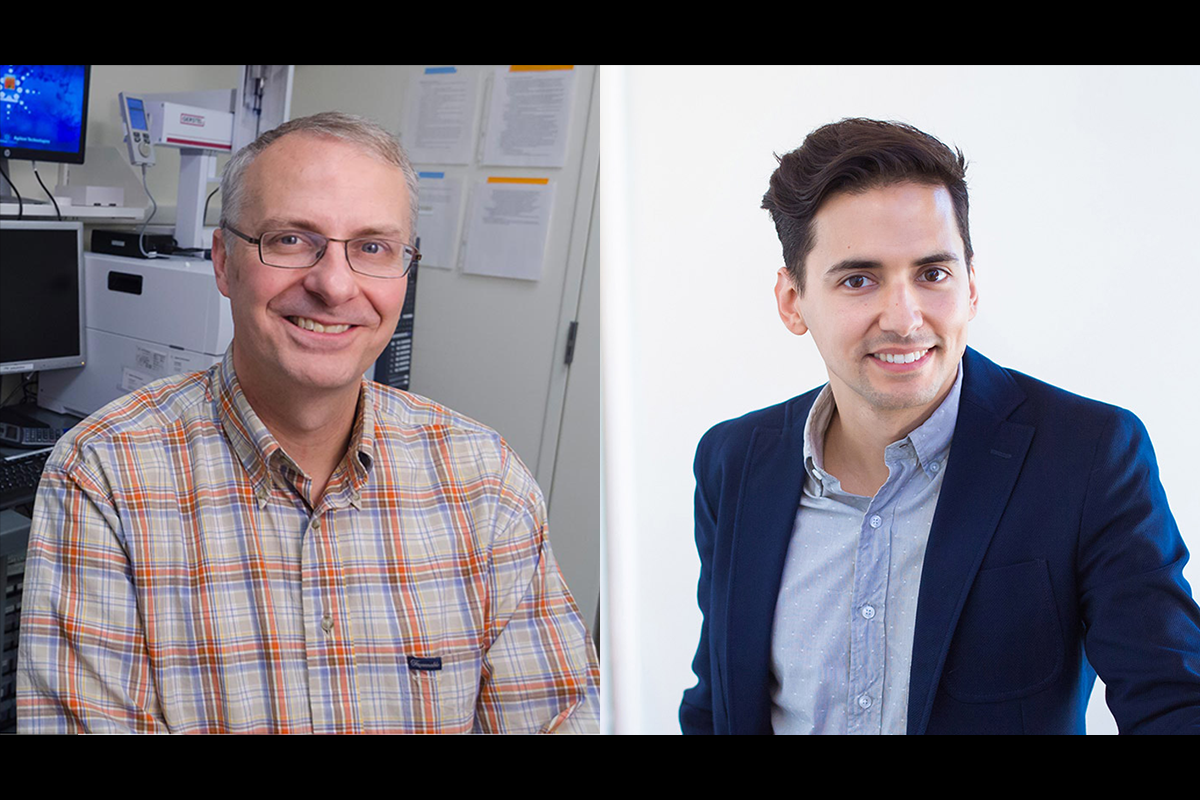
New Whitehead Institute director Ruth Lehmann and new Member Yukiko Yamashita study opposite sides of the germ cell life cycle. Yamashita’s work in male germ cells shows how the cells are formed and maintained; Lehmann studies female germ cells to understand their fates. At the Whitehead Institute, they join Member and former director David Page in painting a fuller picture of how these seemingly immortal cell lines pass instructions uninterrupted from generation to generation.

All other cells in the body — neurons, muscle cells, the stem cells that replenish other tissue types — are made anew in each embryo and go away when organisms die. But not the germ cells. “The germ cell passes its DNA to the next generation, then that DNA is used to build up to a new germ cell,” says Yamashita. “That means that germ cells never cease to exist.”

In this way, an unbroken chain of germ cells stretches back to our most distant ancestor. Scientists study this never-ending link for insights into the fundamentals of biology and evolution. Yamashita began studying germ cells as a model to investigate other questions, but as her research progressed, she grew more and more intrigued by the cells’ special properties.

“This is one thing Ruth and I have in common,” Yamashita says. “There are many biologists that study germ cells, but not many are acutely interested or fascinated by this immortality. We want to know, where does it come from?”

Yamashita, also an Investigator of the Howard Hughes Medical Institute, joined Whitehead Institute in September. Work in her laboratory at Whitehead Institute will focus on two areas, using the fruit fly Drosophila melanogaster as a model. First, she will continue her focus from previous projects on the mechanics of asymmetric cell division using male germline stem cells. These cells, like other stem cells in the body, must undergo a series of asymmetric divisions — instead of simply dividing into two identical daughter cells, the cells must create daughters with different cell fates and programming.

“This balance — maintaining the stem cell number while making some differentiating cells — is considered to be a very important process,” she says. “If you end up making too many stem cells, it can become cancerous; but if you commit too much to the differentiation, you lose the stem cell count, and that means you cannot continue sperm production.”

A newer project in her lab centers on the long sequences of nucleotides within organisms’ genomes that don’t code for any genes. They’re often nonsensical, gibberish combinations or long strings of certain bases. This “genomic junk” has long been dismissed as meaningless filler between essential genes, but Yamashita proposes that the junk is essential for the overall structure of the genome. Much like the binding of a book holds together its contents in an organized fashion, the genomic junk may provide a blueprint for how genetic material is held together and eventually read.

Ultimately, it is the germline cells that are responsible for maintaining this DNA framework. Yamashita hypothesizes that slow changes in junk DNA could provide some explanation for why different species are reproductively incompatible.

“If you look at the chimpanzee genome and the human genome, the protein coding regions are, like, 98 percent, 99 percent identical,” she says. “But the junk DNA part is very, very different. We think this divergence might explain what happens when one species splits into two.”

Yamashita’s research team will share lab space with Lehmann’s group. Both researchers use fruit flies for their experiments, but Lehmann’s research focuses on egg cells, not sperm. “Germ cells are special; you don’t need them for survival, but you need them to keep the species going,” she says. “How are they initially specified and set aside? What makes them different, how are they set aside from somatic cells, and how do they maintain their cell fate?”

One project Lehmann is carrying over from her work at New York University’s Skirball Institute of Biomolecular Medicine involves phase transition condensates — small, membraneless granules that bring together the components needed for complex cellular functions. Lehmann studies a specific type of condensate called a germ granule, an aggregation of small RNAs and RNA binding proteins found only in germline cells, which helps determine the cells’ fate.

Lehmann is also investigating the female germline cells’ role in maternal inheritance. After fertilization, the maternal cell imparts not only its nuclear DNA but also components of its cytoplasm, including mitochondria, RNAs, and even bacteria. “This whole idea of cytoplasmic inheritance and the transgenerational continuum of the cytoplasm is something I’m just starting to think about,” she says.

Yamashita and Lehmann share a large open space on the third floor of the Institute, with researchers from each lab integrated throughout. They will also share a fly room and computational room. The researchers hope the communal setup will allow a flow of ideas between their labs. “By sharing this kind of basic space, we are hoping to let our people interact with each other and for discussions to happen,” Yamashita says.

“This is a new concept for Whitehead, and we’ll see how it works,” Lehmann says. “It’s an exciting experiment in lab sociology.”

“Selfish” DNA helps bacteria cheat and grow in densely-packed microbial communities
Raleigh McElvery
March 12, 2021

Scientists have a term for genes that spread themselves throughout a population at any cost: “selfish” DNA. One way that these genes transmit through bacterial communities is via a type of bacterial sex called conjugation. When one bacterium makes contact with another, DNA from the host cell can be injected into a recipient cell.

Alan Grossman’s lab at the MIT Department of Biology studies a small but selfish chunk of DNA called ICEBs1. His group has identified several ways in which this so-called mobile genetic element actually benefits its host bacterium as it fights to spread. Building off this body of work, Grossman’s lab collaborated with colleagues at Tel Aviv University on a new study recently published in eLife. The international team found that ICEBs1 contains one gene in particular, which allows the host cell to continue dividing in densely-packed microbial communities. This helps the host to grow in conditions where nutrients are scarce, while also potentially helping ICEBs1 to propagate.

“Mobile genetic elements like ICEBs1 are found in the chromosomes of many different types of bacteria,” says Grossman, department head and co-senior author on the study. “Studying these elements — how they spread and how they affect their host cells — is critical for understanding the evolution of bacteria, engineering some types of bacteria to do useful things, and possibly preventing the deleterious effects caused by harmful bacteria.”

Like many DNA segments on the move, ICEBs1 includes genes that encode the molecular machinery required to transfer itself from one cell to the next. But mobile genetic elements can also contain “cargo” genes that bestow the host bacterium with new traits, such as antibiotic resistance. However, in many cases, the properties a cargo gene will endow are hard to predict.

“The host cell can get a lot of new genes in a hurry through mobile genetic elements like ICEBs1, and there’s a lot we still don’t know about the types of phenotypes cargo genes confer,” says the study’s first author, Joshua Jones PhD ’20. “The array of possible traits is probably a lot more diverse than we currently appreciate.”

To investigate the changes that ICEBs1 triggers in the host cell, Jones and colleagues examined large microbial communities called biofilms. These form when many bacteria aggregate on a surface and secrete a slimy “glue” made of sugar, proteins, and DNA that encases the population. Common examples of biofilms include dental plaque, the sludge that coats the inside of pipes, or the deleterious infections that form on surgical implants in patients’ bodies.

Because there are so many bacteria in close contact, biofilms are hot spots for exchanging mobile genetic elements like ICEBs1. However, secreting the materials needed to produce the slimy glue can rapidly deplete resources. As a result, bacteria in a biofilm do not always have the capacity to grow, divide, and potentially spread ICEBs1. Instead, certain types of rod-shaped bacteria begin to produce spores that are analogous to plant seeds. This process, called sporulation, enables these bacteria to become dormant and survive extreme conditions.

Jones found that Bacillus subtilis bacteria containing ICEBs1 were delayed in contributing to the biofilm glue, and also delayed in producing dormant spores. As a result, these bacteria could continue dividing for longer than bacteria without ICEBs1 — increasing the number of bacteria with ICEBs1 and the likelihood that ICEBs1 would spread. The researchers were able to pinpoint one ICEBs1 cargo gene in particular, called Development Inhibitor (devI), that triggered this delay in both biofilm development and sporulation.

“In a way, the cells with ICEBs1 are ‘cheating’ by delaying sporulation and not contributing to the greater good of the biofilm community,” Jones says. But, he explains, they can get away with it because the devI pathway only initiates when ICEBs1-containing cells are the minority in a microbial population. In order to spread as widely as possible, it’s best for ICEBs1 to transfer to new cells that don’t already contain existing copies. Furthermore, accumulating duplicate copies can have detrimental effects on ICEBs1 itself.

“It’s a very clever system for assessing the situation around the cell, and deciding whether it’s worthwhile for ICEBs1 to attempt to transfer,” Jones adds.

Next, the Grossman lab plans to determine precisely how devI exerts its effects on biofilm formation and sporulation. They suspect that other ICEBs1-like elements may also use genes analogous to devI to execute similar propagation strategies. Probing such “cheating” tactics orchestrated by selfish genes will help scientists better understand microbial evolution and, eventually, perhaps even inspire drugs to disrupt harmful biofilms, like those that form around surgical implants.

Members of MIT Biology came together with alumni, industry representatives, and supporters to review the department’s challenges and accomplishments.

March 9, 2021
Grace Johnson earns Harold M. Weintraub Graduate Student Award
March 3, 2021

Grace Johnson, a graduate student in Gene-Wei Li’s lab, has received a 2021 Harold M. Weintraub Graduate Student Award from the Fred Hutchinson Cancer Research Center. Johnson is one of 13 recipients who were honored for the quality, originality, and significance of their work in the biological sciences.

The Weintraub Graduate Student Award was established in 2000, and since then more than 300 graduate students have been recognized for their research contributions. The 2021 awardees study a spectrum of topics, including immunology, molecular biology, neurobiology, and cancer.

Johnson’s research focuses on bacterial gene expression. In bacteria such as Escherichia coli — a widely-studied model organism — the RNA polymerase, which transcribes DNA into RNA, is followed in close pursuit by the ribosome, which translates the RNA into proteins. However, Johnson recently helped to show that the Bacillus subtilis bacterium does not display this common “coupled” transcription-translation. She demonstrated that, rather than working in tandem with the ribosome, the polymerase in B. subtilis speeds ahead. This system of “runaway” transcription creates alternative mechanisms for RNA quality control, and provides insights into the range of molecular processes present in bacteria.

“To me, this work is really exciting because it provides a glimpse into how differences in basic biological properties can shape the evolution of diverse bacteria,” Johnson says. “I was extremely humbled when I heard I had received the Weintraub Award and recognized alongside 12 other graduate students. It is always great to learn that others find my work as exciting as I do.”

“Grace’s thesis work, in collaboration with physics graduate student Jean-Benoît Lalanne, provides an excellent example of how interdisciplinary approaches can generate new knowledge and challenge our understanding of biological mechanisms,” says Li, Johnson’s advisor. “What’s remarkable about Grace is not just her science, but also her deep devotion to make research institutions safer and more inclusive places.”

The Weintraub Award is supported by the Weintraub/Groudine Fellowship for Science and Human Disease, which was established to foster intellectual exchange by promoting programs for graduate students, fellows, and visiting scholars.

Cells are known by the company they keep
Eva Frederick
March 2, 2021

In the paper, published online March 1 in the journal Cell Metabolism, researchers at Whitehead Institute and the Morgridge Institute for Research performed CRISPR-based genetic screens of cells cultured in either traditional media or a new physiologic medium previously designed in the Sabatini Lab at Whitehead Institute designed to more closely reflect the nutrient composition of human blood. The screen revealed that different genes became essential for survival and reproduction in the various conditions.

“This work underscores the importance of using more human-like, physiologically relevant media for culturing human cancer cell lines,” said Whitehead Institute Member and co-senior author David Sabatini, who is also a professor of biology at the Massachusetts Institute of Technology and an investigator of the Howard Hughes Medical Institute. “The information we can learn from screens in human plasma-like media — or media designed to mimic other fluids throughout the body — may inform new therapeutic methods down the line.”

The widespread use of a human plasma-like medium could open the door for many researchers to conduct experiments in the lab that could have more relevance to human disease, but without complicated methods or prohibitive costs.

“Medium composition is both relatively accessible and quite flexible,” said co-senior author Jason Cantor, an Investigator at the Morgridge Institute for Research and an assistant professor of biochemistry at the University of Wisconsin-Madison, and a former postdoc in Sabatini’s lab. “Not all researchers have access to specialized tissue culture incubators, nor can everyone easily pursue some of the more complex 3D and co-culture methods, but most can get their hands on a bottle of media.”

The big screen

The idea that different environmental conditions may lead to different genes being essential is not a new one. “People have done this in microorganisms, and shown that if you throw [bacteria] into different growth conditions — the contributions of different genes to cell fitness can change,” Cantor said.

With this reasoning in mind — that medium composition could affect which genes become necessary for human cells to grow — the researchers set up screens to identify essential genes in a single leukemia cell line in different kinds of culture media. One batch was grown in a traditional medium, and another cultured in the lab’s new medium called Human Plasma-Like Medium, or HPLM, which has a metabolic composition more reflective of that in human blood.

The approach they used — called a CRISPR screen —  takes advantage of CRISPR-Cas9 gene editing technology to systematically snip and knock out genes across the genome, with the goal of creating a population of cells in which every possible gene knockout is represented. The expression of some genes is essential to survival, and cells grow substantially slower or die when those genes are deleted. Other cells may have trouble functioning, and some may grow even faster. Once the pooled cells have had a chance to grow and proliferate, researchers sequence the genetic material of the entire population to determine which genes were critical for survival within the given screen.

Once they completed the initial screens, the researchers identified hundreds of genes that were conditionally essential — that is, necessary for cell growth in one medium versus another. Interestingly, these medium-dependent essential genes collectively had roles in a number of different biological processes.

To determine how much these genes were dependent on the type of cells studied, the researchers then ran similar screens across a panel of human blood cancer cell lines, and then pursued follow-up work to understand why certain genes were identified as conditionally essential.

Ultimately, they uncovered the underlying gene-nutrient interactions, and specifically for these hit genes, traced the effects to availability of certain metabolites — the nutrients and small molecules necessary for metabolism — that are uniquely defined in HPLM versus the traditional media.

The next steps

CRISPR screens can help scientists identify potential drug targets and map out important cellular interactions to inform cancer therapies. “There are so many ways that people use CRISPR screens right now,” said Cantor. “What this study is showing is that the availability metabolites can have a major impact on which genes are important for cell growth, and so I think there are a lot of implications here in terms of how these types of screens could be performed in the future in order to potentially increase the fidelity of what we see in the lab and what might happen in the body.”

The research also establishes more nuanced relationships between cells’ genes and their environment. “What this allows us to do down the line, theoretically, is to tune how important a gene is — how important the encoded protein is — by manipulating metabolite levels in the blood,” said Cantor. “That’s one of our bigger-picture ideas.”

In the future, these relationships could inform cancer treatments. For example, if scientists could “tune” the importance of a specific gene for cancer cell growth, then the protein encoded by that gene could become a more promising drug target — in effect, tricking cancer cells into revealing possible context-dependent vulnerabilities. “The idea of targeting metabolites to treat cancer isn’t itself new — in fact, it [underlies] a well-established anti-cancer therapeutic enzyme still in use today — but I think our work maybe enables us to look for ways to couple this type of approach with other targeted therapies.”

“At our core, we are a basic cell biology lab,” added Nicholas Rossiter, a technician in Cantor’s lab and the first author of the study. “But whenever you’re studying basic cell biology, there’s the potential to translate it into therapeutic strategy. Our plan is just to keep on chugging along in our lab and studying how exactly cell biology can be influenced by these environmental factors. We do the basics, and then there will hopefully be some auspicious findings that can be carried on into therapeutics.”

Seychelle Vos investigates how the genome is organized so it can fit inside the cell — and how that careful organization affects gene expression.

February 24, 2021
Vander Heiden and Lourido receive promotions
February 18, 2021

Effective July 1, Matthew Vander Heiden and Sebastian Lourido will be promoted to Full Professor and Associate Professor (Without Tenure), respectively.

Vander Heiden is Associate Director of the Koch Institute for Integrative Cancer Research, a member of the MIT Center for Precision Cancer Medicine, a member of the Ludwig Center for Molecular Oncology, and a member of the Broad Institute. He joined the department in 2010 and earned tenure in 2017. His work focuses on the biochemical pathways cells use and how they are regulated to meet the metabolic requirements of cells in different physiological situations. His lab investigates the role of metabolism in cancer, particularly how metabolic pathways support cell proliferation. They aim to translate their understanding of cancer cell metabolism into novel cancer therapies. His promotion to full professor reflects his international standing in his field, his excellent and dedicated teaching, and his service to the department and the broader scientific community.

Lourido is a member of the Whitehead Institute and Latham Family Career Development Professor. He joined the department and Whitehead Institute in 2017. His lab is interested in the molecular events that enable apicomplexan parasites to remain widespread and deadly, infectious agents. They study many important human pathogens, including Toxoplasma gondii, to model features conserved throughout the phylum. They seek to expand our understanding of eukaryotic diversity and identify specific features that can be targeted to treat parasite infections.

Posted: 2.18.21