School of Science appoints 11 faculty members to named professorships

Those selected for these positions receive additional support to pursue their research and develop their careers.

School of Science
November 2, 2021

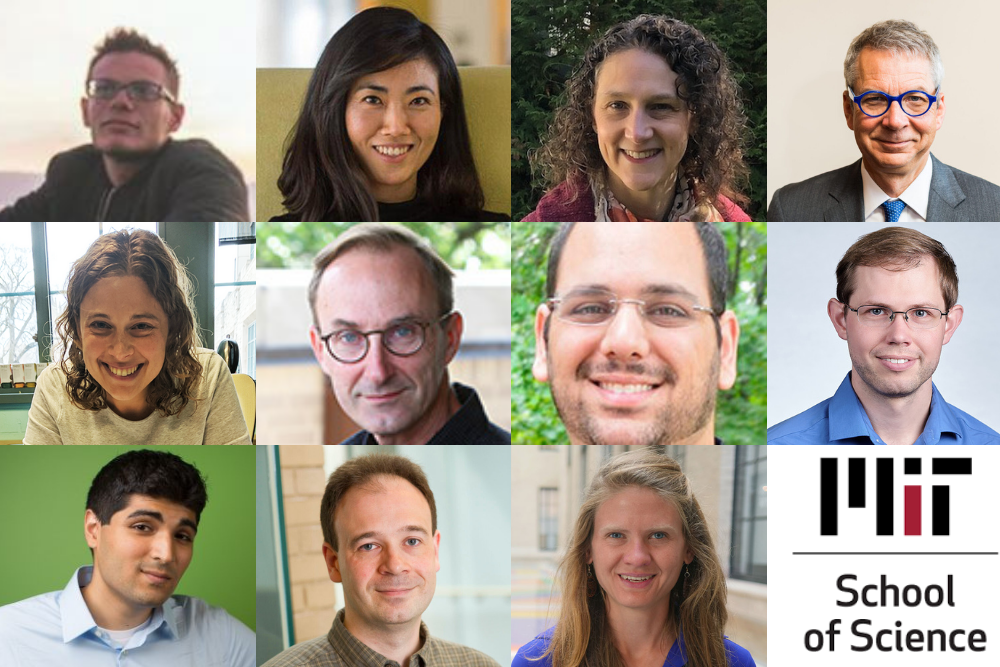
The School of Science has announced that 11 faculty members have been appointed to named professorships. These positions offer additional support to professors to advance their research and develop their careers.

Andrew Babbin was named a Cecil and Ida Green Career Development Professor. A marine biogeochemist, Babbin studies the processes that return fixed nitrogen in the ocean back to nitrogen gas, exploring marine nitrogen’s control on life in the ocean and its effects on climate. His research sheds light on the ocean’s potential to sustain life and store carbon. Babbin earned a BS degree from Columbia University in 2008 and a PhD from Princeton University in 2014. He came to MIT in 2014 as a postdoc in the Department of Civil and Environmental Engineering before joining the Department of Earth, Atmospheric and Planetary Sciences in January 2017.

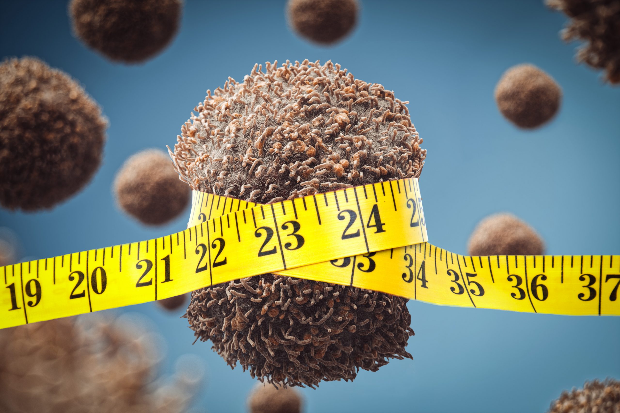
Gloria Choi was selected as the Mark Hyman Jr. Career Development Professor. Choi, an associate professor in the Department of Brain and Cognitive Sciences and an investigator with the Picower Institute, examines the interaction of the immune system with the brain and the effects of that interaction on neurodevelopment, behavior, and mood. She also studies how social behaviors are regulated according to sensory stimuli, context, internal state, and physiological status, and how these factors modulate neural circuit function via a combinatorial code of classic neuromodulators and immune-derived cytokines. She received her bachelor’s degree from the University of California at Berkeley, and her PhD from Caltech, where she studied with David Anderson. She was a postdoc in the laboratory of Richard Axel at Columbia University. Choi joined the MIT faculty as an assistant professor in 2013.

Arlene Fiore joined MIT as the inaugural Peter H. Stone and Paola Malanotte Stone Professor in Earth, Atmospheric and Planetary Sciences in July 2021. Her research encompasses air pollution, chemistry-climate connections, trends and variability in atmospheric constituents, and biosphere-atmosphere interactions. Fiore’s group investigates regional meteorology and climate feedbacks due to aerosols versus greenhouse gases, future air pollution responses to climate change, as well as the factors controlling the oxidizing capacity of the atmosphere. After earning a bachelor’s degree and PhD from Harvard University, Fiore held a research scientist position at the Geophysical Fluid Dynamics Laboratory and was appointed as an associate professor with tenure at Columbia University in 2011.

Peter H. Fisher is now the Thomas A. Frank (1977) Professor of Physics. His interests include the detection of dark matter, development of new particle detectors, compact energy supplies, and wireless energy transmission. Currently serving as the head of the Department of Physics, Fisher also holds appointments in the Institute for Soldier Nanotechnologies, the Laboratory for Nuclear Science, and the Kavli Institute. He is involved in CERN’s Alpha Magnetic Spectrometer experiment to make high-precision measurements of cosmic rays and the development of new ideas for dark matter. After receiving a BS in engineering physics from the University of California at Berkeley in 1983 and a PhD in nuclear physics from Caltech in 1988, Fisher was at the Johns Hopkins University from 1989 to 1994 and joined MIT in 1994.

Danna Freedman has been named the Frederick George Keyes Professor of Chemistry. Freedman leverages inorganic chemistry to solve problems in physics. Her research focuses on creating spin-based quantum bits and synthesizing new emergent materials. Freedman received her bachelor’s degree from Harvard University and her PhD from the University of California at Berkeley, then conducted postdoctoral research at MIT before joining the faculty at Northwestern University as an assistant professor in 2012, where she was promoted to associate professor in 2018 and full professor in 2020. Freedman returned to MIT’s Department of Chemistry in 2021.

Michel Goemans has been named the RSA Professor of Mathematics. Goemans has been head of the Department of Mathematics since July 1, 2018, following a year as interim head. He received his undergraduate degree in applied mathematics from Université Catholique de Louvain in 1987 and completed his PhD at MIT in 1990. He has been on the faculty since 1992, receiving tenure in 1999, and held the Leighton Family Professorship from 2007 to 2017. The RSA cryptosystem is the brainchild of Ron Rivest, Adi Shamir, and Len Adleman, whose fruitful collaboration spanned the Laboratory for Computer Science — today the Computer Science and Artificial Intelligence Laboratory (CSAIL) — and the Department of Mathematics. Goemans is also a member of the Theory of Computation Group of CSAIL, and recently joined the Computing Council of the MIT Schwarzman College of Computing. Goemans’ research interests include combinatorics, optimization and algorithms. In particular, his pioneering use of semidefinite optimization and other techniques for designing approximation algorithms for hard combinatorial optimization problems has been rewarded with several awards, such as the Fulkerson, Farkas and Dantzig prizes.

Or Hen was named the Class of 1956 Career Development Associate Professor of Physics. He investigates quantum chromodynamic effects in the nuclear medium and the interplay between partonic and nucleonic degrees of freedom in nuclei. Specifically, Hen utilizes high-energy scattering of electron, neutrino, photon, proton, and ion off atomic nuclei to study short-range correlations: temporal fluctuations of high-density, high-momentum, nucleon clusters in nuclei with important implications for nuclear, particle, atomic, and astrophysics. He received his undergraduate degree in physics and computer engineering from the Hebrew University and earned his PhD in experimental physics at Tel-Aviv University. Hen was an MIT Pappalardo Fellow in Physics from 2015 to 2017 before joining the physics faculty in July 2017.

Brett McGuire is now the Class of 1943 Career Development Assistant Professor of Chemistry. He uses the tools of physical chemistry, molecular spectroscopy, and observational astrophysics to understand how the chemical ingredients for life evolve with and help shape the formation of stars and planets. His group aims to detect more new molecules in space and to better understand their significance, advancing the field of astrochemistry. McGuire obtained a bachelor’s degree from the University of Illinois at Urbana-Champaign in 2009, a master’s degree from Emory University in 2011, and a PhD from Caltech in 2015. McGuire joined the Department of Chemistry in 2020.

Iain W. Stewart has been selected for the Otto (1939) and Jane Morningstar Professorship in Science. Stewart is a professor of physics and the director of the Center for Theoretical Physics. His research interests involve theoretical nuclear and particle physics. In particular, he focuses upon the development and application of effective field theories to answer fundamental questions about interactions between elementary particles. Stewart earned a bachelor’s degree in physics and mathematics and a master’s degree in physics from the University of Manitoba in Canada. He then received his PhD from Caltech in 1999. Stewart joined the physics faculty at MIT in 2003, was promoted to associate professor with tenure in 2009, and became a full professor in 2013.

Ankur Moitra, a theoretical computer scientist, is now the Norbert Wiener Professor of Mathematics. The aim of his work is to bridge the gap between theoretical computer science and machine learning by developing algorithms with provable guarantees and foundations for reasoning about their behavior. Moitra received his bachelor’s degree in electrical and computer engineering from Cornell University in 2007 and his master’s degree and PhD from MIT in computer science in 2009 and 2011, respectively, then spent two years as a fellow at the Institute for Advanced Study and Princeton University. Moitra returned to MIT in 2013 as a professor in applied mathematics and a principal investigator in CSAIL.

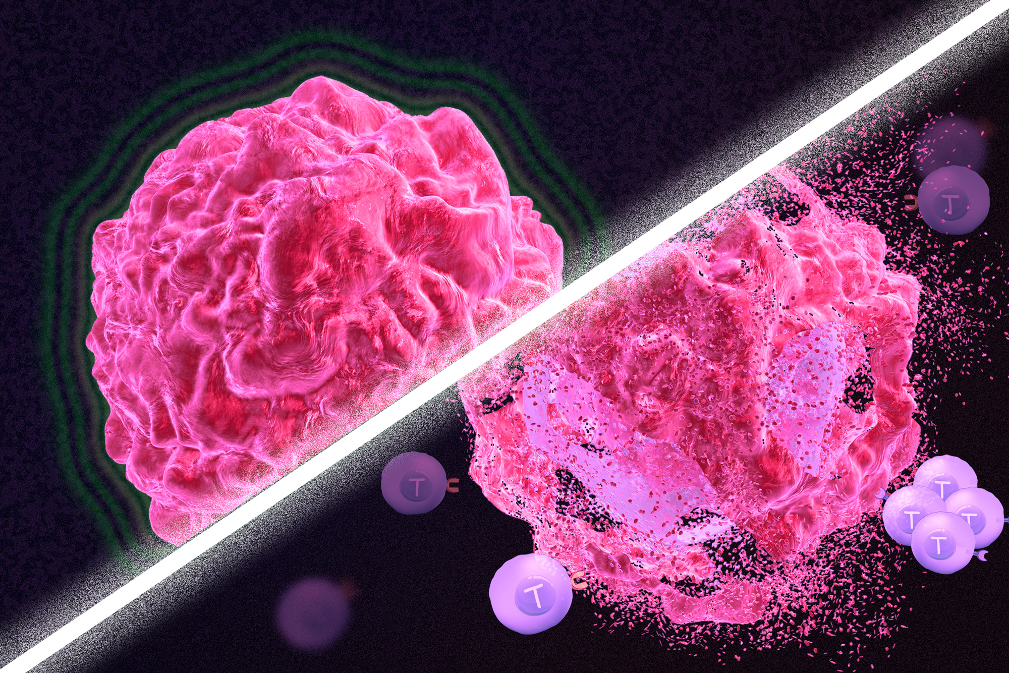
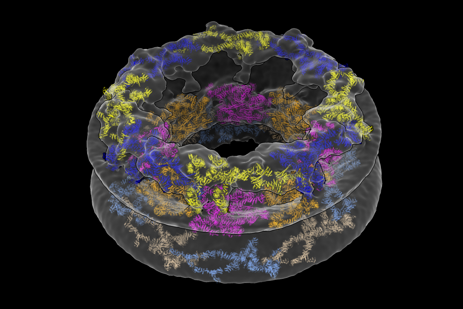
Seychelle M. Vos has been named a Robert A. Swanson (1969) Career Development Professor of Life Sciences. Vos examines the interplay of genome organization and gene expression to gain insight into how the organization of a cell affects what it becomes. Vos’ lab examines these pieces at a molecular scale using varied approaches from single-particle cryo-electron microscopy to X-ray crystallography, biochemistry to genetics. This work can help to build a biological understanding of diseases such as developmental disorders or cancers. She received her BS in genetics in 2008 from the University of Georgia and her PhD in molecular and cell biology in 2013 from the University of California at Berkeley. Vos joined the Department of Biology in 2019.

How diet affects tumors

A new study finds cutting off cells’ supplies of lipids can slow the growth of tumors in mice.

Anne Trafton | MIT News Office
October 20, 2021

In recent years, there has been some evidence that dietary interventions can help to slow the growth of tumors. A new study from MIT, which analyzed two different diets in mice, reveals how those diets affect cancer cells, and offers an explanation for why restricting calories may slow tumor growth.

The study examined the effects of a calorically restricted diet and a ketogenic diet in mice with pancreatic tumors. While both of these diets reduce the amount of sugar available to tumors, the researchers found that only the calorically restricted diet reduced the availability of fatty acids, and this was linked to a slowdown in tumor growth.

The findings do not suggest that cancer patients should try to follow either of these diets, the researchers say. Instead, they believe the findings warrant further study to determine how dietary interventions might be combined with existing or emerging drugs to help patients with cancer.

“There’s a lot of evidence that diet can affect how fast your cancer progresses, but this is not a cure,” says Matthew Vander Heiden, director of MIT’s Koch Institute for Integrative Cancer Research and the senior author of the study. “While the findings are provocative, further study is needed, and individual patients should talk to their doctor about the right dietary interventions for their cancer.”

MIT postdoc Evan Lien is the lead author of the paper, which appears today in Nature.

Metabolic mechanism

Vander Heiden, who is also a medical oncologist at Dana-Farber Cancer Institute, says his patients often ask him about the potential benefits of various diets, but there is not enough scientific evidence available to offer any definitive advice. Many of the dietary questions that patients have focus on either a calorie-restricted diet, which reduces calorie consumption by 25 to 50 percent, or a ketogenic diet, which is low in carbohydrates and high in fat and protein.

Previous studies have suggested that a calorically restricted diet might slow tumor growth in some contexts, and such a diet has been shown to extend lifespan in mice and many other animal species. A smaller number of studies exploring the effects of a ketogenic diet on cancer have produced inconclusive results.

“A lot of the advice or cultural fads that are out there aren’t necessarily always based on very good science,” Lien says. “It seemed like there was an opportunity, especially with our understanding of cancer metabolism having evolved so much over the past 10 years or so, that we could take some of the biochemical principles that we’ve learned and apply those concepts to understanding this complex question.”

Cancer cells consume a great deal of glucose, so some scientists had hypothesized that either the ketogenic diet or calorie restriction might slow tumor growth by reducing the amount of glucose available. However, the MIT team’s initial experiments in mice with pancreatic tumors showed that calorie restriction has a much greater effect on tumor growth than the ketogenic diet, so the researchers suspected that glucose levels were not playing a major role in the slowdown.

To dig deeper into the mechanism, the researchers analyzed tumor growth and nutrient concentration in mice with pancreatic tumors, which were fed either a normal, ketogenic, or calorie-restricted diet. In both the ketogenic and calorie-restricted mice, glucose levels went down. In the calorie-restricted mice, lipid levels also went down, but in mice on the ketogenic diet, they went up.

Lipid shortages impair tumor growth because cancer cells need lipids to construct their cell membranes. Normally, when lipids aren’t available in a tissue, cells can make their own. As part of this process, they need to maintain the right balance of saturated and unsaturated fatty acids, which requires an enzyme called stearoyl-CoA desaturase (SCD). This enzyme is responsible for converting saturated fatty acids into unsaturated fatty acids.

Both calorie-restricted and ketogenic diets reduce SCD activity, but mice on the ketogenic diet had lipids available to them from their diet, so they didn’t need to use SCD. Mice on the calorie-restricted diet, however, couldn’t get fatty acids from their diet or produce their own. In these mice, tumor growth slowed significantly, compared to mice on the ketogenic diet.

“Not only does caloric restriction starve tumors of lipids, it also impairs the process that allows them to adapt to it. That combination is really contributing to the inhibition of tumor growth,” Lien says.

Dietary effects

In addition to their mouse research, the researchers also looked at some human data. Working with Brian Wolpin, an oncologist at Dana-Farber Cancer Institute and an author of the paper, the team obtained data from a large cohort study that allowed them to analyze the relationship between dietary patterns and survival times in pancreatic cancer patients. From that study, the researchers found that the type of fat consumed appears to influence how patients on a low-sugar diet fare after a pancreatic cancer diagnosis, although the data are not complete enough to draw any conclusions about the effect of diet, the researchers say.

Although this study showed that calorie restriction has beneficial effects in mice, the researchers say they do not recommend that cancer patients follow a calorie-restricted diet, which is difficult to maintain and can have harmful side effects. However, they believe that cancer cells’ dependence on the availability of unsaturated fatty acids could be exploited to develop drugs that might help slow tumor growth.

One possible therapeutic strategy could be inhibition of the SCD enzyme, which would cut off tumor cells’ ability to produce unsaturated fatty acids.

“The purpose of these studies isn’t necessarily to recommend a diet, but it’s to really understand the underlying biology,” Lien says. “They provide some sense of the mechanisms of how these diets work, and that can lead to rational ideas on how we might mimic those situations for cancer therapy.”

The researchers now plan to study how diets with a variety of fat sources — including plant or animal-based fats with defined differences in saturated, monounsaturated, and polyunsaturated fatty acid content — alter tumor fatty acid metabolism and the ratio of unsaturated to saturated fatty acids.

The research was funded by the Damon Runyon Cancer Research Foundation, the National Institutes of Health, the Lustgarten Foundation, the Dana-Farber Cancer Institute Hale Family Center for Pancreatic Cancer Research, Stand Up to Cancer, the Pancreatic Cancer Action Network, the Noble Effort Fund, the Wexler Family Fund, Promises for Purple, the Bob Parsons Fund, the Emerald Foundation, the Howard Hughes Medical Institute, the MIT Center for Precision Cancer Medicine, and the Ludwig Center at MIT.

New cancer treatment may reawaken the immune system

By combining chemotherapy, tumor injury, and immunotherapy, researchers show that the immune system can be re-engaged to destroy tumors in mice.

Anne Trafton | MIT News Office
October 20, 2021

Immunotherapy is a promising strategy to treat cancer by stimulating the body’s own immune system to destroy tumor cells, but it only works for a handful of cancers. MIT researchers have now discovered a new way to jump-start the immune system to attack tumors, which they hope could allow immunotherapy to be used against more types of cancer.

Their novel approach involves removing tumor cells from the body, treating them with chemotherapy drugs, and then placing them back in the tumor. When delivered along with drugs that activate T cells, these injured cancer cells appear to act as a distress signal that spurs the T cells into action.

“When you create cells that have DNA damage but are not killed, under certain conditions those live, injured cells can send a signal that awakens the immune system,” says Michael Yaffe, who is a David H. Koch Professor of Science, the director of the MIT Center for Precision Cancer Medicine, and a member of MIT’s Koch Institute for Integrative Cancer Research.

In mouse studies, the researchers found that this treatment could completely eliminate tumors in nearly half of the mice.

Yaffe and Darrell Irvine, who is the Underwood-Prescott Professor with appointments in MIT’s departments of Biological Engineering and Materials Science and Engineering, and an associate director of the Koch Institute, are the senior authors of the study, which appears today in Science Signaling. MIT postdoc Ganapathy Sriram and Lauren Milling PhD ’21 are the lead authors of the paper.

T cell activation

One class of drugs currently used for cancer immunotherapy is checkpoint blockade inhibitors, which take the brakes off of T cells that have become “exhausted” and unable to attack tumors. These drugs have shown success in treating a few types of cancer but do not work against many others.

Yaffe and his colleagues set out to try to improve the performance of these drugs by combining them with cytotoxic chemotherapy drugs, in hopes that the chemotherapy could help stimulate the immune system to kill tumor cells. This approach is based on a phenomenon known as immunogenic cell death, in which dead or dying tumor cells send signals that attract the immune system’s attention.

Several clinical trials combining chemotherapy and immunotherapy drugs are underway, but little is known so far about the best way to combine these two types of treatment.

The MIT team began by treating cancer cells with several different chemotherapy drugs, at different doses. Twenty-four hours after the treatment, the researchers added dendritic cells to each dish, followed 24 hours later by T cells. Then, they measured how well the T cells were able to kill the cancer cells. To their surprise, they found that most of the chemotherapy drugs didn’t help very much. And those that did help appeared to work best at low doses that didn’t kill many cells.

The researchers later realized why this was so: It wasn’t dead tumor cells that were stimulating the immune system; instead, the critical factor was cells that were injured by chemotherapy but still alive.

“This describes a new concept of immunogenic cell injury rather than immunogenic cell death for cancer treatment,” Yaffe says. “We showed that if you treated tumor cells in a dish, when you injected them back directly into the tumor and gave checkpoint blockade inhibitors, the live, injured cells were the ones that reawaken the immune system.”

The drugs that appear to work best with this approach are drugs that cause DNA damage. The researchers found that when DNA damage occurs in tumor cells, it activates cellular pathways that respond to stress. These pathways send out distress signals that provoke T cells to leap into action and destroy not only those injured cells but any tumor cells nearby.

“Our findings fit perfectly with the concept that ‘danger signals’ within cells can talk to the immune system, a theory pioneered by Polly Matzinger at NIH in the 1990s, though still not universally accepted,” Yaffe says.

Tumor elimination

In studies of mice with melanoma and breast tumors, the researchers showed that this treatment eliminated tumors completely in 40 percent of the mice. Furthermore, when the researchers injected cancer cells into these same mice several months later, their T cells recognized them and destroyed them before they could form new tumors.

The researchers also tried injecting DNA-damaging drugs directly into the tumors, instead of treating cells outside the body, but they found this was not effective because the chemotherapy drugs also harmed T cells and other immune cells near the tumor. Also, injecting the injured cells without checkpoint blockade inhibitors had little effect.

“You have to present something that can act as an immunostimulant, but then you also have to release the preexisting block on the immune cells,” Yaffe says.

Yaffe hopes to test this approach in patients whose tumors have not responded to immunotherapy, but more study is needed first to determine which drugs, and at which doses, would be most beneficial for different types of tumors. The researchers are also further investigating the details of exactly how the injured tumor cells stimulate such a strong T cell response.

The research was funded, in part, by the National Institutes of Health, the Mazumdar-Shaw International Oncology Fellowship, the MIT Center for Precision Cancer Medicine, and the Charles and Marjorie Holloway Foundation.

Cellular environments shape molecular architecture

Researchers glean a more complete picture of a complex structure called the nuclear pore complex by studying it directly inside cells.

Raleigh McElvery | Department of Biology
October 13, 2021

Context matters. It’s true for many facets of life, including the tiny molecular machines that perform vital functions inside our cells.

Scientists often purify cellular components, such as proteins or organelles, in order to examine them individually. However, a new study published today in the journal Nature suggests that this practice can drastically alter the components in question.

The researchers devised a method to study a large, donut-shaped structure called the nuclear pore complex (NPC) directly inside cells. Their results revealed that the pore had larger dimensions than previously thought, emphasizing the importance of analyzing complex molecules in their native environments.

“We’ve shown that the cellular environment has a significant impact on large structures like the NPC, which was something we weren’t expecting when we started,” says Thomas Schwartz, the Boris Magasanik Professor of Biology at MIT and the study’s co-senior author. “Scientists have generally thought that large molecules are stable enough to maintain their fundamental properties both inside and outside a cell, but our findings turn that assumption on its head.”

In eukaryotes like humans and animals, most of a cell’s DNA is stored in a rounded structure called the nucleus. This organelle is shielded by the nuclear envelope, a protective barrier that separates the genetic material in the nucleus from the thick fluid filling the rest of the cell. But molecules still need a way to come in and out of the nucleus in order to facilitate important processes, including gene expression. That’s where the NPC comes in. Hundreds — sometimes thousands — of these pores are embedded in the nuclear envelope, creating gateways that allow certain molecules to pass.

The study’s first author, former MIT postdoc Anthony Schuller, compares NPCs to gates at a sports stadium. “If you want to access the game inside, you have to show your ticket and go through one of these gates,” he explains.

The NPC may be tiny by human standards, but it’s one of the largest structures in the cell. It’s comprised of roughly 500 proteins, which has made its structure challenging to parse. Traditionally, scientists have broken it up into individual components to study it piecemeal using a method called X-ray crystallography. According to Schwartz, the technology required to analyze the NPC in a more natural environment has only recently become available.

Together with researchers from the University of Zurich, Schuller and Schwartz employed two cutting-edge approaches to solve the pore’s structure: cryo-focused ion beam (cryo-FIB) milling and cryo-electron tomography (cryo-ET).

An entire cell is too thick to look at under an electron microscope. But the researchers sliced frozen colon cells into thin layers using the cryo-FIB equipment housed at MIT.nano’s Center for Automated Cryogenic Electron Microscopy and the Koch Institute for Integrative Cancer Research’s Peterson (1957) Nanotechnology Materials Core Facility. In doing so, the team captured cross-sections of the cells that included NPCs, rather than simply looking at the NPCs in isolation.

“The amazing thing about this approach is that we’ve barely manipulated the cell at all,” Schwartz says. “We haven’t perturbed the cell’s internal structure. That’s the revolution.”

What the researchers saw when they looked at their microscopy images was quite different from existing descriptions of the NPC. They were surprised to find that the innermost ring structure, which forms the pore’s central channel, is much wider than previously thought. When it’s left in its natural environment, the pore opens up to 57 nanometers — resulting in a 75 percent increase in volume compared to previous estimates. The team was also able to take a closer look at how the NPC’s various components work together to define the pore’s dimensions and overall architecture.

“We’ve shown that the cellular environment impacts NPC structure, but now we have to figure out how and why,” Schuller says. Not all proteins can be purified, he adds, so the combination of cryo-ET and cryo-FIB will also be useful for examining a variety of other cellular components. “This dual approach unlocks everything.”

“The paper nicely illustrates how technical advances, in this case cryo-electron tomography on cryo-focused ion beam milled human cells, provide a fresh picture of cellular structures,” says Wolfram Antonin, a professor of biochemistry at RWTH Aachen University in Germany who was not involved in the study. The fact that the diameter of the NPC’s central transport channel is larger than previously thought hints that the pore could have impressive structural flexibility. “That may be important for the cell to adapt to increased transport demands,” Antonin explains.

Next, Schuller and Schwartz hope to understand how the size of the pore affects which molecules can pass through. For instance, scientists only recently determined that the pore was big enough to allow intact viruses like HIV into the nucleus. The same principle applies to medical treatments: only appropriately-sized drugs with specific properties will be able to access the cell’s DNA.

Schwartz is especially curious to know whether all NPCs are created equal, or if their structure differs between species or cell types.

“We’ve always manipulated cells and taken the individual components out of their native context,” he says. “Now we know this method may have much bigger consequences than we thought.”

Seven from MIT receive National Institutes of Health awards

Awards support high-risk, high-reward biomedical and behavioral research.

School of Science
October 8, 2021

On Oct. 5, the National Institutes of Health announced the names of 106 scientists who have been awarded grants through the High-Risk, High-Reward Research program to advance highly innovative biomedical and behavioral research. Seven of the recipients are MIT faculty members.

The High-Risk, High-Reward Research program catalyzes scientific discovery by supporting research proposals that, due to their inherent risk, may struggle in the traditional peer-review process despite their transformative potential. Program applicants are encouraged to pursue trailblazing ideas in any area of research relevant to the NIH’s mission to advance knowledge and enhance health.

“The science put forward by this cohort is exceptionally novel and creative and is sure to push at the boundaries of what is known,” says NIH Director Francis S. Collins. “These visionary investigators come from a wide breadth of career stages and show that groundbreaking science can happen at any career level given the right opportunity.”

New innovators

Four MIT researchers received New Innovator Awards, which recognize “unusually innovative research from early career investigators.” They are:

  • Pulin Li is a member at the Whitehead Institute for Biomedical Research and an assistant professor in the Department of Biology. Li combines approaches from synthetic biology, developmental biology, biophysics and systems biology to quantitatively understand the genetic circuits underlying cell-cell communication that creates multicellular behaviors.
  • Seychelle Vos, the Robert A. Swanson (1969) Career Development Professor of Life Sciences in the Department of Biology, studies the interplay of gene expression and genome organization. Her work focuses on understanding how large molecular machineries involved in genome organization and gene transcription regulate each others’ function to ultimately determine cell fate and identity.
  • Xiao Wang, the Thomas D. and Virginia Cabot Assistant Professor of Chemistry and a member of the Broad Institute of MIT and Harvard, aims to develop high-resolution and highly-multiplexed molecular imaging methods across multiple scales toward understanding the physical and chemical basis of brain wiring and function.
  • Alison Wendlandt is a Cecil and Ida Green Career Development Assistant Professor of Chemistry. Wendlandt focuses on the development of selective, catalytic reactions using the tools of organic and organometallic synthesis and physical organic chemistry. Mechanistic study plays a central role in the development of these new transformations.

Transformative researchers

Two MIT researchers have received Transformative Research Awards, which “promote cross-cutting, interdisciplinary approaches that could potentially create or challenge existing paradigms.” The recipients are:

  • Manolis Kellis is a professor of computer science at MIT in the area of computational biology, an associate member of the Broad Institute, and a principal investigator with MIT’s Computer Science and Artificial Intelligence Laboratory. He aims to further our understanding of the human genome by computational integration of large-scale functional and comparative genomics datasets.
  • Myriam Heiman is the Latham Family Career Development Associate Professor of Neuroscience in the Department of Brain and Cognitive Sciences and an investigator in the Picower Institute for Learning and Memory. Heiman studies the selective vulnerability and pathophysiology seen in two neurodegenerative diseases of the basal ganglia, Huntington’s disease, and Parkinson’s disease.

Together, Heiman, Kellis and colleagues will launch a five-year investigation to pinpoint what may be going wrong in specific brain cells and to help identify new treatment approaches for amyotrophic lateral sclerosis (ALS) and frontotemporal lobar degeneration with motor neuron disease (FTLD/MND). The project will bring together four labs, including Heiman and Kellis’ labs at MIT, to apply innovative techniques ranging from computational, genomic, and epigenomic analyses of cells from a rich sample of central nervous system tissue, to precision genetic engineering of stem cells and animal models.

Pioneering researchers

  • Polina Anikeeva received a Pioneer Award, which “challenges investigators at all career levels to pursue new research directions and develop groundbreaking, high-impact approaches to a broad area of biomedical, behavioral, or social science.” Anikeeva is an MIT professor of materials science and engineering, a professor of brain and cognitive sciences, and a McGovern Institute for Brain Research associate investigator. She has established a research program that uniquely combines materials synthesis, device fabrication, neurophysiology, and animal models of behavior. Her group carries out projects that understand, invent, and design materials from the level of atoms to functional devices with applications in fundamental neuroscience.

The program is supported by the NIH Common Fund, which oversees programs that pursue major opportunities and gaps throughout the research enterprise that are of great importance to NIH and require collaboration across the agency to succeed. It issues four awards each year: the Pioneer Award, the New Innovator Award, the Transformative Research Award, and the Early Independence Award.

This year, NIH issued 10 Pioneer awards, 64 New Innovator awards, 19 Transformative Research awards (10 general, four ALS-related, and five Covid-19-related), and 13 Early Independence awards for 2021. Funding for the awards comes from the NIH Common Fund, the National Institute of General Medical Sciences, the National Institute of Mental Health, and the National Institute of Neurological Disorders and Stroke.

3 Questions: Sheena Vasquez and Christian Loyo on communicating science through poetry

PhD students discuss their participation in The Poetry of Science project and the importance of bringing the arts into science communication.

Grace van Deelen | Department of Biology
October 5, 2021

Christian Loyo of the Grossman lab and Sheena Vasquez of the Drennan lab, both graduate students in the Department of Biology, were recently selected to participate in The Poetry of Science. The project, founded by Joshua Sariñana PhD ’11, aims to advance racial justice at the intersection of science and art by bringing together Cambridge, Massachusetts-affiliated poets and scientists of color to create poems about scientific research. These poems will be on public display, along with the scientists’ portraits, at the Massachusetts General Hospital main lobby from Nov. 13 through Nov. 30 and the Rotch Library at MIT during Independent Activities Period (IAP) in January 2022.

For Loyo and Vasquez, The Poetry of Science was the ideal opportunity to create something of impact by combining their personal passions for poetry, science communication, and racial justice. They worked with two poets (Danielle Legros Georges and Luisa Fernanda Apolaya Torres, respectively) to create poems about their research. Loyo and Vasquez sat down to discuss the project.

Q: Both of you are accomplished scientists, but you also spend time working within your communities on various outreach programs. How have these experiences influenced the way you approach your scientific research or your roles as scientists?

Vasquez: I’ve conducted outreach from K-12 to the undergraduate level during my time here at MIT. My most recent outreach is targeted to local community colleges around MIT, including Bunker Hill Community College and Roxbury Community College. Outreach experiences really make me take a step back and think about how to make my science more accessible to the general public. Overall, experiences like these allow me to enhance my mentoring skills. Working with students from different backgrounds shows me how fortunate I am to conduct research at one of the top institutions in the world. If I can make it at an institute like MIT, I feel like anyone else can.

Loyo: For me, it was not easy to get into science. It took a lot of people who became my mentors to teach me what I now know about being a scientist and navigating academia. As an undergrad, I was looking for a research lab and I emailed probably 50 professors; none of them had room. I was about to give up when I finally found one professor who wanted to meet and take a chance on me. This meant a lot to me and is actually in Luisa’s poem. I ended up having a great experience and exploring research questions at a pretty high level for an undergraduate. This opportunity made me realize it’s really important to pay it forward. There are a lot of people who are having a tougher time than I ever did getting into science. Helping them ensures that the future of science is more inclusive.

Q: Science and art are often seen as two distinct disciplines, but The Poetry of Science project is all about bridging that gap. How do you think combining science and the arts can further the goal of advancing racial justice?

Loyo: I think science is about understanding the universe that we live in, and art is about understanding what it means to be human. Because we are human, we all have biases. One of those biases can be racial bias. If you look at who has historically been doing science, it’s mostly white men. That’s not because those people were the best at science; that’s because everyone else was not traditionally allowed to do science. Art gives us an opportunity to share our experiences as people from these historically excluded groups, and to highlight how we became scientists — even if, growing up, we didn’t often see scientists who looked like us.

Vasquez: The Poetry of Science exhibition also offers a chance to create new and positive representations of people of color. More examples of people of color in science helps us break down stereotypes and learn more about the individuals themselves. It allows for more stories to be told in different ways, which creates room for different perspectives. For example — and Danielle included this in her poem — something I really wish people would know is that I’m human, and, just like all scientists, I make mistakes. I am still learning and growing.

Q: What was it like to communicate your research through poetry, and how do you think the arts contribute to scientific literacy?

Vasquez: It was interesting to see what Danielle latched onto when I was explaining my research to her. For example, there’s part of the poem where she writes about how the proteins spiral, and she compares them to a girl’s curly hair. That was the alpha helix I was showing her, and it does spiral like a girl’s hair — like both of our hair. It was neat to see how she made connections between my science and general life.

The arts bring science to life, which helps improve scientific literacy. That’s important because it puts us all on the same page about what’s true and what’s not true. If we didn’t have certain scientific understandings about viruses, for example, we would not have made it this far in combating the pandemic.

Loyo: When you write a poem about science, it becomes much less about the nitty-gritty details, and instead captures the love behind the research. There’s this wonder and awe that we have for the natural world, and when we can discover something about the natural world that we didn’t know before, that feels so good. People really connect to your work when they can feel that same sort of excitement and emotion.

People also take pride in art. For example, I’m of Mexican descent, and I am a big fan of Frida Kahlo and Diego Rivera, who are Mexican painters. Using art can connect people to science even if they don’t really know what the science is about. If they can see that the person doing the experiments, for example, also grew up where they grew up, that can really be beneficial.

David Julius ’77 shares the Nobel Prize in physiology or medicine

MIT alumnus and one other honored for their discoveries in how the nervous system senses temperature and touch.

Anne Trafton | MIT News Office
October 4, 2021

David Julius, a 1977 graduate of MIT, will share the 2021 Nobel Prize in physiology or medicine, the Royal Swedish Academy of Sciences announced this morning in Stockholm.

Julius, a professor at the University of California at San Francisco, shares the prize with Ardem Patapoutian, a professor at the Scripps Research Institute, for their discoveries in how the body senses touch and temperature.

Both scientists helped to answer a fundamental question regarding how the nervous system interprets our environment: How are temperature and mechanical stimuli converted into electrical impulses in the nervous system?

Using capsaicin, a compound that gives chili peppers their distinctive burning sensation, Julius was able to identify a receptor in the nerve endings of skin that responds to heat. His experiments revealed that this receptor, which he called TRPV1, is an ion channel that is activated by painful heat.

“David Julius’ discovery of TRPV1 was the breakthrough that allowed us to understand how differences in temperature can induce electrical signals in the nervous system,” according to today’s announcement by the Nobel committee.

Later, Julius and Patapoutian independently discovered a receptor called TRPM8, which responds to cold. Patapoutian was also honored for his discovery of receptors that respond to mechanical force in the skin and other organs. Their work on how the body senses temperature and mechanical stimuli is now being harnessed to develop treatments for a variety of diseases, including chronic pain.

Julius, who was born in New York, earned his bachelor’s degree in biology from MIT in 1977. He received a PhD in 1984 from University of California at Berkeley and was a postdoc at Columbia University before joining the faculty of the University of California at San Francisco in 1989.

He is the 39th MIT graduate to win a Nobel Prize.

Lindsey Backman: Biochemist, mentor, and advocate

The PhD candidate studies the human microbiome and its proteins, while also championing the Latinx community on MIT’s campus.

Hannah Meiseles | MIT News Office
September 28, 2021

Raised in Tampa, Florida, Lindsey Backman takes pride in her family’s history and its role in the vibrant Cuban American community there. She remembers the weekends she would spend as a kid, getting café con leche with her grandparents and dancing in the studio with her friends. The cultural experiences she shared with friends, family, and  neighbors growing up helped her feel comfortable being herself while growing up, and showed her from an early age how valuable a welcoming community could be to a person’s success.

Backman went on to pursue her BS in chemistry at the University of Florida. Surrounded by a diverse community, she felt supported as she leaned deeper into her interest in science. She was soon nominated to a program that matched students from underrepresented backgrounds in STEM with a university professor to pursue a summer research project. Although Backman was still uncertain about going to another university to do lab research, with encouragement from her department she gave the program a chance.

Backman matched with Professor Catherine Drennan at MIT to work on visualizing structural biology and took part in the MIT Summer Research Program in Biology (MSRP-Bio). The research clicked with her immediately and became a turning point; Backman returned to participate in the lab the following summer and then applied to graduate school.

“Getting nominated to the program changed my life. I certainly wouldn’t have applied to MIT otherwise,” says Backman. “At first, I was convinced I wouldn’t fit in, but soon found myself surrounded by people as passionate about science as I was. I knew I was in the right place.”

Uncovering secrets about the human microbiome

Today, Backman is a graduate student in the Drennan lab and researches the chemistry of the human microbiome, a collection of gut microbes essential to sustaining the body. Backman is interested in how certain bacteria can outcompete other strains by producing unique proteins that process abundant nutrients or repair broken enzymes. Her use of X-ray crystallography has helped her produce atomic models that shed light on the structure of these proteins.

One type of protein Backman and her team have characterized is called a spare part protein. When produced, this protein can help restore a broken enzyme’s ability to catalyze essential reactions. “When fixing a car with a flat tire, you would replace the tire and not the whole car. A similar strategy is being used here. These spare-part proteins act to bind and restore the activity of the enzyme completely,” she says.

Over the years, Backman has seen the depth of questions surrounding the microbiome grow. Scientists have begun to recognize how important the microbiome is to human health. “Ever since my first summer research experience at MIT, I’ve been dedicated to studying this one unique repair mechanism,” says Backman. “We’ve gone from solving the structure of the proteins to now understanding how the mechanism works. But there’s still so much more to learn — we have started to suspect these repair mechanisms speak to a broader motif in other enzymes as well.”

Backman and her team have also been leaders in characterizing how an important enzyme, called hydroxy-L-proline dehydratase (HypD), performs its unusual chemistry. This abundant enzyme takes hydroxyproline, a common nutrient in the gut, and can obtain a competitive advantage by using it as a nutrient and source of energy.

“Only a unique subset of bacteria can process hydroxyproline. On the clinical side, we have seen during infection that virulent bacteria with this ability, such as C. difficile, will start rapidly consuming hydroxyproline to proliferate,” says Backman. “Conversely, we could one day create antibiotics that specifically inhibit HypD without killing our beneficial bacteria.”

Encouraging the future of science

Outside of her research, Backman cares deeply about serving and being a part of the Latinx community on campus. She helped co-found the MIT Latinx Graduate Student Association and has served for four years as a graduate resident assistant for La Casa, the Latinx undergraduate living community at New House. “La Casa is a really tight-knit and familial community,” says Backman. “Some of our original freshmen are now seniors, so it’s been really rewarding to see their whole transition throughout college. I love getting to watch students explore and come to realize what they’re passionate about.”

Backman has also been instrumental in spurring equity initiatives on campus. She is currently a student representative for the MIT Department of Chemistry Diversity, Equity, and Inclusion Committee and has worked to implement programs that support the success of underrepresented groups on campus. Her five years of service as an MIT Chemistry Access Program mentor have encouraged many underrepresented undergraduate students to pursue chemistry graduate programs. For all her hard work at improving MIT’s campus, Backman recently received the Hugh Hampton Young Fellowship.

In the future, Backman aspires to continue researching the microbiome and mentoring students by becoming a professor. She hopes to continue the cycle and inspire more young scientists to recognize their inner potential. “I was never one of those kids that knew I wanted to be a scientist someday. My PI completely changed my life, and I would not be at MIT today without her,” she says. “Having mentors that believe in you at critical points in your life can make all the difference.”

“I think there’s this wrong assumption that diversity initiative work takes away from time that could be spent doing science. In my mind, we need to recognize how these things go hand in hand,” says Backman.

“The only way we’re going to get the best scientists is by creating a healthier, more diverse environment where people of all backgrounds feel welcomed. It’s only when people feel comfortable that they can make their greatest contributions to the field.”

Biologists identify new targets for cancer vaccines

Vaccinating against certain proteins found on cancer cells could help to enhance the T cell response to tumors.

Anne Trafton | MIT News Office
September 16, 2021

Over the past decade, scientists have been exploring vaccination as a way to help fight cancer. These experimental cancer vaccines are designed to stimulate the body’s own immune system to destroy a tumor, by injecting fragments of cancer proteins found on the tumor.

So far, none of these vaccines have been approved by the FDA, but some have shown promise in clinical trials to treat melanoma and some types of lung cancer. In a new finding that may help researchers decide what proteins to include in cancer vaccines, MIT researchers have found that vaccinating against certain cancer proteins can boost the overall T cell response and help to shrink tumors in mice.

The research team found that vaccinating against the types of proteins they identified can help to reawaken dormant T cell populations that target those proteins, strengthening the overall immune response.

“This study highlights the importance of exploring the details of immune responses against cancer deeply. We can now see that not all anticancer immune responses are created equal, and that vaccination can unleash a potent response against a target that was otherwise effectively ignored,” says Tyler Jacks, the David H. Koch Professor of Biology, a member of the Koch Institute for Integrative Cancer Research, and the senior author of the study.

MIT postdoc Megan Burger is the lead author of the new study, which appears today in Cell.

T cell competition

When cells begin to turn cancerous, they start producing mutated proteins not seen in healthy cells. These cancerous proteins, also called neoantigens, can alert the body’s immune system that something has gone wrong, and T cells that recognize those neoantigens start destroying the cancerous cells.

Eventually, these T cells experience a phenomenon known as “T cell exhaustion,” which occurs when the tumor creates an immunosuppressive environment that disables the T cells, allowing the tumor to grow unchecked.

Scientists hope that cancer vaccines could help to rejuvenate those T cells and help them to attack tumors. In recent years, they have worked to develop methods for identifying neoantigens in patient tumors to incorporate into personalized cancer vaccines. Some of these vaccines have shown promise in clinical trials to treat melanoma and non-small cell lung cancer.

“These therapies work amazingly in a subset of patients, but the vast majority still don’t respond very well,” Burger says. “A lot of the research in our lab is aimed at trying to understand why that is and what we can do therapeutically to get more of those patients responding.”

Previous studies have shown that of the hundreds of neoantigens found in most tumors, only a small number generate a T cell response.

The new MIT study helps to shed light on why that is. In studies of mice with lung tumors, the researchers found that as tumor-targeting T cells arise, subsets of T cells that target different cancerous proteins compete with each other, eventually leading to the emergence of one dominant population of T cells. After these T cells become exhausted, they still remain in the environment and suppress any competing T cell populations that target different proteins found on the tumor.

However, Burger found that if she vaccinated these mice with one of the neoantigens targeted by the suppressed T cells, she could rejuvenate those T cell populations.

“If you vaccinate against antigens that have suppressed responses, you can unleash those T cell responses,” she says. “Trying to identify these suppressed responses and specifically targeting them might improve patient responses to vaccine therapies.”

Shrinking tumors

In this study, the researchers found that they had the most success when vaccinating with neoantigens that bind weakly to immune cells that are responsible for presenting the antigen to T cells. When they used one of those neoantigens to vaccinate mice with lung tumors, they found the tumors shrank by an average of 27 percent.

“The T cells proliferate more, they target the tumors better, and we see an overall decrease in lung tumor burden in our mouse model as a result of the therapy,” Burger says.

After vaccination, the T cell population included a type of cells that have the potential to continuously refuel the response, which could allow for long-term control of a tumor.

In future work, the researchers hope to test therapeutic approaches that would combine this vaccination strategy with cancer drugs called checkpoint inhibitors, which can take the brakes off exhausted T cells, stimulating them to attack tumors. Supporting that approach, the results published today also indicate that vaccination boosts the number of a specific type of T cells that have been shown to respond well to checkpoint therapies.

The research was funded by the Howard Hughes Medical Institute, the Ludwig Center at Harvard University, the National Institutes of Health, the Koch Institute Support (core) Grant from the National Cancer Institute, the Bridge Project of the Koch Institute and Dana-Farber/Harvard Cancer Center, and fellowship awards from the Jane Coffin Childs Memorial Fund for Medical Research and the Ludwig Center for Molecular Oncology at MIT.

Professor Emeritus Paul Schimmel donates $50 million to support MIT life sciences enterprise

Schimmel Family Program for Life Sciences will benefit graduate students and research.

School of Science
August 30, 2021

Professor Emeritus Paul Schimmel PhD ’66 and his family recently committed $50 million to support the life sciences at MIT. They provided an initial gift of $25 million to establish the Schimmel Family Program for Life Sciences. This gift matches $25 million secured from other sources in support of the Department of Biology. The remaining $25 million from the Schimmel family will go to support the Schimmel Family Program in the form of matching funds as other gifts are secured over the next five years. Schimmel, who is the John D. and Catherine T. MacArthur Professor of Biochemistry and Biophysics Emeritus, is a lifelong supporter of the Institute in teaching, research, and philanthropy.

“I am tremendously grateful to Paul and his family for their generosity and support, and for their advocacy for our department and the life sciences,” says department head Alan D. Grossman, the Praecis Professor of Biology.

This most recent gift is one among many that Schimmel and his family have provided to MIT during their more than 50-year affiliation with the Institute, which includes Paul’s doctorate and his 30 years of teaching and research in the department. While at MIT, Paul and Cleo, Paul’s wife and philanthropic partner, provided an anonymous donation for the construction of Building 68, the most recent home for the Department of Biology.

“We cannot overstate our gratitude for our MIT experience. It was MIT that provided a ‘frontier of knowledge, which has no bounds’ and introduced us to some of the finest minds and people in the world,” Schimmel says.  

“They educated and uplifted us, and convinced us of MIT’s singular role in making this a better world for all peoples,” says Cleo Schimmel, who was a past chair of the MIT Women’s League and, in her own right, contributed to the endowment of the league and other efforts to support women at MIT.

Currently, Paul Schimmel is the Ernst and Jean Hahn Professor at the Skaggs Institute for Chemical Biology at the Scripps Research Institute. Schimmel formally left MIT in 1997 to join Scripps Research, but he has remained actively involved in supporting the Institute’s research enterprise, specifically MIT graduate students.

Graduate funding for the future

Shortly after Paul left MIT, the Schimmels endowed four graduate fellowships for outstanding women in life sciences. “Since 2000, the Cleo and Paul Schimmel Scholars fellowships have helped the biology department recruit and retain the best talent,” says Grossman. Kristin Knouse PhD ’17 is a former Schimmel Scholar who rejoined the department this past July as an assistant professor.

“The MIT Department of Biology encompasses a remarkable breath of biology within a very close-knit community that places a strong emphasis on graduate training,” says Knouse. “Once in the lab, the resources and collaborations available through MIT provide unparalleled opportunities to accelerate and advance your research.”

Schimmel, who sits on the department’s Visiting Committee, continued to champion graduate student support by helping to endow the Teresa Keng Graduate Teaching Prize to support excellence in graduate student teaching in the department. In 2013, the Schimmel family donated the proceeds from the sale of their La Jolla, California, home for the purpose of training the next generation of MIT graduates in the life sciences. What formally became the department’s Graduate Training Initiative (GTI) was supported by others, including biology alumni Eric Schmidt PhD ’96 and Tracy Smith PhD ’96.

The GTI supports departmental efforts to enhance the graduate student experience in the form of both direct student support, including tuition and stipend, and indirect support, including programmatic activities such as seed funds for student-directed projects, shared computing facilities, and forums related to post-graduation employment.

This new gift to establish the Schimmel Family Program for Life Sciences will support not only the GTI in the Department of Biology, but also graduate students across MIT.

“The life sciences educational enterprise spreads across a dozen departments at MIT,” says Schimmel. “What makes the biology department and the life sciences at MIT so extraordinary is the singular ability to transfer knowledge and inventions to society for its benefit. That is much of why Kendall Square and Boston are what they are.”

To that end, Schimmel has also been an active player in shaping the MIT-Kendall Square innovation ecosystem, including the founding of companies such as Alnylam Pharmaceuticals in 2002. Alnylam — founded by Schimmel along with Institute Professor Phillip Sharp, MIT Professor David Bartel, MIT postdocs Thomas Tuschl and Phillip Zamore, and investors — has been a major player in the biopharma scene. Most recently, Alnylam partnered with Vir Biotechnology to develop therapeutics for coronavirus infections, including Covid-19.

Having a longstanding interest in the applications of basic biomedical research to human health, Schimmel holds numerous patents and is a co-founder or founding director of several biotechnology companies in addition to Alnylam, including aTyr Pharma, Alkermes, Cubist Pharmaceuticals, Metabolon, Repligen, and Sirtris Pharmaceuticals.

“I’ve been talking to the people that I’ve started companies with, reminding them that none of the extensive commercial and residential real estate development, restaurants, hotels, and the founding and locating of major biopharmaceutical enterprises would have happened without the MIT life sciences enterprise,” says Schimmel. “MIT’s Kendall Square is to biopharma what Silicon Valley is to technology. None of the robust economic impact would have occurred if it hadn’t been for MIT’s life sciences.”

The $50 million commitment was a capstone gift to MIT’s Campaign for a Better World, supporting important campaign priorities of human health and discovery science. In addition, Schimmel has future plans to continue supporting the life sciences at MIT through his estate plan with the Institute.

“We are extraordinarily grateful to Paul, Cleo, and the entire family,” says Nergis Mavalvala PhD ’97, the Curtis and Kathleen Marble Professor of Astrophysics and the dean of the MIT School of Science. “Not only do the Schimmels understand, from a firsthand perspective, the need to support graduate students, but they also understand that these young researchers are the future of our life sciences endeavors outside of MIT, in fundamental research, biopharma industries, and beyond.”

Schimmel graduated from Ohio Wesleyan University, earned a doctorate from MIT, and completed postdoc research at Stanford University. His many accomplishments include the publication of more than 500 scientific papers, numerous awards and honorary degrees, and elected membership to the American Academy of Arts and Sciences, the National Academy of Sciences, the American Philosophical Society, the Institute of Medicine (National Academy of Medicine), and National Academy of Inventors.