

To create proteins, DNA is transcribed into RNA, and that RNA is then “translated” into protein. Between the creation of the RNA and the translation to protein is often a step called splicing. During splicing, segments called introns are removed, and the remaining pieces, called exons, are joined together to form the blueprint for translation. By splicing together different exons, the cell can create different proteins from the same section of genetic code. When splicing goes awry, it can lead to diseases and cancers.
New research recently published in Disease Models & Mechanisms from the Calo Lab in the Department of Biology at MIT has identified the mechanism for how cells respond to disruptions in splicing, which involves activating a cellular stress response. The stress response, once activated, causes widespread effects, including changes to cell metabolism.
Researchers have discovered cellular stress responses for other core cellular processes, such as ribosome biogenesis. However, this is the first time researchers have identified how cells respond to perturbing the splicing process.
A particular protein acts as a kind of canary in a coal mine: Mdm2, which responds to a broad range of splicing disruptions. Mdm2 does not cause a stress response by itself. Rather, Mdm2 is itself spliced differently in response to splicing disruptions. Downstream, the alternative splicing of Mdm2 leads to the activation of a protein called p53, which is known to orchestrate a cascade of responses to stress.
Researchers have long wondered why some cell types seem more sensitive to splicing disruptions than others. For example, some disorders caused by mutations in proteins that perform RNA splicing, despite affecting the whole organism, induce more noticeable changes in tissues derived from the neural crest—a collection of stem cells that contributes to the formation of the face, jaw, retinas, limbs, and heart during development. Certain splicing inhibitors have also increased the effectiveness of some cancer treatments, but the mechanism is unknown.
One of the p53-induced stress responses includes changing the metabolism of cells and how they use sugars, which may explain why some cells are more sensitive to splicing disruptions than others. Inhibiting glycolysis, the reactions that extract energy from glucose, can affect how cells divide and migrate.
The way cells divide and migrate is critical during development; in experiments, zebrafish treated with glycolysis inhibitors exhibited similar changes to craniofacial features as those where splicing was disrupted. Cancerous cells, too, are known to require high levels of sugar metabolism and, therefore, may be especially sensitive to treatments that induce changes in the splicing pathway.
The researchers knocked down genes to mimic milder splicing disruptions instead of knocking them out entirely. Splicing is so essential that knocking out the splicing machinery can lead to extreme responses like cell death. In organismal models like zebrafish, those severe phenotypes don’t accurately reflect how splicing disruptions present in human diseases.
First author Jade Varineau, a graduate student in the Calo lab, was drawn to the project because it allowed her to explore what was happening at the RNA and cellular level while also observing how splicing perturbations were affecting the whole organism.
“I think this data can help us reframe the way we think about diseases and cancers that are impacted by splicing—that a treatment that works for one may work for another because all the symptoms may stem from the same cellular response,” Varineau says.
Although the results indicate how cells broadly respond to splicing perturbations, the mechanism for how disruptions in splicing induce the alternate splicing of Mdm2 remains unclear. Senior author Eliezer Calo says the lab is also exploring how splicing mechanisms may be altered for things like cancer. Their work, he says, opens the door for further exploration of cell-type specificity of genetic disorders and improvements in cancer treatments using splicing inhibitors.
“We know that the sensor is encoded in the gene Mdm2—what are the molecules that allow Mdm2 to act as a sensor, and how does the sensor malfunction for things like cancer?” Calo says. “The next step is to find out how the sensor works.”
One of the immune system’s primary roles is to detect and kill cells that have acquired cancerous mutations. However, some early-stage cancer cells manage to evade this surveillance and develop into more advanced tumors.
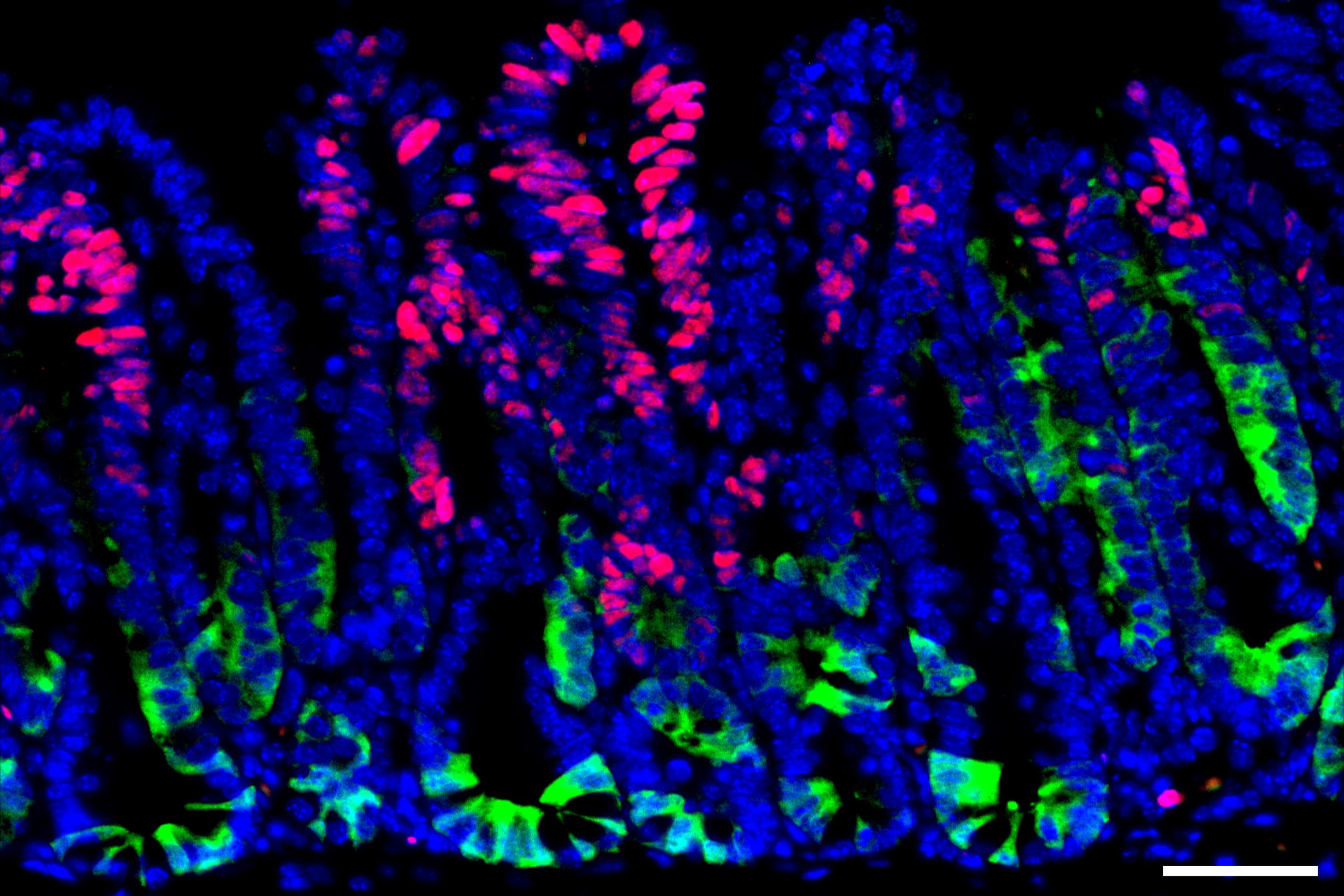
A new study from MIT and Dana-Farber Cancer Institute has identified one strategy that helps these precancerous cells avoid immune detection. The researchers found that early in colon cancer development, cells that turn on a gene called SOX17 can become essentially invisible to the immune system.
If scientists could find a way to block SOX17 function or the pathway that it activates, this may offer a new way to treat early-stage cancers before they grow into larger tumors, the researchers say.
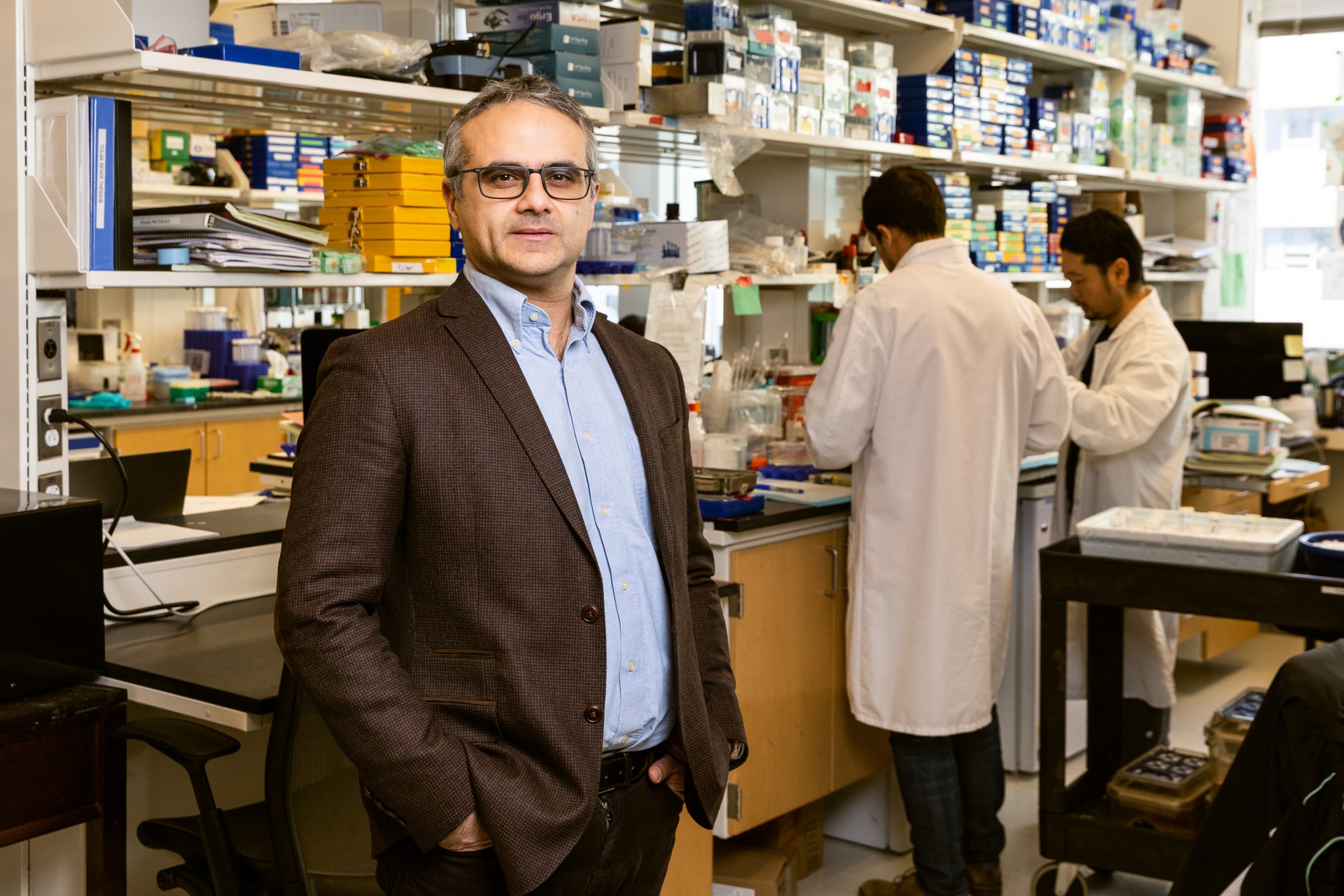
“Activation of the SOX17 program in the earliest innings of colorectal cancer formation is a critical step that shields precancerous cells from the immune system. If we can inhibit the SOX17 program, we might be better able to prevent colon cancer, particularly in patients that are prone to developing colon polyps,” says Omer Yilmaz, an MIT associate professor of biology, a member of MIT’s Koch Institute for Integrative Cancer Research, and one of the senior authors of the study.
Judith Agudo, a principal investigator at Dana-Farber Cancer Institute and an assistant professor at Harvard Medical School, is also a senior author of the study, which appears today in Nature. The paper’s lead author is MIT Research Scientist Norihiro Goto. Other collaborators include Tyler Jacks, a professor of biology and a member of MIT’s Koch Institute; Peter Westcott, a former Jacks lab postdoc who is now an assistant professor at Cold Spring Harbor Laboratory; and Saori Goto, an MIT postdoc in the Yilmaz lab.
Immune evasion
Colon cancer usually arises in long-lived cells called intestinal stem cells, whose job is to continually regenerate the lining of the intestines. Over their long lifetime, these cells can accumulate cancerous mutations that lead to the formation of polyps, a type of premalignant growth that can eventually become metastatic colon cancer.
To learn more about how these precancerous growths evade the immune system, the researchers used a technique they had previously developed for growing mini colon tumors in a lab dish and then implanting them into mice. In this case, the researchers engineered the tumors to express mutated versions of cancer-linked genes Kras, p53, and APC, which are often found in human colon cancers.
Once these tumors were implanted in mice, the researchers observed a dramatic increase in the tumors’ expression of SOX17. This gene encodes a transcription factor that is normally active only during embryonic development, when it helps to control development of the intestines and the formation of blood vessels.
The researchers’ experiments revealed that when SOX17 is turned on in cancer cells, it helps the cells to create an immunosuppressive environment. Among its effects, SOX17 prevents cells from synthesizing the receptor that normally detects interferon gamma, a molecule that is one of the immune system’s primary weapons against cancer cells.
Without those interferon gamma receptors, cancerous and precancerous cells can simply ignore messages from the immune system, which would normally direct them to undergo programmed cell death.
“One of SOX17’s main roles is to turn off the interferon gamma signaling pathway in colorectal cancer cells and in precancerous adenoma cells. By turning off interferon gamma receptor signaling in the tumor cells, the tumor cells become hidden from T cells and can grow in the presence of an immune system,” Yilmaz says.
Without interferon gamma signaling, cancer cells also minimize their production of molecules called MHC proteins, which are responsible for displaying cancerous antigens to the immune system. The cells’ insensitivity to interferon gamma also prevents them from producing immune molecules called chemokines, which normally recruit T cells that would help destroy the cancerous cells.
Targeting SOX17
When the researchers generated colon tumor organoids with SOX17 knocked out, and implanted those into mice, the immune system was able to attack those tumors much more effectively. This suggests that preventing cancer cells from turning off SOX17 could offer a way to treat colon cancer in its earliest stages.
“Just by turning off SOX17 in fairly complex tumors, we were able to essentially obliterate the ability of these tumor cells to persist,” Goto says.
As part of their study, the researchers also analyzed gene expression data from patients with colon cancer and found that SOX17 tended to be highly expressed in early-stage colon cancers but dropped off as the tumors became more invasive and metastatic.
“We think this makes a lot of sense because as colorectal cancers become more invasive and metastatic, there are other mechanisms that create an immunosuppressive environment,” Yilmaz says. “As the colon cancer becomes more aggressive and activates these other mechanisms, then there’s less importance for SOX17.”
Transcription factors such as SOX17 are considered difficult to target using drugs, in part because of their disorganized structure, so the researchers now plan to identify other proteins that SOX17 interacts with, in hopes that it might be easier to block some of those interactions.
The researchers also plan to investigate what triggers SOX17 to turn on in precancerous cells.
The research was funded by the MIT Stem Cell Initiative via Fondation MIT, the National Institutes of Health/National Cancer Institute, and a Koch Institute-Dana Farber Harvard Cancer Center Bridge Project grant.
When Albert E. Almada PhD ’13 embarks on a new project, he always considers two criteria instilled in him during his time as a graduate student in the Department of Biology at MIT.
“If you want to make a big discovery, you have to approach it from a unique perspective — a unique angle,” Almada says. “You also have to be willing to dive into the unknown and go to the leading edge of your field.”
This is not without its challenges — but with an innovative spirit, Almada says, one can find ways to apply technologies and approaches to a new area of research where a roadmap doesn’t yet exist.
Now an assistant professor of orthopedic surgery and stem cell biology and regenerative medicine at the Keck School of Medicine of the University of Southern California (USC), Almada studies the mechanics of how stem cells rebuild tissues after trauma and how stem cell principles are dysregulated and drive conditions like degenerative disease and aging, exploring these topics through an evolutionary lens.
He’s also trying to solve a mystery that has intrigued scientists for centuries: Why can some vertebrate species like fish, salamanders, and lizards regenerate entire body parts, but mammals cannot? Almada’s laboratory at USC tackles these critical questions in the musculoskeletal system.
Almada’s fascination with muscle development and regeneration can be traced back to growing up in southern California. Almada’s brother had a degenerative muscle disease called Duchenne muscular dystrophy — and, while Almada grew stronger and stronger, his brother grew weaker and weaker. Last summer, Almada’s brother, unfortunately, lost his battle with his disorder at the age of 41.
“Watching his disease progress in those early years is what inspired me to become a scientist,” Almada recalls. “Sometimes science can be personal.”
Almada went to the University of California at Irvine for his undergraduate degree, majoring in biological sciences. During his summers, he participated in the Undergraduate Research Program (URP) at the Cold Spring Harbor Laboratory and the MIT Summer Research Program-Bio (now the Bernard S. and Sophie G. Gould MIT Summer Research Program in Biology, BSG-MSRP-Bio), where he saw the passion, rigor, and drive that solidified his desire to pursue a PhD.
Despite his interest in clinical applications, skeletal muscle, and regenerative biology, Almada was drawn to the Department of Biology at MIT, which is focused on basic fundamental research.
“I was willing to bet that it all came down to understanding basic cellular processes and things going wrong with the cell and how it interacts with its environment,” he says. “The MIT biology program really helped me define an identity for myself and gave me a template for how to tackle clinical problems from a molecular perspective.”
Almada’s PhD thesis work was based on a curious finding that Phillip Sharp, Institute Professor emeritus, professor emeritus of biology, and intramural faculty at the Koch Institute for Integrative Cancer Research, had made in 2007 — that transcription, the process of copying DNA into a messenger molecule called RNA, can occur in both directions at gene promoters. In one direction, it was long understood that fully formed mRNA is transcribed and can be used as a blueprint to make a protein. The transcription Sharp observed, in the opposite direction, results in a very short RNA that is not used as a gene product blueprint.
Almada’s project dug into what those short RNA molecules are — their structure and sequence, and why they’re not produced the same way that coding messenger RNA is. In two papers published in PNAS and Nature, Almada and colleagues discovered that a balance between splicing and transcription termination signals controls the length of an RNA. This finding has wider implications because toxic RNAs are produced and can build up in several degenerative diseases; being able to splice out or shorten RNAs to remove the harmful segments could be a potential therapeutic treatment.
“That experience convinced me that if I want to make big discoveries, I have to focus on basic science,” he says. “It also gave me the confidence that if I can succeed at MIT, I can succeed just about anywhere and in any field of biology.”
At the time Almada was in graduate school, there was a lot of excitement about transcription factor reprogramming. Transcription factors are the proteins responsible for turning on essential genes that tell a cell what to be and how to behave; a subset of them can even theoretically turn one cell type into another.
Almada began to wonder whether a specialized set of transcription factors instructs stem cells to rebuild tissues after trauma. After MIT, Almada moved on to a postdoctoral position in the lab of Amy Wagers, a leader in muscle stem cell biology at Harvard University, to immerse himself in this problem.
In many tissues in our bodies, a population of stem cells typically exists in an inactive, non-dividing state called quiescence. Once activated, these stem cells interact with their environment, sense damage signals, and turn on programs of proliferation and differentiation, as well as self-renewal, which is critical to maintaining a pool of stem cells in the tissue.
One of the biggest mysteries in the field of regenerative biology is how stem cells transition from dormancy into that activated, highly regenerative state. The body’s ability to turn on stem cells, including those in the skeletal muscle system, declines as we age and is often dysregulated in degenerative diseases — diseases like the one Almada’s brother suffered from.
In a study Almada published in Cell Reports several years ago, he identified a family of transcription factors that work together to turn on a critical regenerative gene program within hours of muscle trauma. This program drives muscle stem cells out of quiescence and speeds up healing.
“Now my lab is studying this regenerative program and its potential dysregulation in aging and degenerative muscle diseases using mouse and human models,” Almada says. “We’re also drawing parallels with super-healing species like salamanders and lizards.”
Recently, Almada has been working on characterizing the molecular and functional properties of stem cells in lizards, attempting to understand how the genes and pathways differ from mammalian stem cells. Lizards can regenerate massive amounts of skeletal muscle from scratch — imagine if human muscle tissue could be regrown as seamlessly as a lizard’s tail can. He is also exploring whether the tail is unique, or if stem cells in other tissues in lizards can regenerate faster and better than the tail, by comparing analogous injuries in a mouse model.
“This is a good example of approaching a problem from a new perspective: We believe we’re going to discover new biology in lizards that we can use to enhance skeletal muscle growth in vulnerable human populations, including those that suffer from deadly muscle disorders,” Almada says.
In just three years of starting his faculty position at USC, his work and approach have already received recognition in academia, with junior faculty awards from the Baxter Foundation and the Glenn Foundation/American Federation of Aging Research. He also received his first RO1 award from the National Institutes of Health with nearly $3 million in funding. Almada and his first graduate student, Alma Zuniga Munoz, were also awarded the HHMI Gilliam Fellowship last summer. Zuniga Munoz is the first to be recognized with this award at USC; fellowship recipients, student and advisor pairs, are selected with the goal of preparing students from underrepresented groups for leadership roles in science.
Almada himself is a second-generation Mexican American and has been involved in mentoring and training throughout his academic career. He was a graduate resident tutor for Spanish House at MIT and currently serves as the chair of the Diversity, Equity, and Inclusion Committee in the Department of Stem Cell Biology and Regenerative Medicine at USC; more than half of his lab members identify as members of the Hispanic community.
“The focus has to be on developing good scientists,” Almada says. “I learned from my past research mentors the importance of putting the needs of your students first and providing a supportive environment for everyone to excel, no matter where they start.”
As a mentor and researcher, Almada knows that no question and no challenge is off limits — foundations he built in Cambridge, where his graduate studies focused on teaching him to think, not just do.
“Digging deep into the science is what MIT taught me,” he says. “I’m now taking all of my knowledge in molecular biology and applying it to translationally oriented questions that I hope will benefit human health and longevity.”
Germline stem cells are the pool of stem cells capable of becoming eggs or sperm. They divide asymmetrically, such that one of the cells resulting from a division is another stem cell and the other is a differentiated cell, which has progressed one step further down the path towards becoming an egg or sperm. Researchers have thought that this asymmetrical division served to replenish the pool of stem cells—making sperm or eggs, but also making more stem cells to produce future sperm or eggs. However, the germline has another way to replenish itself: cells that have differentiated only one or two steps down the path to becoming eggs or sperm are capable of reverting into stem cells. Why, then, do stem cells divide asymmetrically?
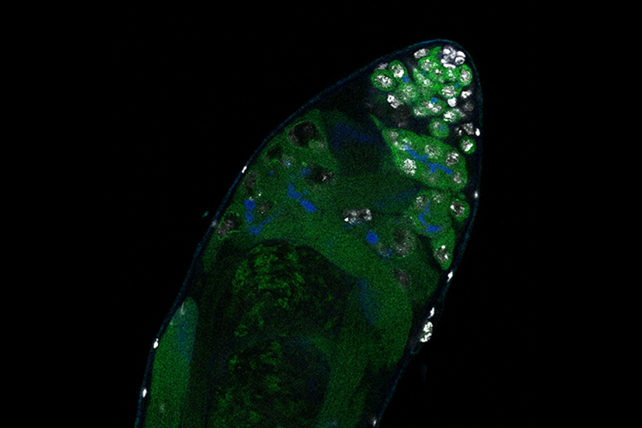
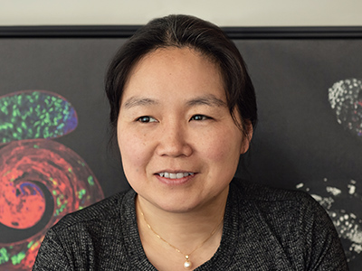
New research from Whitehead Institute Member Yukiko Yamashita, who is also a professor of biology at the Massachusetts Institute of Technology and an HHMI Investigator, and former postdoc in her lab Jonathan Nelson shows that asymmetrical division in germline stem cells serves a different but equally important purpose in male fruit flies (Drosophila melanogaster), a common model animal for germline research. The work, published in the journal Proceedings of the National Academy of Sciences (PNAS) on November 13, suggests that in flies, germline stem cells divide asymmetrically in order to unequally split a certain kind of DNA, called ribosomal DNA (rDNA), between the two dividing cells and then keep the cell with more rDNA in the stem cell pool. This is necessary in order to keep the germline viable over generations of cell divisions, and so to keep individual flies fertile and capable of reproduction. The researchers show that only germline stem cells, and not other types of germ cells, drive this process, and explain why stem cells’ asymmetric divisions make them uniquely suited to maintaining rDNA.
Ribosomal DNA is critical to maintain in the germline because it contains the instructions for making a major part of ribosomes, the cellular machines that build proteins from genetic instructions. Proteins are the main workhorses of the cell, and so cells need to make many ribosomes in order to build all of the proteins that they need. Consequently, rDNA exists as many copies repeated in a row of the code for components of the ribosome. All of these repeats make it easy for the cell to mass produce ribosomes, but they also come with a risk: repetitive DNA is prone to losing repeats during cell division. When the cell’s rDNA is copied, it’s easy for a few of the many identical repeats to get cut out, so that the resulting copy of the genome has fewer rDNA repeats than the original.
Most cells can afford to lose a few rDNA repeats without too many negative effects, but the germline cannot. Whereas other cells die with the body they are in, germ cells produce eggs and sperm that will form a new body, which produces new germ cells, and so on. The germ cell lineage is effectively immortal. Over the course of its endless cycle of cell division, the loss of rDNA repeats would add up until the cells became dysfunctional and then died. This would make the individual bearing those germ cells infertile, and so cause their lineage to go extinct.
Researchers have known that germ cells have some way to regain rDNA repeats when the number gets too low—if germ cells couldn’t do this, none of us would exist—but the details of how cells achieve this have been largely mysterious. One proposed model was that when a germ cell divides, sometimes it might divide up its rDNA unequally between the two resulting cells, so that one cell would gain rDNA repeats. Yamashita and Nelson have previously found evidence that this model is correct, and they discovered some of the specific mechanisms that enable it to happen. In a 2023 PNAS paper, the researchers showed that a retrotransposon, a “selfish” genetic element whose function is to make more copies of itself, actually helps germ cells maintain rDNA. During cell division, the retrotransposon R2 slices open one copy of the chromosome containing rDNA in its quest to insert extra copies of itself into the genome. The cell tries to repair the break using the copy on the other intact chromosome, but the tricky nature of repetitive DNA can cause the cell to lose its place, so that it stitches a stretch of rDNA repeats from one copy of the chromosome into the other copy instead.
Through this process, the germline can boost the level of rDNA in a cell—but only by as much as another cell loses. How does this win-lose exchange lead to an overall increase in rDNA levels across the germline cell population to compensate for lost rDNA? In this latest work, Yamashita and Nelson show through mathematical modeling that in cells that divide symmetrically, it would not. Gains and losses in rDNA through this form of exchange would occur essentially at random and cancel each other out over time.
Now consider an asymmetric division. After a germline stem cell divides, the cell that differentiates will go through a few more divisions and ultimately create a specific number of sperm cells–the number happens to be sixty-four. If this daughter cell gets the chromosome with more rDNA repeats, then that would lead to sixty-four sperm with more rDNA repeats—but that would be it, as the sperm have exited the pool of replicating germline stem cells.
However, the daughter cell that remains a germline stem cell will divide again to create a differentiated cell (which will become sixty-four sperm) and another stem cell, which will divide again, leading to another sixty-four sperm and another stem cell—and so on. All of these cells, including many sperm, would inherit the higher number of rDNA repeats. Furthermore, at each division, there would be an opportunity for another unequal split of rDNA. As long as the stem cell always gets the boost in rDNA, then the cumulative number of rDNA repeats would keep growing in the overall population over time—and Yamashita and colleagues’ past work shows that the germline can ensure this. A 2022 Science Advances paper from Yamashita and then-postdoc in her lab George Watase showed that when a germline stem cell divides, the DNA strand with more rDNA repeats is tagged with a protein that the researchers named Indra, which helps mark it to stay in the daughter cell that will become another stem cell. Yamashita and Nelson’s new paper includes mathematical modeling by second author Tomohiro Kumon, a postdoc in Yamashita’s lab, that proves that this is not only sufficient to restore the level of rDNA repeats over time, but that it is the most effective and efficient way for the germline to do so.
“There was this problem with the unequal exchange model of rescuing rDNA, because every cell that gained rDNA did so at the expense of another that was losing it,” Nelson says. “What we show here is that the reason why there’s a bias towards gain in the germline is because this process is happening within these asymmetrically dividing germline stem cells that can gain and gain and gain, while the cells that lose rDNA exit the cycle and so have a limited effect.”
The researchers complemented their mathematical modeling with evidence that the process to increase rDNA repeats occurs primarily or solely in germline stem cells. They found that when the number of rDNA repeats got low enough, then expression of R2 and the presence of double-stranded DNA breaks both increased in germline stem cells, but not significantly in other germ cell types.
Yamashita and Nelson propose that the different cell types in the germline take on different functions to create a pipeline for maximizing the health of future sperm. Germ cells that are one or two steps down the path of differentiation from stem cells are essentially identical to them, to the point that they can be difficult to tell apart in testing, but they divide symmetrically. They are also much more sensitive to DNA damage; the researchers found that R2 exposure kills these cells.
Germline stem cells, with their asymmetrical division and ability to tolerate R2 expression, serve to restore rDNA levels when they get too low. Then the differentiated germ cells serve to weed out mutations—including those introduced during R2 expression in the earlier stem cell stage—by killing off cells with DNA damage. The different strengths of the different types of germ cells creates an effective pipeline to produce the largest number of sperm cells with high rDNA repeat number and low DNA damage.
Eventually, this new understanding of the details of how cells maintain their rDNA could lead to medical therapies. For example, cancer cells are, like germ cells, an essentially immortal cell line, and so must have a way to maintain their rDNA. If researchers could someday find a way to prevent them from doing so, that could be a good treatment strategy. The work also may have implications for research on aging, as rDNA decreases with age in other cell types. In the meantime, Yamashita and Nelson are excited to have solved several long-standing mysteries in their field, including how germ cells can restore rDNA at a population level when each division creates an equal loss and gain of rDNA, and why germline stem cells divide asymmetrically.
“Typically, when you publish a paper, you feel like you’ve fit two puzzle pieces together, but in this case, I feel like we fit a bunch of puzzle pieces together,” Yamashita says. “It’s been immensely satisfying to find answers to multiple questions and see how they all fit together to explain the mechanisms of this process that’s necessary for germline immortality.”
Every three to five days, all of the cells lining the human intestine are replaced. That constant replenishment of cells helps the intestinal lining withstand the damage caused by food passing through the digestive tract.
This rapid turnover of cells relies on intestinal stem cells, which give rise to all of the other types of cells found in the intestine. Recent research has shown that those stem cells are heavily influenced by diet, which can help keep them healthy or stimulate them to become cancerous.
“Low-calorie diets such as fasting and caloric restriction can have antiaging effects and antitumor effects, and we want to understand why that is. On the other hand, diets that lead to obesity can promote diseases of aging, such as cancer,” says Omer Yilmaz, the Eisen and Chang Career Development Associate Professor of Biology at MIT.
For the past decade, Yilmaz has been studying how different diets and environmental conditions affect intestinal stem cells, and how those factors can increase the risk of cancer and other diseases. This work could help researchers develop new ways to improve gastrointestinal health, either through dietary interventions or drugs that mimic the beneficial effects of certain diets, he says.
“Our findings have raised the possibility that fasting interventions, or small molecules that mimic the effects of fasting, might have a role in improving intestinal regeneration,” says Yilmaz, who is also a member of MIT’s Koch Institute for Integrative Cancer Research.
A clinical approach
Yilmaz’s interest in disease and medicine arose at an early age. His father practiced internal medicine, and Yilmaz spent a great deal of time at his father’s office after school, or tagging along at the hospital where his father saw patients.
“I was very interested in medicines and how medicines were used to treat diseases,” Yilmaz recalls. “He’d ask me questions, and many times I wouldn’t know the answer, but he would encourage me to figure out the answers to his questions. That really stimulated my interest in biology and in wanting to become a doctor.”
Knowing that he wanted to go into medicine, Yilmaz applied and was accepted to an eight-year, combined bachelor’s and MD program at the University of Michigan. As an undergraduate, this gave him the freedom to explore areas of interest without worrying about applying to medical school. While majoring in biochemistry and physics, he did undergraduate research in the field of protein folding.
During his first year of medical school, Yilmaz realized that he missed doing research, so he decided to apply to the MD/PhD program at the University of Michigan. For his PhD research, he studied blood-forming stem cells and identified new markers that allowed such cells to be more easily isolated from the bone marrow.
“This was important because there’s a lot of interest in understanding what makes a stem cell a stem cell, and how much of it is an internal program versus signals from the microenvironment,” Yilmaz says.
After finishing his PhD and MD, he thought about going straight into research and skipping a medical residency, but ended up doing a residency in pathology at Massachusetts General Hospital. During that time, he decided to switch his research focus from blood-forming stem cells to stem cells found in the gastrointestinal tract.
“The GI tract seemed very interesting because in contrast to the bone marrow, we knew very little about the identity of GI stem cells,” Yilmaz says. “I knew that once GI stem cells were identified, there’d be a lot of interesting questions about how they respond to diet and how they respond to other environmental stimuli.”
Dietary questions
To delve into those questions, Yilmaz did postdoctoral research at the Whitehead Institute, where he began investigating the connections between stem cells, metabolism, diet, and cancer.
Because intestinal stem cells are so long-lived, they are more likely to accumulate genetic mutations that make them susceptible to becoming cancerous. At the Whitehead Institute, Yilmaz began studying how different diets might influence this vulnerability to cancer, a topic that he carried into his lab at MIT when he joined the faculty in 2014.
One question his lab has been exploring is why low-calorie diets often have protective effects, including a boost in longevity — a phenomenon that has been seen in many studies in animals and humans.
In a 2018 study, his lab found that a 24-hour fast dramatically improves stem cells’ ability to regenerate. This effect was seen in both young and aged mice, suggesting that even in old age, fasting or drugs that mimic the effects of fasting could have a beneficial effect.
On the flip side, Yilmaz is also interested in why a high-fat diet appears to promote the development of cancer, especially colorectal cancer. In a 2016 study, he found that when mice consume a high-fat diet, it triggers a significant increase in the number of intestinal stem cells. Also, some non-stem-cell populations begin to resemble stem cells in their behavior. “The upshot of these changes is that both stem cells and non-stem-cells can give rise to tumors in a high-fat diet state,” Yilmaz says.
To help with these studies, Yilmaz’s lab has developed a way to use mouse or human intestinal stem cells to generate miniature intestines or colons in cell culture. These “organoids” can then be exposed to different nutrients in a very controlled setting, allowing researchers to analyze how different diets affect the system.
Recently, his lab adapted the system to allow them to expand their studies to include the role of immune cells, fibroblasts, and other supportive cells found in the microenvironment of stem cells. “It would be remiss of us to focus on just one cell type,” Yilmaz says. “We’re looking at how these different dietary interventions impact the entire stem cell neighborhood.”
While Yilmaz spends most of his time running his lab at MIT, he also devotes six to eight weeks per year to his work at MGH, where he is an associate pathologist focusing on gastrointestinal pathology.
“I enjoy my clinical work, and it always reminds me about the importance of the research we do,” he says. “Seeing colon cancer and other GI cancers under the microscope, and seeing their complexity, reminds me of the importance of our mission to figure out how we can prevent these cancers from forming.”
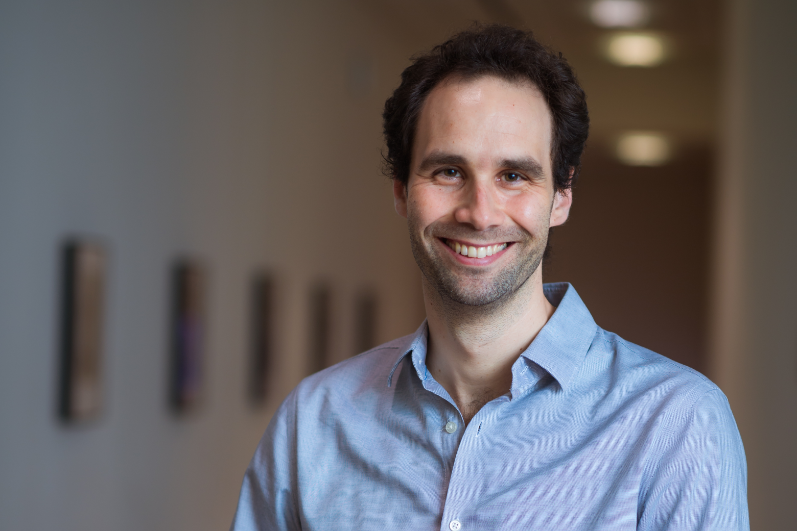
Whitehead Institute Member Siniša Hrvatin has been named as one of the 15 researchers to be selected as 2023 Searle Scholars. The Searle Scholars Program supports the research of exceptional young faculty in the biomedical sciences and chemistry.
Chosen by an advisory board of eminent scientists, Searle Scholars are considered among the most creative researchers pursuing careers in academic research. Their investigations address challenging research questions and can lead to new insights that fundamentally change their fields—and to opportunities for translating discoveries into new therapeutics and diagnostics.
“I am truly grateful for the support of the Searle Scholar Program as we embark on this ambitious project,” says Hrvatin, who joined the Institute in 2021 and is also an assistant professor of biology at Massachusetts Institute of Technology. The three-year grant accompanying the award will support his work developing a new animal model for the study of hibernation.
“The ability to maintain nearly constant body temperature is a defining feature of mammalian and avian evolution; but, when challenged by harsh environments, many species decrease body temperature and metabolic rate and initiate energy-conserving states of torpor and hibernation,” Hrvatin notes. “Science has not yet answered the fundamental questions of how mammals initiate, regulate, and survive these extraordinary hypometabolic and hypothermic states.
“However, those answers could have profound medical applications,” he explains. “For example, harnessing the mechanisms behind hibernation might provide new approaches to protect neurons from ischemic injury and to preserve tissues and organs for transplantation.”
In the Searle-supported study, Hrvatin aims to discover a control center in the brain that regulates distinct stages of hibernation in the Syrian hamster. His lab will start by identifying the brain regions active during the deep torpor stage of hibernation and, using molecular profiling techniques, will then identify the specific neuronal populations and molecular pathways involved. Finally, the team will develop new tools to determine specific activities in those neural populations that are necessary for natural hibernation—and that may be sufficient to induce a synthetic state of hibernation.
“Taken together,” Hrvatin says, “I believe that our discoveries and the tools we build will help establish the first controllable animal model of hibernation.”
Since 1981, 677 scientists have been named Searle Scholars and the Program has awarded more than $152 million in support for Scholars’ research. To date, 85 Searle Scholars have been inducted into the National Academy of Sciences, 20 have been recognized with a MacArthur Fellowship, and two have been awarded the Nobel Prize for Chemistry.
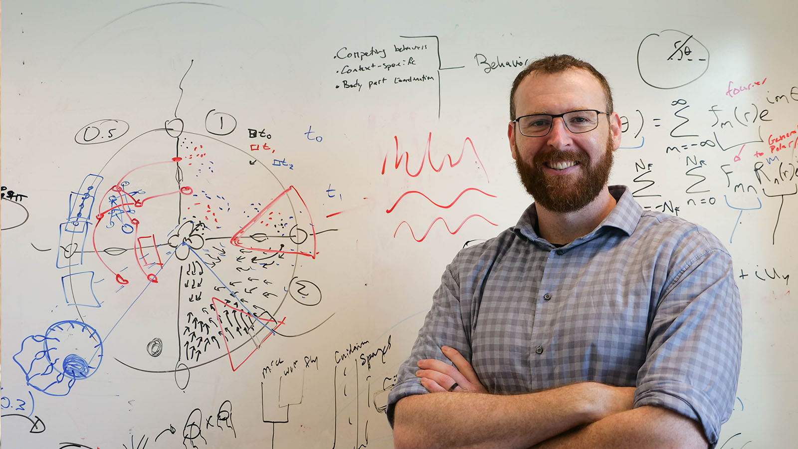
How does animal behavior emerge from networks of connected neurons? How are these incredible nervous systems and behaviors actually generated by evolution? Are there principles shared by all nervous systems or is evolution constantly innovating? What did the first nervous system look like that gave rise to the incredible diversity of life that we see around us?
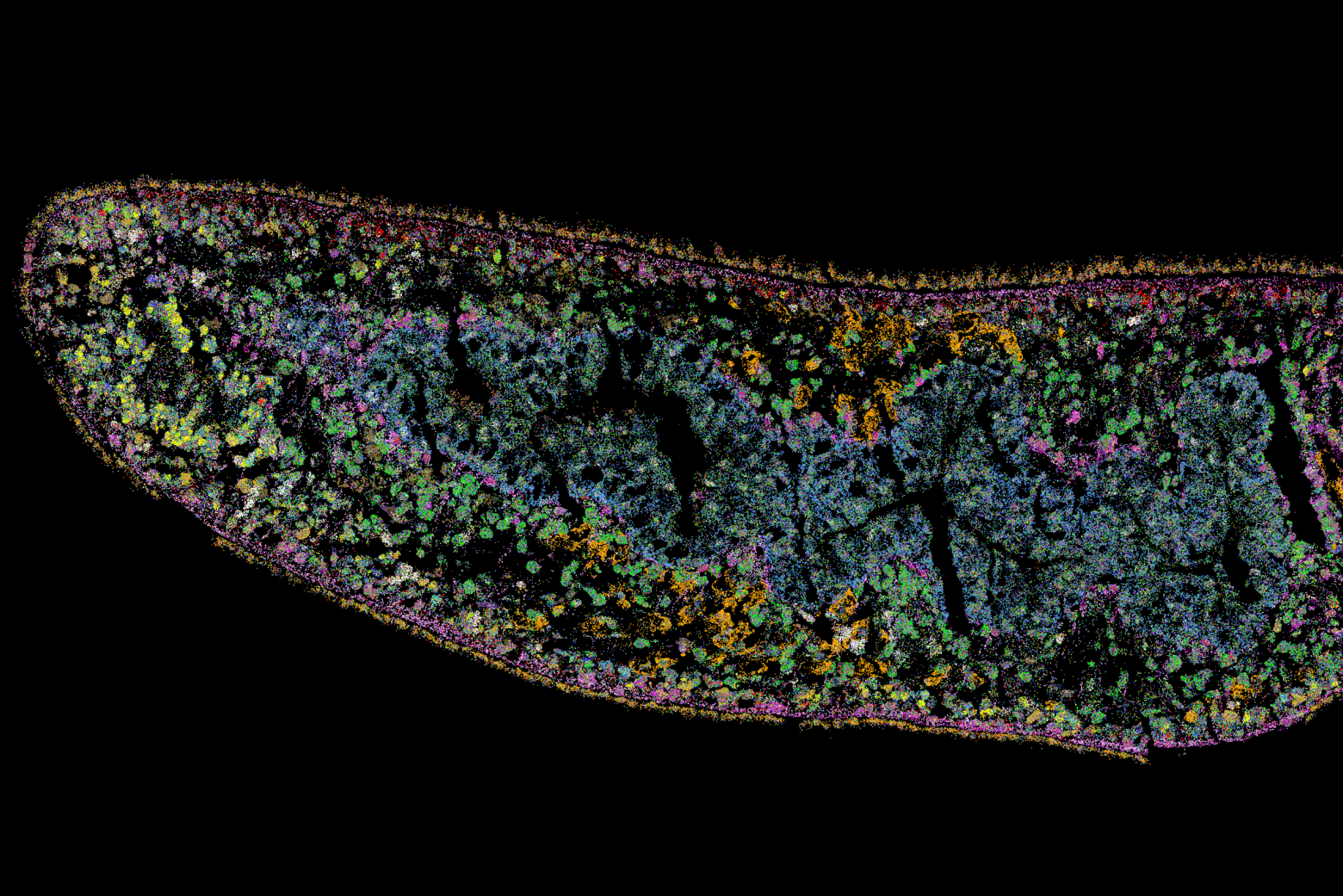
Combining the study of animal behavior with studies of nervous system form, function, and evolution, Brady Weissbourd, a new faculty member in the Department of Biology and investigator in The Picower Institute for Learning and Memory, uses the tiny, transparent jellyfish Clytia hemisphaerica, a new neuroscience model.
Q: In your work, you developed a new model organism for neuroscience research, the transparent jellyfish Clytia hemisphaerica. How do these jellyfish answer questions about neuroscience, the nervous system, and evolution in ways that other models cannot?
A: First, I believe in the importance of more broadly understanding the natural world and diversifying the organisms that we deeply study. One reason is to find experimentally tractable organisms to identify generalizable biological principles – for example, we understand the basis of how neurons “fire” from studies of the squid giant axon. Another reason is that transformative breakthroughs have come from identifying evolutionary innovations that already exist in nature – for example, green fluorescent protein (GFP, from jellyfish) or CRISPR (from bacteria). In both ways, this jellyfish is a valuable complement to existing models.
I have always been interested in the intersection of two types of problems: how nervous systems generate our behaviors; and how these incredible systems were actually created by evolution.
On the systems neuroscience side, ever since working on the serotonin system during my PhD I have been fascinated by the problem of how animals control all of their behaviors simultaneously in a flexible and context-dependent manner, and how behavioral choices depend not just on incoming stimuli but on how those stimuli interact with constantly changing states of the nervous system and body. These are extremely complex and difficult problems, with the particular challenge of interactions across scales, from chemical signaling and dynamic cell biology to neural networks and behavior.
To address these questions, I wanted to move into a model organism with exceptional experimental tractability.
There have been exciting breakthroughs in imaging techniques for neuroscience, including these incredible ways in which we can actually watch and manipulate neuronal activity in a living animal. So, the first thing I wanted was a small and transparent organism that would allow for this kind of optical approach. These jellyfish are a few millimeters in diameter and perfectly transparent, with interesting behaviors but relatively compact nervous systems. They have thousands of neurons where we have billions, which also puts them at a nice intermediate complexity compared to other transparent models that are widely used – for example, C. elegans have 302 neurons and larval zebrafish have something like 100,000 in the brain alone. These features will allow us to look at the activity of the whole nervous system in behaving animals to try to understand how that activity gives rise to behaviors and how that activity itself arises from networks of neurons.
On the evolution side of our work, we are interested in the origins of nervous systems, what the first nervous systems looked like, and broadly what the options are for how nervous systems are organized and functioning: to what extent there are principles versus interesting and potentially useful innovations, and if there are principles, whether those are optimal or somehow constrained by evolution. Our last common ancestor with jellyfish and their relatives (the cnidarians) was something similar to the first nervous system, so by comparing what we find in cnidarians with work in other models we can make inferences about the origins and early evolution of nervous systems. As we further explore these highly divergent animals, we are also finding exciting evolutionary innovations: specifically, they have incredible capabilities for regenerating their nervous systems. In the future, it will be exciting to better understand how these neural networks are organized to allow for such robustness.
Q: What work is required to develop a new organism as a model, and why did you choose this particular species of jellyfish?
A: If you’re choosing a new animal model, it’s not just about whether it has the right features for the questions you want to ask, but also whether it technically lets you do the right experiments. The model we’re using was first developed by a research group in France, who spent many years doing the really hard work of figuring out how to culture the whole life cycle in the lab, injecting eggs, and developing other key resources. For me, the big question was whether we’d be able to use the genetic tools that I was describing earlier for looking at neural activity. Working closely with collaborators in France, our first step was figuring out how to insert things into the jellyfish genome. If we couldn’t figure that out, I was going to switch back to working with mice. It took us about two years of troubleshooting, but now we can routinely generate genetically modified jellyfish in the lab.
Switching to a new animal model is tough – I have a mouse neuroscience background and joined a postdoc lab that used mice and flies; I was the only person working with jellyfish but had no experience. For example, building an aquaculture system and figuring out how to keep jellyfish healthy is not trivial, particularly now that we’re trying to do genetics. One of my goals is now to optimize and simplify this whole process so that when other labs want to start working with jellyfish we have a simple aquaculture platform to get them started, even if they have no experience.
In addition to the fact that these things are tiny and transparent, the main reason that we chose this particular species is because it has an amazing life cycle that makes it an exciting laboratory animal.
They have separate sexes that spawn daily with the fertilized eggs developing into larvae that then metamorphose into polyps. We grow these polyps on microscope slides, where they form colonies that are thought to be immortal. These colonies are then constantly releasing jellyfish, which are all genetically identical “clones” that can be used for experiments. That means that once you create a genetically modified strain, like a transgenic line or a knockout, you can keep it forever as a polyp colony – and since the animals are so small, we can culture them in large numbers in the lab.
There’s still a huge amount of foundational work to do, like characterizing their behavioral repertoire and nervous system organization. It’s shocking how little we know about the basics of jellyfish biology – particularly considering that they kill more people per year than sharks and stingrays combined – and the more we look into it the more questions there are.
Q: What drew you to a faculty position at MIT?
A: I wanted to be in a department that does fundamental research, is enthusiastic about basic science, is open-minded, and is very diverse in what people work on and think about. My goal is also to be able to ultimately link mechanisms at the molecular and cellular level to organismal behavior, which is something that MIT Biology is particularly strong at doing. It’s been an exciting first few months! MIT Biology is such an amazing place to do science and it’s been wonderful how enthusiastic and supportive everyone in the department has been.
I was additionally drawn to MIT by the broader community and have already found it so easy to start collaborations with people in neuroscience, engineering, and math. I’m also thrilled to have recently become a member of The Picower Institute for Learning and Memory, which further enables these collaborations in a way that I believe will be transformational for the work in my lab.
It’s a new lab. It’s a new organism. There isn’t a huge, well-established field that is taking these approaches. There’s so much we don’t know, and so much that we have to establish from scratch. My goal is for my lab to have a sense of adventure and fun, and I’m really excited to be doing that here in MIT Biology.
Eight MIT faculty members are among more than 250 leaders from academia, the arts, industry, public policy, and research elected to the American Academy of Arts and Sciences, the academy announced April 19.
One of the nation’s most prestigious honorary societies, the academy is also a leading center for independent policy research. Members contribute to academy publications, as well as studies of science and technology policy, energy and global security, social policy and American institutions, the humanities and culture, and education.
Those elected from MIT in 2023 are:
“With the election of these members, the academy is honoring excellence, innovation, and leadership and recognizing a broad array of stellar accomplishments. We hope every new member celebrates this achievement and joins our work advancing the common good,” says David W. Oxtoby, president of the academy.
Since its founding in 1780, the academy has elected leading thinkers from each generation, including George Washington and Benjamin Franklin in the 18th century, Maria Mitchell and Daniel Webster in the 19th century, and Toni Morrison and Albert Einstein in the 20th century. The current membership includes more than 250 Nobel and Pulitzer Prize winners.