Pursuing the secrets of a stealthy parasite

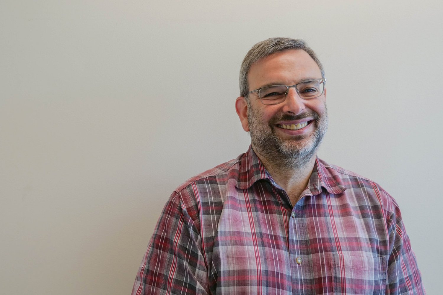
By unraveling the genetic pathways that help Toxoplasma gondii persist in human cells, Sebastian Lourido hopes to find new ways to treat toxoplasmosis.

Anne Trafton | MIT News
August 25, 2024

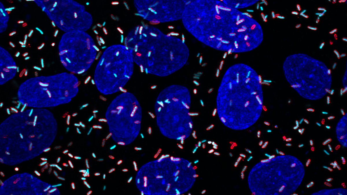
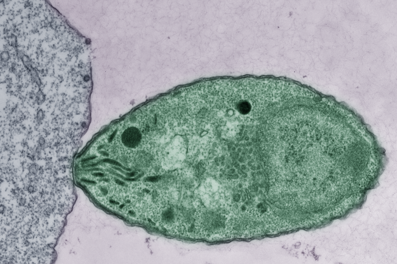
Toxoplasma gondii, the parasite that causes toxoplasmosis, is believed to infect as much as one-third of the world’s population. Many of those people have no symptoms, but the parasite can remain dormant for years and later reawaken to cause disease in anyone who becomes immunocompromised.

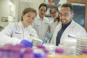
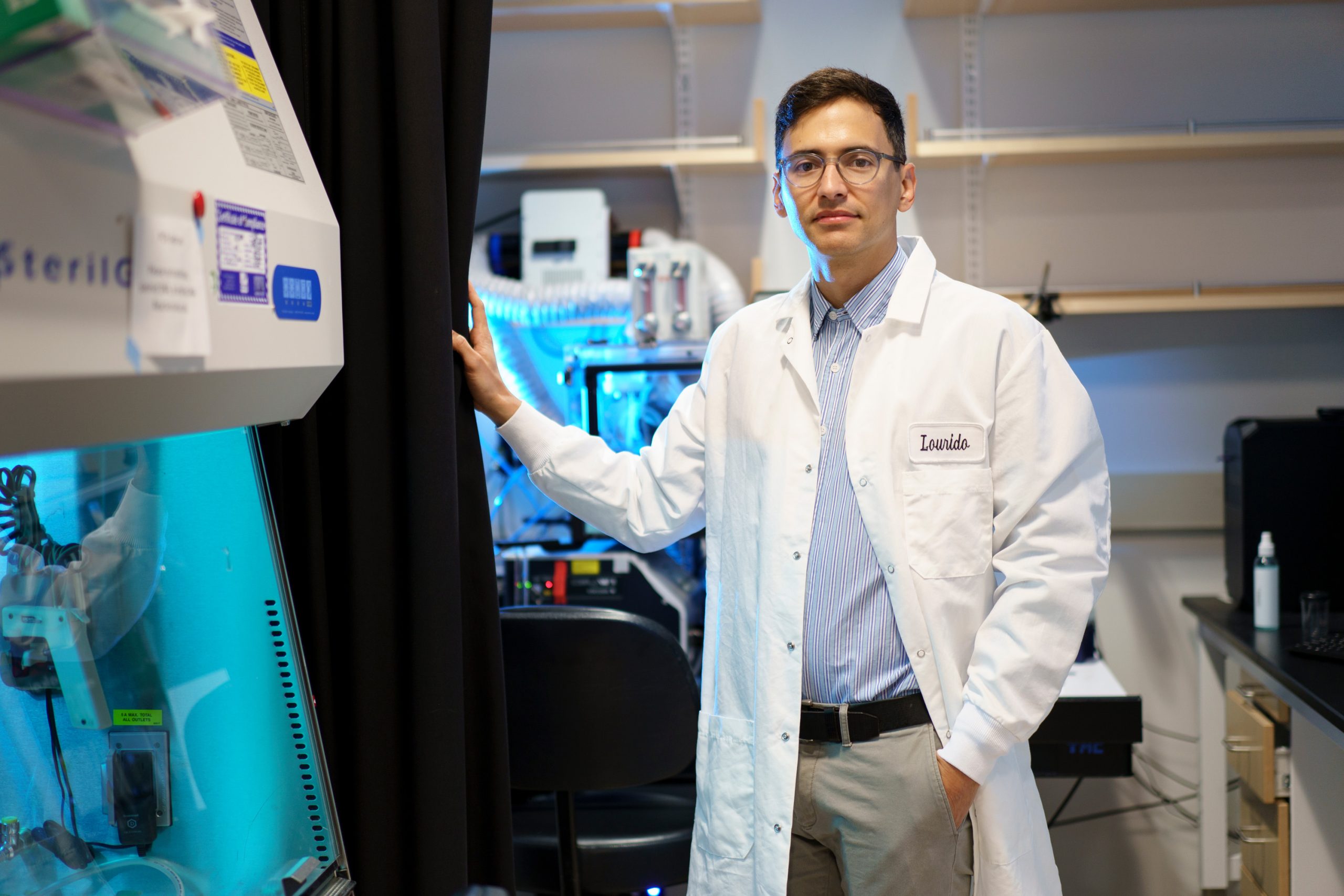
Why this single-celled parasite is so widespread, and what triggers it to reemerge, are questions that intrigue Sebastian Lourido, an associate professor of biology at MIT and member of the Whitehead Institute for Biomedical Research. In his lab, research is unraveling the genetic pathways that help to keep the parasite in a dormant state, and the factors that lead it to burst free from that state.

“One of the missions of my lab to improve our ability to manipulate the parasite genome, and to do that at a scale that allows us to ask questions about the functions of many genes, or even the entire genome, in a variety of contexts,” Lourido says.

There are drugs that can treat the acute symptoms of Toxoplasma infection, which include headache, fever, and inflammation of the heart and lungs. However, once the parasite enters the dormant stage, those drugs don’t affect it. Lourido hopes that his lab’s work will lead to potential new treatments for this stage, as well as drugs that could combat similar parasites such as a tickborne parasite known as Babesia, which is becoming more common in New England.

“There are a lot of people who are affected by these parasites, and parasitology often doesn’t get the attention that it deserves at the highest levels of research. It’s really important to bring the latest scientific advances, the latest tools, and the latest concepts to the field of parasitology,” Lourido says.

A fascination with microbiology

As a child in Cali, Colombia, Lourido was enthralled by what he could see through the microscopes at his mother’s medical genetics lab at the University of Valle del Cauca. His father ran the family’s farm and also worked in government, at one point serving as interim governor of the state.

“From my mom, I was exposed to the ideas of gene expression and the influence of genetics on biology, and I think that really sparked an early interest in understanding biology at a fundamental level,” Lourido says. “On the other hand, my dad was in agriculture, and so there were other influences there around how the environment shapes biology.”

Lourido decided to go to college in the United States, in part because at the time, in the early 2000s, Colombia was experiencing a surge in violence. He was also drawn to the idea of attending a liberal arts college, where he could study both science and art. He ended up going to Tulane University, where he double-majored in fine arts and cell and molecular biology.

As an artist, Lourido focused on printmaking and painting. One area he especially enjoyed was stone lithography, which involves etching images on large blocks of limestone with oil-based inks, treating the images with chemicals, and then transferring the images onto paper using a large press.

“I ended up doing a lot of printmaking, which I think attracted me because it felt like a mode of expression that leveraged different techniques and technical elements,” he says.

At the same time, he worked in a biology lab that studied Daphnia, tiny crustaceans found in fresh water that have helped scientists learn about how organisms can develop new traits in response to changes to their environment. As an undergraduate, he helped develop ways to use viruses to introduce new genes into Daphnia. By the time he graduated from Tulane, Lourido had decided to go into science rather than art.

“I had really fallen in love with lab science as an undergrad. I loved the freedom and the creativity that came from it, the ability to work in teams and to build on ideas, to not have to completely reinvent the entire system, but really be able to develop it over a longer period of time,” he says.

After graduating from college, Lourido spent two years in Germany, working at the Max Planck Institute for Infection Biology. In Arturo Zychlinksy’s lab, Lourido studied two bacteria known as Shigella and Salmonella, which can cause severe illnesses, including diarrhea. His studies there helped to reveal how these bacteria get into cells and how they modify the host cells’ own pathways to help them replicate inside cells.

As a graduate student at Washington University in St. Louis, Lourido worked in several labs focusing on different aspects of microbiology, including virology and bacteriology, but eventually ended up working with David Sibley, a prominent researcher specializing in Toxoplasma.

“I had not thought much about Toxoplasma before going to graduate school,” Lourido recalls. “I was pretty unaware of parasitology in general, despite some undergrad courses, which honestly very superficially treated the subject. What I liked about it was here was a system where we knew so little — organisms that are so different from the textbook models of eukaryotic cells.”

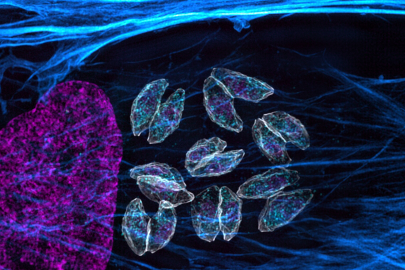
Toxoplasma gondii belongs to a group of parasites known as apicomplexans — a type of protozoans that can cause a variety of diseases. After infecting a human host, Toxoplasma gondii can hide from the immune system for decades, usually in cysts found in the brain or muscles. Lourido found the organism especially intriguing because as a 17-year-old, he had been diagnosed with toxoplasmosis. His only symptom was swollen glands, but doctors found that his blood contained antibodies against Toxoplasma.

“It is really fascinating that in all of these people, about a quarter to a third of the world’s population, the parasite persists. Chances are I still have live parasites somewhere in my body, and if I became immunocompromised, it would become a big problem. They would start replicating in an uncontrolled fashion,” he says.

A transformative approach

One of the challenges in studying Toxoplasma is that the organism’s genetics are very different from those of either bacteria or other eukaryotes such as yeast and mammals. That makes it harder to study parasitic gene functions by mutating or knocking out the genes.

Because of that difficulty, it took Lourido his entire graduate career to study the functions of just a couple of Toxoplasma genes. After finishing his PhD, he started his own lab as a fellow at the Whitehead Institute and began working on ways to study the Toxoplasma genome at a larger scale, using the CRISPR genome-editing technique.

With CRISPR, scientists can systematically knock out every gene in the genome and then study how each missing gene affects parasite function and survival.

“Through the adaptation of CRISPR to Toxoplasma, we’ve been able to survey the entire parasite genome. That has been transformative,” says Lourido, who became a Whitehead member and MIT faculty member in 2017. “Since its original application in 2016, we’ve been able to uncover mechanisms of drug resistance and susceptibility, trace metabolic pathways, and explore many other aspects of parasite biology.”

Using CRISPR-based screens, Lourido’s lab has identified a regulatory gene called BFD1 that appears to drive the expression of genes that the parasite needs for long-term survival within a host. His lab has also revealed many of the molecular steps required for the parasite to shift between active and dormant states.

“We’re actively working to understand how environmental inputs end up guiding the parasite in one direction or another,” Lourido says. “They seem to preferentially go into those chronic stages in certain cells like neurons or muscle cells, and they proliferate more exuberantly in the acute phase when nutrient conditions are appropriate or when there are low levels of immunity in the host.”

News Brief: Lamason Lab uncovers seven novel effectors in Rickettsia parkeri infection

The enemy within: new research reveals insights into the arsenal Rickettsia parkeri uses against its host

Lillian Eden | Department of Biology
July 29, 2024

Identifying secreted proteins is critical to understanding how obligately intracellular pathogens hijack host machinery during infection, but identifying them is akin to finding a needle in a haystack.

For then-graduate student Allen Sanderlin, PhD ’24, the first indication that a risky, unlikely project might work was cyan, tic tac-shaped structures seen through a microscope — proof that his bacterial pathogen of interest was labeling its own proteins.  

Sanderlin, a member of the Lamason Lab in the Department of Biology at MIT, studies Rickettsia parkeri, a less virulent relative of the bacterial pathogen that causes Rocky Mountain Spotted Fever, a sometimes severe tickborne illness. No vaccine exists and definitive tests to diagnose an infection by Rickettsia are limited.

Rickettsia species are tricky to work with because they are obligately intracellular pathogens whose entire life cycles occur exclusively inside cells. Many approaches that have advanced our understanding of other bacterial infections and how those pathogens interact with their host aren’t applicable to Rickettsia because they can’t be grown on a plate in a lab setting. 

In a paper recently published in Nature Communications, the Lamason Lab outlines an approach for labeling and isolating R. parkeri proteins released during infection. This research reveals seven previously unknown secreted factors, known as effectors, more than doubling the number of known effectors in R. parkeri. 

Better-studied bacteria are known to hijack the host’s machinery via dozens or hundreds of secreted effectors, whose roles include manipulating the host cell to make it more susceptible to infection. However, finding those effectors in the soup of all other materials within the host cell is akin to looking for a needle in a haystack, with an added twist that researchers aren’t even sure what those needles look like for Rickettsia.  

Approaches that worked to identify the six previously known secreted effectors are limited in their scope. For example, some were found by comparing pathogenic Rickettsia to nonpathogenic strains of the bacteria, or by searching for proteins with domains that overlap with effectors from better-studied bacteria. Predictive modeling, however, relies on proteins being evolutionarily conserved. 

“Time and time again, we keep finding that Rickettsia are just weird — or, at least, weird compared to our understanding of other bacteria,” says Sanderlin, the paper’s first author. “This labeling tool allows us to answer some really exciting questions about rickettsial biology that weren’t possible before.”

The cyan tic tacs

To selectively label R. parkeri proteins, Sanderlin used a method called cell-selective bioorthogonal non-canonical amino acid tagging. BONCAT was first described in research from the Tirrell Lab at Caltech. The Lamason Lab, however, is the first group to use the tool successfully in an obligate intracellular bacterial pathogen; the thrilling moment when Sanderlin saw cyan tic-tac shapes indicated successfully labeling only the pathogen, not the host. 

Sanderlin next used an approach called selective lysis, carefully breaking open the host cell while leaving the pathogen, filled with labeled proteins, intact. This allowed him to extract proteins that R. parkeri had released into its host because the only labeled proteins amid other host cell material were effectors the pathogen had secreted. 

Sanderlin had successfully isolated and identified seven needles in the haystack, effectors never before identified in Rickettsia biology. The novel secreted rickettsial factors are dubbed SrfA, SrfB, SrfC, SrfD, SrfE, SrfF, and SrfG. 

“Every grad student wants to be able to name something,” Sanderlin says. “The most exciting — but frustrating — thing was that these proteins don’t look like anything we’ve seen before.”

Special delivery

Theoretically, Sanderlin says, once the effectors are secreted, they work independently from the bacteria — a driver delivering a pizza does not need to check back in with the store at every merge or turn.

Since SrfA-G didn’t resemble other known effectors or host proteins the pathogen could be mimicking during infection, Sanderlin then tried to answer some basic questions about their behavior. Where the effectors localize, meaning where in the cell they go, could hint at their purpose and what further experiments could be used to investigate it. 

To determine where the effectors were going, Sanderlin added the effectors he’d found to uninfected cells by introducing DNA that caused human cell lines to express those proteins. The experiment succeeded: he discovered that different Srfs went to different places throughout the host cells.  

SrfF and SrfG are found throughout the cytoplasm, whereas SrfB localizes to the mitochondria. That was especially intriguing because its structure is not predicted to interact with or find its way to the mitochondria, and the organelle appears unchanged despite the presence of the effector. 

Further, SrfC and SrfD found their way to the endoplasmic reticulum. The ER would be especially useful for a pathogen to appropriate, given that it is a dynamic organelle present throughout the cell and has many essential roles, including synthesizing proteins and metabolizing lipids. 

Aside from where effectors localize, knowing what they may interact with is critical. Sanderlin showed that SrfD interacts with Sec61, a protein complex that delivers proteins across the ER membrane. In keeping with the theme of the novelty of Sanderlin’s findings, SrfD does not resemble any proteins known to interact with the ER or Sec61. 

With this tool, Sanderlin identified novel proteins whose binding partners and role during infection can now be studied further. 

“These results are exciting but tantalizing,” Sanderlin says. “What Rickettsia secrete — the effectors, what they are, and what they do is, by and large, still a black box.” 

There are very likely other effectors in the proverbial cellular haystack. Sanderlin found that SrfA-G are not found in every species of Rickettsia, and his experiments were solely conducted with Rickettsia at late stages of infection — earlier windows of time may make use of different effectors. This research was also carried out in human cell lines, so there may be an entirely separate repertoire of effectors in ticks, which are responsible for spreading the pathogen.

Expanding Tool Development

Becky Lamason, the senior author of the Nature Communications paper, noted that this tool is one of a few avenues the lab is exploring to investigate R. parkeri, including a paper in the Journal of Bacteriology on conditional genetic manipulation. Characterizing how the pathogen behaves with or without a particular effector is leaps and bounds ahead of where the field was just a few years ago when Sanderlin was Lamason’s first graduate student to join the lab.

“What I always hoped for in the lab is to push the technology, but also get to the biology. These are two of what will hopefully be a suite of ways to attack this problem of understanding how these bacteria rewire and manipulate the host cell,” Lamason says. “We’re excited, but we’ve only scratched the surface.”

A genome-wide screen in live hosts reveals new secrets of parasite infection

Researchers in the Lourido Lab performed the first genome-wide screen of Toxoplasma gondii in live hosts, revealing genes that are important for infection but previously undetected in cell culture experiments. 

Greta Friar | Whitehead Institute
July 8, 2024

Apicomplexan parasites are a common cause of disease, infecting hundreds of millions of people each year. They are responsible for spreading malaria; cryptosporidiosis – a severe childhood diarrheal disease; and toxoplasmosis – a disease that endangers immune compromised people and fetuses, and is the reason why pregnant women are told to avoid changing cat litter. Apicomplexan parasites are very good at infecting humans and many other animals, and persisting inside of them. The more that researchers can learn about how apicomplexans infect hosts, the better they will be able to develop effective treatments against the parasites.

To this end, researchers in Whitehead Institute Member Sebastian Lourido’s lab, led by graduate student Christopher Giuliano, have now completed a genome-wide screen of the apicomplexan parasite Toxoplasma gondii (T. gondii), which causes toxoplasmosis, during its infection of mice. This screen shows how important each gene is for the parasite’s ability to infect a host, providing clues to genes’ functions. In the journal Nature Microbiology on July 8, the researchers share their approach for tracing lineages of parasites in a live host, and some specific findings of interest—including a possible anti-parasitic drug target.

From dish to animal

Researchers in Lourido’s lab previously developed a screen to test the function of every T. gondii gene in cells in a dish in 2016. They used CRISPR gene editing technology to make mutant parasites in which each lineage had one gene inactivated. The researchers could then assess the importance of each gene to a parasite’s fitness, or ability to thrive, based on how well the mutants missing that gene did. If a mutant died off, this implied that its inactivated gene is essential for the parasite’s survival.

This screen taught the researchers a lot about T. gondii’s biology but faced a common limitation: the parasites were studied in a dish rather than a live host. Cell culture provides an easier way to study parasites, but the conditions are not the same as what parasites face in an animal host. A host’s body is a more complex and dynamic environment, so it may require parasites to rely on genes that they don’t need in the artificial setting of cell culture.

To overcome this limitation, researchers in Lourido’s lab figured out how to repeat the T. gondii genome-wide screen, which their colleagues in the lab had previously done in cell culture, in live mice. This was a massive undertaking, which required solving various technical challenges and running a large number of parallel experiments. T. gondii has around eight thousand genes, so the researchers performed pooled experiments, with each mouse getting infected by many different mutants—but not so many as to overwhelm the mouse. This meant that the researchers needed a way to more closely monitor the trajectories of mutants in the mouse. They needed to track the lineages of parasites that carried the same mutation over time, as this would allow them to see how different replicate lineages of a particular mutant performed.

“This is an outstanding resource,” says Lourido. “The results of the screen reveal such a broader spectrum of ways in which the parasites are interacting with hosts, and enrich our perception of the parasites’ abilities and vulnerabilities.”
The researchers added barcodes to the CRISPR tools that inactivated a gene of interest in the parasite. When they harvested the parasites’ descendants, the barcode would identify the lineage, distinguishing replicate parasites that had been mutated in the same way. This allowed the researchers to use a population-based analytical approach to rule out false results and decrease experimental noise. Then they could draw conclusions about how well each lineage did. Lineage tracing allowed them to map how different populations of parasites traveled throughout the host’s body, and whether some populations grew better in one organ versus another.

The researchers found 237 genes that contribute to the parasite’s fitness more in a live host than in cell culture. Many of these were not previously known to be important for the parasite’s fitness. The genes identified in the current screen are active in different parts of the parasite, and affect diverse aspects of its interactions with a host. The researchers also found instances in which parasite fitness in a live host increased when a gene was inactivated; these genes may be, for example, related to signals that the host immune system uses to detect the parasites. Next, the researchers followed up on several of the fitness-improving genes that stuck out as of particular interest.

Genes that make the difference in a live host

One gene that stuck out was GTP cyclohydrolase I (GCH), which codes for an enzyme involved in the production of the essential nutrient folate. Apicomplexans rely on folate, and so the researchers wanted to understand GCH’s role in securing it for the parasite. Cell culture media contains high levels of folate, and in this nutrient-rich environment, GCH is not essential. However, in a live mouse, the parasite must both scavenge folate and synthesize it using the metabolic pathway containing GCH. Lourido and Giuliano uncovered new details of how that pathway works.

Although previously GCH’s role was not fully understood, the importance of folate for apicomplexans is a well-known vulnerability that has been used to design anti-parasitic therapies. The anti-folate drug pyrimethamine was commonly used to treat malaria, but many parasites have developed resistance to it.

Some drug-resistant apicomplexans have increased the number of GCH gene copies that they have, suggesting that they may be using GCH-mediated folate synthesis to overcome pyrimethamine. The researchers found that combining a GCH inhibitor with pyrimethamine increased the efficacy of the drug against the parasites. The GCH inhibitor was also effective on its own. Unfortunately, the currently available GCH inhibitor targets mammalian as well as parasitic folate pathways, and so is not safe for use in animals. Giuliano and colleagues are working on developing a GCH inhibitor that is parasite-specific as a possible therapy.

“There was an entire half of the folate metabolism pathway that previously looked like it wasn’t important for parasites, simply because people add so much folate to cell culture media,” Giuliano says. “This is a good example of what can be missed in cell culture experiments, and what’s particularly exciting is that the finding has led us to a new drug candidate.”

Another gene of interest was RASP1. The researchers determined that RASP1 is not involved in initial infection attempts, but is needed if the parasites fail and need to mount a second attempt. They found that RASP1 is needed to reload an organelle of the parasites called a rhoptry that the parasites use to breach and reprogram host cells. Without RASP1, the parasites could only deploy one set of rhoptries, and so could only attempt one invasion.

Identifying the function of RASP1 in infection also demonstrated the importance of studying how parasites interact with different cell types. In cell culture, researchers typically culture parasites in fibroblasts, a connective tissue cell. The researchers found that parasites could invade fibroblasts with or without RASP1, suggesting that this cell type is easy for them to invade. However, when the parasites tried to invade macrophages, an immune cell, those without RASP1 often failed, suggesting that macrophages present the parasites with more of a challenge, requiring multiple attempts. The screen uncovered other probable cell-type specific pathways, which would not have been found using only model cell types in a dish.

The screen also highlighted a previously unnamed gene that the researchers are calling GRA72. Previous studies suggested that this gene plays a role in the vacuole or protective envelope that the parasite forms around itself. The Lourido lab researchers confirmed this, and discovered additional details of how the absence of GRA72 disrupts the parasite vacuole.

A rich resource for the future

Lourido, Giuliano, and colleagues hope that their findings will provide new insights into parasite biology and, especially in the case of GCH, lead to new therapies. They intend to continue pulling from the treasure trove of results—their screen identified many other genes of interest that require follow-up—to learn more about apicomplexan parasites and their interactions with mammalian hosts. Lourido says that other researchers in his lab have already used the results of the screen to guide them towards relevant genes and pathways in their own projects.

“This is an outstanding resource,” says Lourido, who is also an associate professor of biology at MIT. “The results of the screen reveal such a broader spectrum of ways in which the parasites are interacting with hosts, and enrich our perception of the parasites’ abilities and vulnerabilities.”

News brief: Davis Lab

Exploring the cellular neighborhood

Alison Biester | Department of Biology
March 12, 2024

New software allows scientists to model shapeshifting proteins in native cellular environments

Cells rely on complex molecular machines composed of protein assemblies to perform essential functions such as energy production, gene expression, and protein synthesis. To better understand how these machines work, scientists capture snapshots of them by isolating proteins from cells and using various methods to determine their structures. However, isolating proteins from cells also removes them from the context of their native environment, including protein interaction partners and cellular location.

Recently, cryogenic electron tomography (cryo-ET) has emerged as a way to observe proteins in their native environment by imaging frozen cells at different angles to obtain three-dimensional structural information. This approach is exciting because it allows researchers to directly observe how and where proteins associate with each other, revealing the cellular neighborhood of those interactions within the cell.

With the technology available to image proteins in their native environment, graduate student Barrett Powell wondered if he could take it one step further: what if molecular machines could be observed in action? In a paper published today in Nature Methods, Powell describes the method he developed, called tomoDRGN, for modeling structural differences of proteins in cryo-ET data that arise from protein motions or proteins binding to different interaction partners. These variations are known as structural heterogeneity. 

Although Powell had joined the Davis Lab as an experimental scientist, he recognized the potential impact of computational approaches in understanding structural heterogeneity within a cell. Previously, the Davis Lab developed a related methodology named cryoDRGN to understand structural heterogeneity in purified samples. As Powell and Associate Professor of Biology Joey Davis saw cryo-ET rising in prominence in the field, Powell took on the challenge of reimagining this framework to work in cells. 

When solving structures with purified samples, each particle is imaged only once. By contrast, cryo-ET data is collected by imaging each particle more than 40 times from different angles. That meant tomoDRGN needed to be able to merge the information from more than 40 images, which was where the project hit a roadblock: the amount of data led to an information overload.

To address the information overload, Powell successfully rebuilt the cryoDRGN model to prioritize only the highest-quality data. When imaging the same particle multiple times, radiation damage occurs. The images acquired earlier, therefore, tend to be of higher quality because the particles are less damaged.

“By excluding some of the lower quality data, the results were actually better than using all of the data–and the computational performance was substantially faster,” Powell says.

Just as Powell was beginning work on testing his model, he had a stroke of luck: the authors of a groundbreaking new study that visualized, for the first time, ribosomes inside cells at near-atomic resolution, shared their raw data on the Electric Microscopy Public Image Archive (EMPIAR). This dataset was an exemplary test case for Powell, through which he demonstrated that tomoDRGN could uncover structural heterogeneity within cryo-ET data. 

According to Powell, one exciting result is what tomoDRGN found surrounding a subset of ribosomes in the EMPIAR dataset. Some of the ribosomal particles were associated with a bacterial cell membrane and engaged in a process called cotranslational translocation. This occurs when a protein is being simultaneously synthesized and transported across a membrane. Researchers can use this result to make new hypotheses about how the ribosome functions with other protein machinery integral to transporting proteins outside of the cell, now guided by a structure of the complex in its native environment. 

After seeing that tomoDRGN could resolve structural heterogeneity from a structurally diverse dataset, Powell was curious: how small of a population could tomoDRGN identify? For that test, he chose a protein named apoferritin which is a commonly used benchmark for cryo-ET and is often treated as structurally homogeneous. Ferritin is a protein used for iron storage and is referred to as apoferritin when it lacks iron.

Surprisingly, in addition to the expected particles, tomoDRGN revealed a minor population of ferritin particles–with iron bound–making up just 2% of the dataset that was not previously reported. This result further demonstrated tomoDRGN’s ability to identify structural states that occur so infrequently that they would be averaged out with traditional analysis tools. 

Powell and other members of the Davis Lab are excited to see how tomoDRGN can be applied to further ribosomal studies and to other systems. Davis works on understanding how cells assemble, regulate, and degrade molecular machines, so the next steps include exploring ribosome biogenesis within cells in greater detail using this new tool.

“What are the possible states that we may be losing during purification?” Davis says. “Perhaps more excitingly, we can look at how they localize within the cell and what partners and protein complexes they may be interacting with.” 

CLAMP complex helps parasites enter human cells

Apicomplexan parasites are responsible for several serious and prevalent diseases, including malaria & toxoplasmosis. New work from the Lourido Lab identifies the CLAMP protein complex, which plays a key role in helping apicomplexan parasites invade new cells.

Greta Friar | Whitehead Institute
October 27, 2023
3 Questions: Daniel Lew on what we can learn about cells from yeast

New professor of biology uses budding yeast to address fundamental questions in cell biology.

Lillian Eden | Department of Biology
September 28, 2023

Sipping a beer on an early autumn evening, one might not consider that humans and yeast have been inextricably linked for thousands of years; winemaking, baking, and brewing all depend on budding yeast. Outside of baking and fermentation, researchers also use Saccharomyces cerevisiae, classified as a fungus, to study fundamental questions of cell biology.

Budding yeast gets its name from the way it multiplies. A daughter cell forms first as a swelling, protruding growth on the mother cell. The daughter cell projects further and further from the mother cell until it detaches as an independent yeast cell.

How do cells decide on a front and back? How do cells decode concentration gradients of chemical signals to orient in useful directions, or sense and navigate around physical obstacles? New Department of Biology faculty member Daniel “Danny” Lew uses the model yeast S. cerevisiae, and a non-model yeast with an unusual pattern of cell division, to explore these questions. 

Q: Why is it useful to study yeast, and how do you approach the questions you hope to answer?

A: Humans and yeast are descended from a common ancestor, and some molecular mechanisms developed by that ancestor have been around for so long that yeast and mammals often use the same mechanisms. Many cells develop a front and migrate or grow in a particular direction, like the axons in our nervous system, using similar molecular mechanisms to those of yeast cells orienting growth towards the bud.

When I started my lab, I was working on cell cycle control, but I’ve always been interested in morphogenesis and the cell biology of how cells change shape and decide to do different things with different parts of themselves. Those mechanisms turn out to be conserved between yeast and humans.

But some things are very different about fungal and animal cells. One of the differences is the cell wall and what fungal cells have to do to deal with the fact that they have a cell wall.

Fungi are inflated by turgor pressure, which pushes their membranes against the rigid cell wall. This means they’ll die if there is any hole in the cell wall, which would be expected to happen often as cells remodel the wall in order to grow. We’re interested in understanding how fungi sense when any weak spots appear in the wall and repair them before those weak spots become dangerous.

Yeast cells, like most fungi, also mate by fusing with a partner. To succeed, they must do the most dangerous thing in the fungal life cycle: get rid of the cell wall at the point of contact to allow fusion. That means they must be precise about where and when they remove the wall. We’re fascinated to understand how they know it is safe to remove the wall there, and nowhere else.

We take an interdisciplinary approach. We’ve used genetics, biochemistry, cell biology, and computational biology to try and solve problems in the past. There’s a natural progression: observation and genetic approaches tend to be the first line of attack when you know nothing about how something works. As you learn more, you need biochemical approaches and, eventually, computational approaches to understand exactly what mechanism you’re looking at.

I’m also passionate about mentoring, and I love working with trainees and getting them fascinated by the same problems that fascinate me. I’m looking to work with curious trainees who love addressing fundamental problems.

Q: How does yeast decide to orient a certain way — toward a mating partner, for example?

A: We are still working on questions of how cells analyze the surrounding environment to pick a direction. Yeast cells have receptors that sense pheromones that a mating partner releases. What is amazing about that is that these cells are incredibly small, and pheromones are released by several potential partners in the neighborhood. That means yeast cells must interpret a very confusing landscape of pheromone concentrations. It’s not apparent how they manage to orient accurately toward a single partner.

That got me interested in related questions. Suppose the cell is oriented toward something that isn’t a mating partner. The cell seems to recognize that there’s an obstacle in the way, and it can change direction to go around that obstacle. This is how fungi get so good at growing into things that look very solid, like wood, and some fungi can even penetrate Kevlar vests.

If they recognize an obstacle, they have to change directions and go around it. If they recognize a mating partner, they have to stick with that direction and allow the cell wall to get degraded. How do they know they’ve hit an obstacle? How do they know a mating partner is different from an obstacle? These are the questions we’d like to understand.

Q: For the last couple of years, you’ve also been studying a budding yeast that forms multiple buds when it reproduces instead of just one. How did you come across it, and what questions are you hoping to explore?

A: I spent several years trying to figure out why most yeasts make one bud and only one bud, which I think is related to the question of why migrating cells make one and only one front. We had what we thought was a persuasive answer to that, so seeing a yeast completely disobey that and make as many buds as it felt like was a shock, which got me intrigued.

We started working on it because my colleague, Amy Gladfelter, had sampled the waters around Woods Hole, Massachusetts. When she saw this specimen under a microscope, she immediately called me and said, “You have to look at this.”

A question we’re very intrigued by is if the cell makes five, seven, or 12 buds simultaneously, how do they divide the mother cell’s material and growth capacity five, seven, or 12 ways? It looks like all of the buds grow at the same rate and reach about the same size. One of our short-term goals is to check whether all the buds really get to exactly the same size or whether they are born unequal.

And we’re interested in more than just growth rate. What about organelles? Do you give each bud the same number of mitochondria, nuclei, peroxisomes, and vacuoles? That question will inevitably lead to follow-up questions. If each bud has the same number of mitochondria, how does the cell measure mitochondrial inheritance to do that? If they don’t have the same amount, then buds are each born with a different complement and ratio of organelles. What happens to buds if they have very different numbers of organelles?

As far as we can tell, every bud gets at least one nucleus. How the cell ensures that each bud gets a nucleus is a question we’d also very much like to understand.

We have molecular candidates because we know a lot about how model yeasts deliver nuclei, organelles, and growth materials from the mother to the single bud. We can mutate candidate genes and see if similar molecular pathways are involved in the multi-budding yeast and, if so, how they are working.

It turns out that this unconventional yeast has yet to be studied from the point of view of basic cell biology. The other thing that intrigues me is that it’s a poly-extremophile. This yeast can survive under many rather harsh conditions: it’s been isolated in Antarctica, from jet engines, from all kinds of plants, and of course from the ocean as well. An advantage of working with something so ubiquitous is we already know it’s not toxic to us under almost any circumstances. We come into contact with it all the time. If we learn enough about its cell biology to begin to manipulate it, then there are many potential applications, from human health to agriculture.

MIT alum filling in the gaps in urology research

Now an assistant professor at UT Dallas, Nicole De Nisco draws on love of problem solving and interdisciplinary skills honed as an undergraduate and graduate student at MIT

Lillian Eden | Department of Biology
June 12, 2023

There were early signs that Nicole De Nisco, SB ‘07, PhD ‘13, might become a scientist. She ran out of science classes to take in high school and fondly remembers the teacher that encouraged her to pursue science instead of the humanities. But she ended up at MIT, in part, out of spite. 

“I applied because my guidance counselor told me I wouldn’t get in,” she said. The rest, as they say, is history for the first-generation college student from Los Angeles. 

Now, she’s an assistant professor of biological sciences at UT Dallas studying urinary tract infections (UTIs) and the urinary microbiome in postmenopausal women. 

De Nisco has already made some important advancements in the field: she developed a new technique for visualizing bacteria in the bladder and used it to demonstrate that bacteria form reservoirs in human bladder tissue, leading to chronic or recurrent UTIs. 

It was known that in mice, bacteria are able to create communities within the bladder tissue, forming reservoirs and staying there long term—but no one had shown that occurring in human tissue before. 

People in lab coats looking at something Nicole De Nisco is holding in her hand.
De Nisco says MIT prepared her well for the type of interdisciplinary work she does every day at UT Dallas, where all research buildings are fully integrated. She works closely with mathematicians, chemists, and engineers. Photo provided by The University of Texas at Dallas

De Nisco found that reservoirs of tissue-resident bacteria exist in human patients with recurring UTIs, a condition which may ultimately lead to women needing to have their bladder removed. De Nisco now mostly works with postmenopausal women who have been suffering from decades of recurring UTIs. 

There was a big gap in the field, De Nisco explained, so entering the field of urology was also an opportunity to make new discoveries and find new ways to treat those recurring infections.  

De Nisco said she’s in the minority, both as a woman studying urology and as someone studying diseases that affect female patients. Most researchers in the urology field are men, and most focus on the prostate. 

But things are changing. 

“I think there are a lot of women in the field who are now pushing back, and I actually collaborate with a lot of other female investigators in the field. We’re trying to support each other so that we can survive and, hopefully, actually advance the science—instead of it being in the same place it was 15 years ago,” De Nisco says.

De Nisco first fell in love with biomedical research as an undergrad doing a UROP in Catherine Drennan’s lab, back when Drennan was still located in the chemistry building. 

“Cathy herself was incredibly encouraging, and is probably the main reason I decided to pursue a career in science—or felt that I could,” De Nisco said. 

De Nisco became fascinated with the dialogue between a microbe and a host organism during an undergraduate course in microbial physiology with Graham Walker, which led to De Nisco’s decision to remain at MIT for her PhD work and to perform her doctoral research in rhizobia legume symbiosis in Walker’s lab. 

De Nisco said during her time at MIT, Drennan and Walker gave her a lot of encouragement and “room to do my own thing,” fostering a love of discovery and problem solving. It’s a mentoring style she’s using now with her own graduate students; she currently has eight working in her lab. 

“Every student is different: some just want a project and they want to know what they’re doing, and some want to explore,” she said. “I was the type that wanted to do my own thing and so they gave me the room and the patience to be able to explore and find something new that I was interested in and excited about.” 

As a low-income student sending financial help home, she also pursued teaching opportunities outside of her usual duties; Walker was very supportive of pursuing other teaching opportunities. De Nisco was a graduate student tutor for Next House watching over 40 undergrads, served as a teaching fellow with the Harvard Extension School, and worked with Eric Lander to help launch the course 7.00x Introduction to Biology – The Secret of Life for EdX, one of the most highly rated MOOCs of all time.  

She said MIT definitely prepared her for a life as a professor, teacher, and mentor; the most important thing about graduate school isn’t choosing “the most cutting-edge research project,” but making sure you have a good training experience and an advisor who can provide that. 

“You don’t need to start building your name in the field when you’re a grad student. The lab environment is much more important than the topic. It’s easy to get burned out or to be turned off to a career in academia altogether if you have the wrong advisor,” she said. “You need to learn how to be a scientist, and you have plenty of time later in your career to follow whatever path you want to follow.”

She knows this from experience: her current research is somewhat parallel but unrelated to her previous research experience. 

“I think my motivation for being a scientist is rooted in my desire to help people doing something I enjoy,” she said. “I was not doing this kind of research as a graduate student, and that doesn’t mean that I wasn’t able to end up at this point in my career where I’m doing research that is focused on improving the lives of women, specifically.”

She did her postdoctoral work at UT Southwestern Medical Center studying Vibrio parahaemolyticus, a human pathogen that causes gastroenteritis. The work was a marriage of her interests in biochemistry and host-microbiome interactions.

She said MIT prepared her well for the type of interdisciplinary work that she does every day: At UT Dallas, all the research buildings are fully integrated, with engineers, chemists, physicists, and biologists sharing lab spaces in the same building. Her closest collaborators are mathematicians, chemists, and engineers. 

Although she may not be fully literate, she has a common language with the people she works with thanks to MIT’s undergraduate course requirements in many different topics and MIT’s focus on interdisciplinary research, which is “how real advancement is made.” 

Ultimately, De Nisco said she is glad to this day that she attended MIT. 

“Getting that acceptance letter to attend MIT probably changed the trajectory of my life,” she said. “You never know, on paper, what someone is going to achieve eventually, and what kind of force they’re going to be. I’m always grateful to whoever was on the admissions committees that made the decision to accept me—twice.”