MIT professor and intensivist/trauma surgeon explains the new challenges that Covid-19 brings to treating patients in acute respiratory distress.
Bendta Schroeder | Koch Institute
April 30, 2020
During the Covid-19 pandemic, frontline health care workers have had to adapt rapidly to treating patients with lung failure, not only because of shortages of equipment such as ventilators often used to treat severe cases, but also because such approaches are not always effective due to the unique and still imperfectly understood pathology of Covid-19 infections.
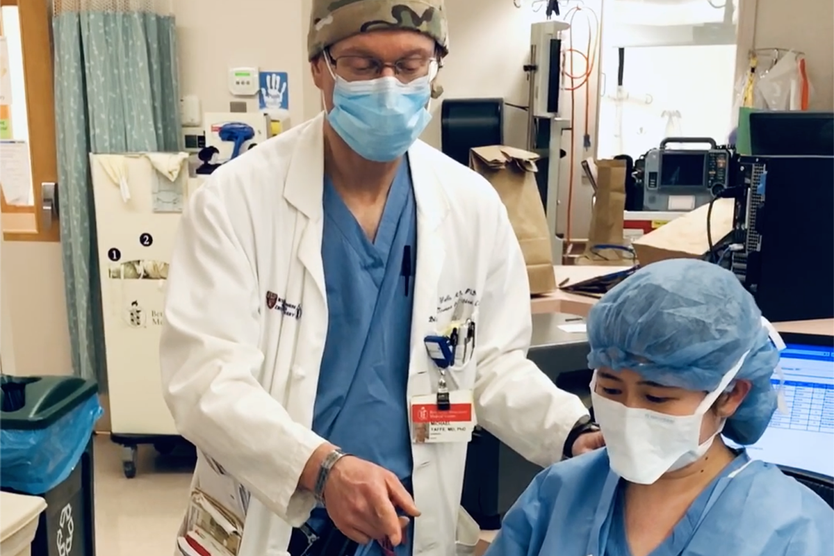
Michael Yaffe, the David H. Koch Professor in Science, normally divides his time among his roles as a researcher and professor of biology and biological engineering at MIT, an intensivist/trauma surgeon at Beth Israel Deaconess Medical Center (BIDMC), and a colonel in the U.S. Army Reserve Medical Corps. Currently, he is developing treatments for Covid-19 infections in his laboratory at the Koch Institute for Integrative Cancer Research at MIT. Additionally, he runs one of the Covid-19 Intensive Care Units at BIDMC and serves as co-director of the acute care and ICU section of Boston Hope, the 500-bed pop-up hospital organized by the City of Boston, Massachusetts in the Boston Convention and Exposition Center. Yaffe shares how he is working to improve outcomes for Covid-19 patients and offers his perspective on how emergency care for acute respiratory distress will need to evolve during this crisis and beyond.
Q: What are the special considerations for Covid-19 patients receiving treatment for respiratory failure?
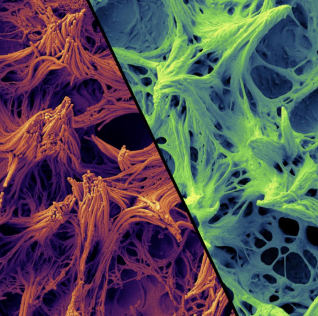
A: We have known about acute respiratory distress syndrome (ARDS) for decades. It was first recognized in battlefield casualties during the Vietnam War, and was initially called “Da-Nang Lung,” but later was understood to be the result of many different diseases. In ARDS, fluid builds up in the tiny air sacs, or alveoli, preventing the lungs from filling up with enough air, and in severe cases is treated by putting patients on ventilators or other devices that support breathing.
The type of lung injury we are seeing in Covid-19 patients behaves very differently from the traditional type of ARDS, and seems to involve early damage to the cells that line the lungs, followed by intense inflammation. The inflammation leads to a massive increase in blood clotting that affects all of the blood vessels in the body, but particularly the blood vessels in the lungs. As a consequence, even if we can force air into the lungs, it does not get delivered very efficiently into the bloodstream.
In ICUs in Boston, New York, and Colorado, we have started a clinical trial using a clot-busting drug called tPA that we think will help rescue patients whose lungs are failing despite maximal support with a mechanical ventilator. This approach has gathered a lot of attention from other hospitals, both nationally and internationally, who are also trying this approach. The work has now led to FDA approval for this drug as an Investigational New Drug, meaning that it is now approved for use in Covid-19 ARDS in the setting of clinical trials.
Q: How has your wide-ranging expertise equipped you to address new challenges that you face in the ICU?
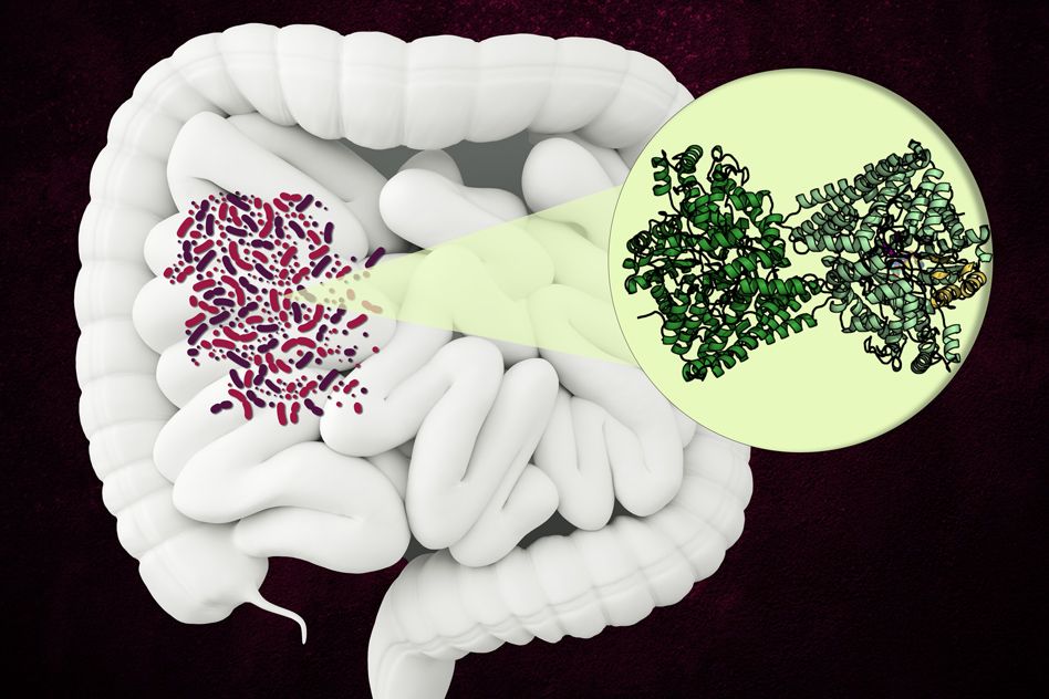
A: I have been very fortunate to be well-prepared to help out in this crisis. First, my training as an intensive care physician and trauma surgeon makes me comfortable in a crisis situation. The clinical problems that we are dealing with here — ARDS, kidney failure, etc. — are exactly within the scope of my regular clinical practice. Second, my Army deployment experience as a surgeon and critical care doctor in Afghanistan and in Central America has made me very comfortable having to make decisions in resource-limited situations. Finally, it has been incredibly fortuitous that much of my lab’s work has been in the area of cell injury, particularly cancer treatment-related cell injury, but also in the setting of a condition called systemic inflammatory response syndrome, which is essentially exactly what Covid-19 is. In this area, my lab has been studying the link between inflammation and blood clotting for over a decade, and the basic science insights from that work have now become central to our understanding of Covid-19 lung failure, which no one could have foreseen when we first started that research.
Q: What implications do you think the Covid-19 pandemic will have for emergency care after it is over?
A: I think the implications of Covid-19 for the future are immense. First, I hope the lessons learned from this pandemic lead to a complete re-thinking of our national public health policy (or lack of one, really) and a re-engagement with World Health Organization officials for monitoring the outbreak of emerging diseases.
Second, I think that this crisis may fuel additional research funding in the area of critical care medicine. Before the Covid-19 crisis, very few people had heard of ARDS, or even critical care as a field of medicine, since it does not have the glamour of conditions like cancer medicine or cardiovascular disease. Historically, research in this area has been underfunded, but now that ARDS has taken the spotlight in the news, I am hopeful that the recognition that some patients with Covid-19 are dying because of critical illness and lung failure will lead to new efforts to better understand the link between inflammation, lung function, and innate immunity, including blood coagulation. The Covid-19 crisis will not end when this first wave subsides, but will re-visit us again in the fall. Additionally, other coronavirus diseases as well as viral epidemics are likely to continue to plague us in the future.
One final lesson we are learning from this terrible pandemic is how important it is to treat all of the different parts of the body as a complex interacting unit, and to apply what we know from systems biology and other fields of study to understand how those parts are integrated into one coherent system. The lung failure, kidney failure, and inflammation of the heart that are the hallmarks of Covid-19 critical illness directly reflect how different inflammatory molecules in the blood alter the function of each of these different organ systems. Our traditional medical approach of having separate specialists in infectious disease, pulmonary medicine, renal medicine, and hematology does not work well when all the organ systems are cross-talking to each other. The job of the intensive care physician is to integrate all of the relevant basic biology and pathology of these organs into a comprehensive holistic treatment approach for the patient. Covid-19 has made that need to think across multiple disciplines and connect basic science to clinical care even more apparent.