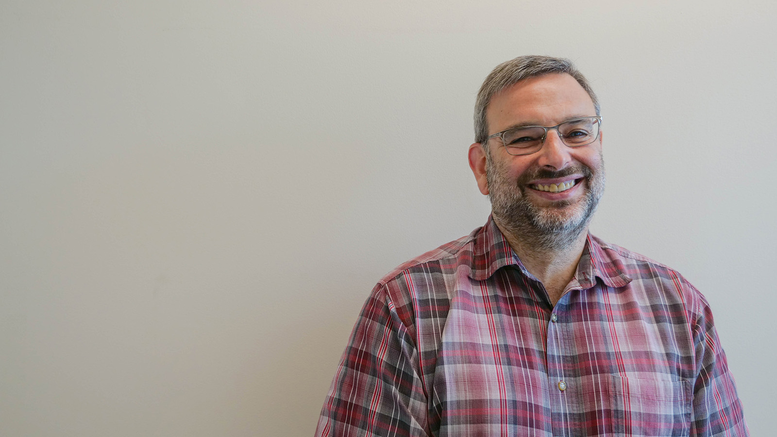
Matt Shoulders will lead an interdisciplinary team to improve RuBisCO — the photosynthesis enzyme thought to be the holy grail for improving agricultural yield.
Carolyn Blais | Abdul Latif Jameel Water and Food Systems Lab
May 10, 2023
According to MIT’s charter, established in 1861, part of the Institute’s mission is to advance the “development and practical application of science in connection with arts, agriculture, manufactures, and commerce.” Today, the Abdul Latif Jameel Water and Food Systems Lab (J-WAFS) is one of the driving forces behind water and food-related research on campus, much of which relates to agriculture. In 2022, J-WAFS established the Water and Food Grand Challenge Grant to inspire MIT researchers to work toward a water-secure and food-secure future for our changing planet. Not unlike MIT’s Climate Grand Challenges, the J-WAFS Grand Challenge seeks to leverage multiple areas of expertise, programs, and Institute resources. The initial call for statements of interests returned 23 letters from MIT researchers spanning 18 departments, labs, and centers. J-WAFS hosted workshops for the proposers to present and discuss their initial ideas. These were winnowed down to a smaller set of invited concept papers, followed by the final proposal stage.
Today, J-WAFS is delighted to report that the inaugural J-WAFS Grand Challenge Grant has been awarded to a team of researchers led by Professor Matt Shoulders and research scientist Robert Wilson of the Department of Chemistry. A panel of expert, external reviewers highly endorsed their proposal, which tackles a longstanding problem in crop biology — how to make photosynthesis more efficient. The team will receive $1.5 million over three years to facilitate a multistage research project that combines cutting-edge innovations in synthetic and computational biology. If successful, this project could create major benefits for agriculture and food systems worldwide.
“Food systems are a major source of global greenhouse gas emissions, and they are also increasingly vulnerable to the impacts of climate change. That’s why when we talk about climate change, we have to talk about food systems, and vice versa,” says Maria T. Zuber, MIT’s vice president for research. “J-WAFS is central to MIT’s efforts to address the interlocking challenges of climate, water, and food. This new grant program aims to catalyze innovative projects that will have real and meaningful impacts on water and food. I congratulate Professor Shoulders and the rest of the research team on being the inaugural recipients of this grant.”
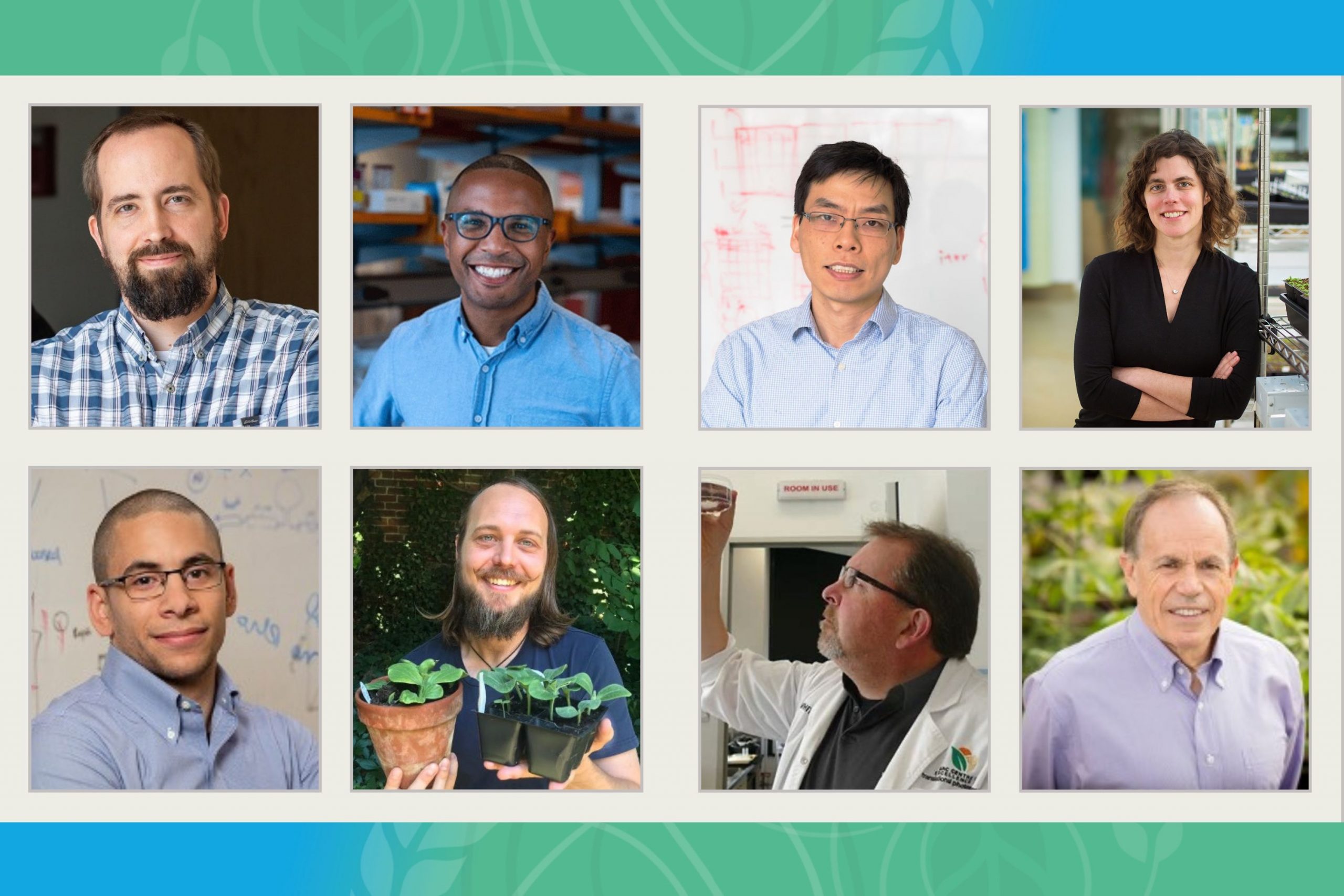
Shoulders will work with Bryan Bryson, associate professor of biological engineering, as well as Bin Zhang, associate professor of chemistry, and Mary Gehring, a professor in the Department of Biology and the Whitehead Institute for Biomedical Research. Robert Wilson from the Shoulders lab will be coordinating the research effort. The team at MIT will work with outside collaborators Spencer Whitney, a professor from the Australian National University, and Ahmed Badran, an assistant professor at the Scripps Research Institute. A milestone-based collaboration will also take place with Stephen Long, a professor from the University of Illinois at Urbana-Champaign. The group consists of experts in continuous directed evolution, machine learning, molecular dynamics simulations, translational plant biochemistry, and field trials.
“This project seeks to fundamentally improve the RuBisCO enzyme that plants use to convert carbon dioxide into the energy-rich molecules that constitute our food,” says J-WAFS Director John H. Lienhard V. “This difficult problem is a true grand challenge, calling for extensive resources. With J-WAFS’ support, this long-sought goal may finally be achieved through MIT’s leading-edge research,” he adds.
RuBisCO: No, it’s not a new breakfast cereal; it just might be the key to an agricultural revolution
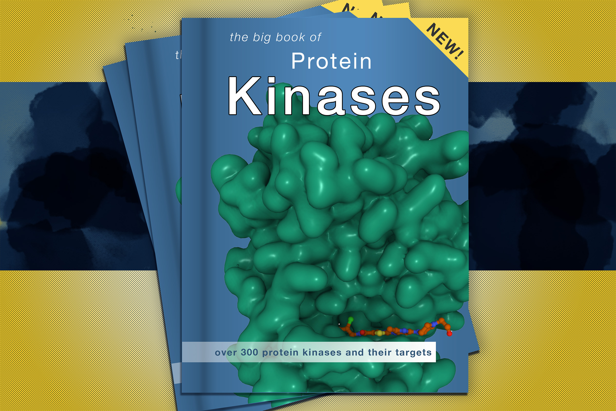
A growing global population, the effects of climate change, and social and political conflicts like the war in Ukraine are all threatening food supplies, particularly grain crops. Current projections estimate that crop production must increase by at least 50 percent over the next 30 years to meet food demands. One key barrier to increased crop yields is a photosynthetic enzyme called Ribulose-1,5-Bisphosphate Carboxylase/Oxygenase (RuBisCO). During photosynthesis, crops use energy gathered from light to draw carbon dioxide (CO2) from the atmosphere and transform it into sugars and cellulose for growth, a process known as carbon fixation. RuBisCO is essential for capturing the CO2 from the air to initiate conversion of CO2 into energy-rich molecules like glucose. This reaction occurs during the second stage of photosynthesis, also known as the Calvin cycle. Without RuBisCO, the chemical reactions that account for virtually all carbon acquisition in life could not occur.
Unfortunately, RuBisCO has biochemical shortcomings. Notably, the enzyme acts slowly. Many other enzymes can process a thousand molecules per second, but RuBisCO in chloroplasts fixes less than six carbon dioxide molecules per second, often limiting the rate of plant photosynthesis. Another problem is that oxygen (O2) molecules and carbon dioxide molecules are relatively similar in shape and chemical properties, and RuBisCO is unable to fully discriminate between the two. The inadvertent fixation of oxygen by RuBisCO leads to energy and carbon loss. What’s more, at higher temperatures RuBisCO reacts even more frequently with oxygen, which will contribute to decreased photosynthetic efficiency in many staple crops as our climate warms.
The scientific consensus is that genetic engineering and synthetic biology approaches could revolutionize photosynthesis and offer protection against crop losses. To date, crop RuBisCO engineering has been impaired by technological obstacles that have limited any success in significantly enhancing crop production. Excitingly, genetic engineering and synthetic biology tools are now at a point where they can be applied and tested with the aim of creating crops with new or improved biological pathways for producing more food for the growing population.
An epic plan for fighting food insecurity
The 2023 J-WAFS Grand Challenge project will use state-of-the-art, transformative protein engineering techniques drawn from biomedicine to improve the biochemistry of photosynthesis, specifically focusing on RuBisCO. Shoulders and his team are planning to build what they call the Enhanced Photosynthesis in Crops (EPiC) platform. The project will evolve and design better crop RuBisCO in the laboratory, followed by validation of the improved enzymes in plants, ultimately resulting in the deployment of enhanced RuBisCO in field trials to evaluate the impact on crop yield.
Several recent developments make high-throughput engineering of crop RuBisCO possible. RuBisCO requires a complex chaperone network for proper assembly and function in plants. Chaperones are like helpers that guide proteins during their maturation process, shielding them from aggregation while coordinating their correct assembly. Wilson and his collaborators previously unlocked the ability to recombinantly produce plant RuBisCO outside of plant chloroplasts by reconstructing this chaperone network in Escherichia coli (E. coli). Whitney has now established that the RuBisCO enzymes from a range of agriculturally relevant crops, including potato, carrot, strawberry, and tobacco, can also be expressed using this technology. Whitney and Wilson have further developed a range of RuBisCO-dependent E. coli screens that can identify improved RuBisCO from complex gene libraries. Moreover, Shoulders and his lab have developed sophisticated in vivo mutagenesis technologies that enable efficient continuous directed evolution campaigns. Continuous directed evolution refers to a protein engineering process that can accelerate the steps of natural evolution simultaneously in an uninterrupted cycle in the lab, allowing for rapid testing of protein sequences. While Shoulders and Badran both have prior experience with cutting-edge directed evolution platforms, this will be the first time directed evolution is applied to RuBisCO from plants.
Artificial intelligence is changing the way enzyme engineering is undertaken by researchers. Principal investigators Zhang and Bryson will leverage modern computational methods to simulate the dynamics of RuBisCO structure and explore its evolutionary landscape. Specifically, Zhang will use molecular dynamics simulations to simulate and monitor the conformational dynamics of the atoms in a protein and its programmed environment over time. This approach will help the team evaluate the effect of mutations and new chemical functionalities on the properties of RuBisCO. Bryson will employ artificial intelligence and machine learning to search the RuBisCO activity landscape for optimal sequences. The computational and biological arms of the EPiC platform will work together to both validate and inform each other’s approaches to accelerate the overall engineering effort.
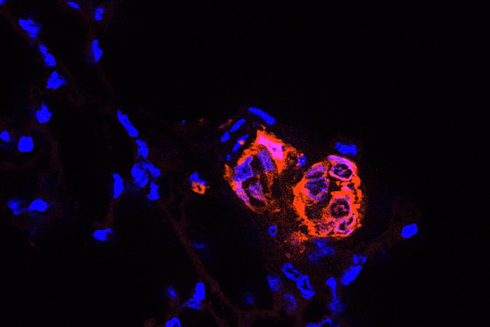
Shoulders and the group will deploy their designed enzymes in tobacco plants to evaluate their effects on growth and yield relative to natural RuBisCO. Gehring, a plant biologist, will assist with screening improved RuBisCO variants using the tobacco variety Nicotiana benthamianaI, where transient expression can be deployed. Transient expression is a speedy approach to test whether novel engineered RuBisCO variants can be correctly synthesized in leaf chloroplasts. Variants that pass this quality-control checkpoint at MIT will be passed to the Whitney Lab at the Australian National University for stable transformation into Nicotiana tabacum (tobacco), enabling robust measurements of photosynthetic improvement. In a final step, Professor Long at the University of Illinois at Urbana-Champaign will perform field trials of the most promising variants.
Even small improvements could have a big impact
A common criticism of efforts to improve RuBisCO is that natural evolution has not already identified a better enzyme, possibly implying that none will be found. Traditional views have speculated a catalytic trade-off between RuBisCO’s specificity factor for CO2 / O2 versus its CO2 fixation efficiency, leading to the belief that specificity factor improvements might be offset by even slower carbon fixation or vice versa. This trade-off has been suggested to explain why natural evolution has been slow to achieve a better RuBisCO. But Shoulders and the team are convinced that the EPiC platform can unlock significant overall improvements to plant RuBisCO. This view is supported by the fact that Wilson and Whitney have previously used directed evolution to improve CO2 fixation efficiency by 50 percent in RuBisCO from cyanobacteria (the ancient progenitors of plant chloroplasts) while simultaneously increasing the specificity factor.
The EPiC researchers anticipate that their initial variants could yield 20 percent increases in RuBisCO’s specificity factor without impairing other aspects of catalysis. More sophisticated variants could lift RuBisCO out of its evolutionary trap and display attributes not currently observed in nature. “If we achieve anywhere close to such an improvement and it translates to crops, the results could help transform agriculture,” Shoulders says. “If our accomplishments are more modest, it will still recruit massive new investments to this essential field.”
Successful engineering of RuBisCO would be a scientific feat of its own and ignite renewed enthusiasm for improving plant CO2 fixation. Combined with other advances in photosynthetic engineering, such as improved light usage, a new green revolution in agriculture could be achieved. Long-term impacts of the technology’s success will be measured in improvements to crop yield and grain availability, as well as resilience against yield losses under higher field temperatures. Moreover, improved land productivity together with policy initiatives would assist in reducing the environmental footprint of agriculture. With more “crop per drop,” reductions in water consumption from agriculture would be a major boost to sustainable farming practices.
“Our collaborative team of biochemists and synthetic biologists, computational biologists, and chemists is deeply integrated with plant biologists and field trial experts, yielding a robust feedback loop for enzyme engineering,” Shoulders adds. “Together, this team will be able to make a concerted effort using the most modern, state-of-the-art techniques to engineer crop RuBisCO with an eye to helping make meaningful gains in securing a stable crop supply, hopefully with accompanying improvements in both food and water security.”